

The Technique of Thyroid Cartilage Scaffold Support Formation for Extrusion-Based Bioprinting
- PMID: 33997436
- PMCID: PMC8114092
- DOI: 10.18063/ijb.v7i2.348
The Technique of Thyroid Cartilage Scaffold Support Formation for Extrusion-Based Bioprinting
Abstract
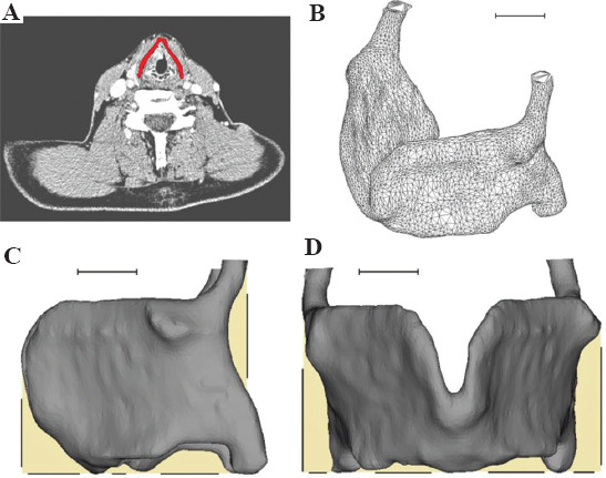
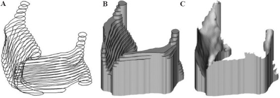
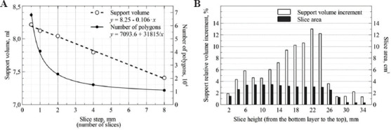
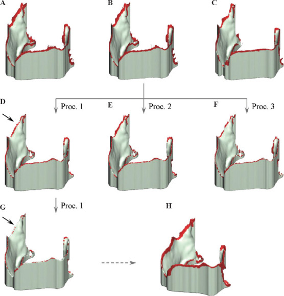
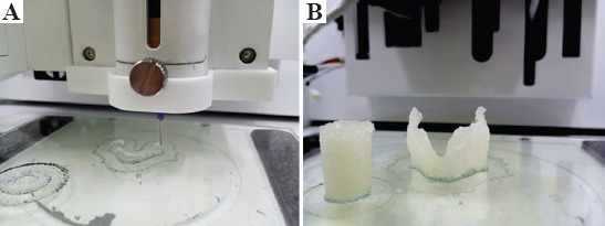
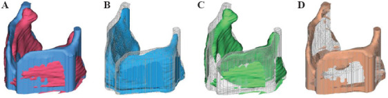
During biofabrication, a tissue scaffold may require temporary support. The aim of this study was to develop an approach of human thyroid cartilage scaffold temporal support formation. The scaffold 3D-model was based on DICOM images. XY plane projections were used to form scaffold supporting part. To verify the technique, collagen hydrogel was chosen as the main scaffold component. Gelatin was applied for the supporting part. To test the applicability of the approach, a model of thyroid cartilage scaffold with the support was printed. The scaffold corresponded to a given model, although some discrepancy in geometry was observed during verification by computed tomography.
Keywords: 3D-bioprinting; Cartilage; Collagen; Computer-aided design/Computer-aided manufacturing; Gelatin.
Copyright: © 2021 Arguchinskaya, et al.
Conflict of interest statement
No potential conflict of interest was reported by the authors.
Figures
References
-
- Ng WL, Chua CK, Shen YF. Print Me An Organ!Why We Are Not There Yet. Prog Polym Sci. 2019;97:101145. https://doi.org/10.1016/j.progpolymsci.2019.101145.
-
- Lee JM, Ng WL, Yeong WY. Resolution and Shape in Bio-Printing:Strategizing Towards Complex Tissue and Organ Printing. Appl Phys Rev. 2019;6:11307. https://doi.org/10.1063/1.5053909.
-
- Engler AJ, Sen S, Sweeney HL, et al. Matrix Elasticity Directs Stem Cell Lineage Specification. Cell. 2006;126:677–89. https://doi.org/10.1016/j.cell.2006.06.044. - PubMed
-
- Hadden WJ, Young JL, Holle AW, et al. Stem Cell Migration and Mechanotransduction on Linear Stiffness Gradient Hydrogels. Proc Natl Acad Sci. 2017;114:5647–52. https://doi.org/10.1073/pnas.1618239114. - PMC - PubMed
-
- Ke D, Yi H, Est-Witte S, et al. Bioprinted Trachea Constructs With Patient-Matched Design, Mechanical and Biological Properties. Biofabrication. 2019;12:15022. https://doi.org/10.1088/1758-5090/ab5354. - PubMed
LinkOut - more resources
Full Text Sources
Other Literature Sources