Case Reports
. 2021 Apr 21:22:101098.
doi: 10.1016/j.ajoc.2021.101098. eCollection 2021 Jun.

Delayed dark adaptation in central serous chorioretinopathy

Affiliations
Case Reports

Delayed dark adaptation in central serous chorioretinopathy

Archana Nigalye et al. Am J Ophthalmol Case Rep. .

Abstract

Purpose: To evaluate the effect of central serous chorioretinopathy (CSCR) on retinal function using dark adaptation in a human subject, and to follow it through resolution of the disease.

Patients: Single patient, 50 years old male patient, with acute CSCR in one eye and resolved old CSCR in the other eye.

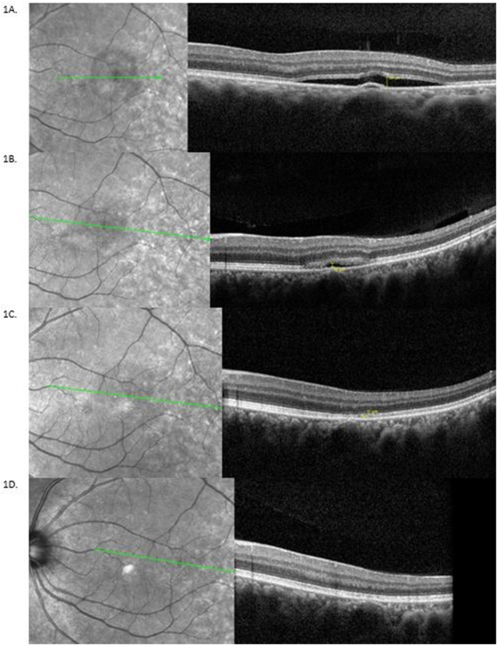
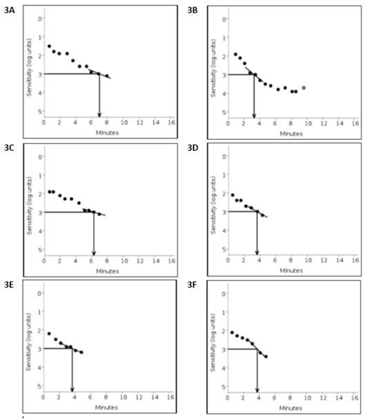
Observations: Observational study in patient with CSCR followed through resolution of the subretinal fluid (52 days). Dark adaptation was assessed using the AdaptDx® (Maculogix Inc.) measured by Rod Intercept time (RIT) in minutes. A normal retinal locus of the same eye on the opposite side of the fovea was used as control. Retinal separation (microns) was measured using Spectralis Optical Coherence Tomography (Spectralis®, HRA + OCT, Heidelberg engineering). Change in time to dark adapt, were correlated with retinal separation measured in microns, during the course of CSCR.The Rod Intercept time was delayed in the area of detached retina compared to the normal region (control) on presentation with retinal separation (RS) of 104 μm. The Rod Intercept time returned to normal as the retinal separation from retinal pigment epithelium decreased and eventually resolved.

Conclusions: This case shows that delay in dark adaptation is proportional to the amount of separation of neurosensory retina from retinal pigment epithelium in CSCR, this may offer a potential of using DA to characterize visual function in CSCR. The association of dark adaptation response with the state of retinal pigment epithelial function and its ability to predict the recurrence of CSCR needs further evaluation.

Keywords: Central serous chorioretinopathy; Dark adaptation.


Conflict of interest statement

The following authors have no financial disclosures.

Figures

Fig. 1
Fig. 1
Comparison of Infrared fundus image and SDOCT B-scan findings at the initial presentation and serial follow-ups for the left eye: A. Near infrared fundus image and spectral domain Optical Coherence Tomography (SDOCT) B-scan at initial presentation to the clinic. The left eye (OS) shows a well-circumscribed lesion just temporal to fovea corresponding to the serous detachment of the neurosensory retina, with RS measurement of 104μm. B. Infrared fundus images and comparative SDOCT B-scans of the macular area for the left eye at day 16. The macula shows gradually resolving central serous retinopathy (CSCR), with RS of 42μm. B-scans for subsequent follow-up visits were taken at a tilted angle and narrower degree view to ensure capturing the whole extent of CSCR to be able to measure any residual SRF. It was ensured that all the measurements are done on same horizontal raphe C. Infrared fundus images and comparative SDOCT B-scans at day 31 with RS of 11μm. D. The final visit shows a normal appearing macula. SDOCT B-scans of the left eye at day 52 shows resolved fluid and no measurable RS. Retinal separation measurement was performed on the same line scan at maximum fluid height on follow-up visits.
Fig. 2
Fig. 2
Dark adaptation trend for left eye CSCR and Control region and retinal separation height: DA Rod intercept time (RIT) and the separation of retina measurements for the CSCR region starting from initial presentation to day 52. Measurements were taken at 5° horizontal offset from fovea on either side for the left eye. The CSCR region temporal to the fovea, and the normal retina nasal to the fovea for control. In the Right eye RIT was measured on the day 16 in temporal macula 5° from fovea to demonstrate RIT for the area of long resolved CSCR (Represented in the graph by single solid blue block as OD Control). RIT measurements for the nasal field were down-trending, corresponding to resolution of SRF in the CSCR location. By day 52, the retina appears normal without fluid on OCT and the nasal region RIT is similar to the temporal region RIT. The temporal region RITs are consistent with the normal range of RIT described for Adapt Dx throughout the serial dark adaptation testing with minimal inter-test variation (average ± std. RIT: 3.7 ± 0.3 minutes). (For interpretation of the references to color in this figure legend, the reader is referred to the Web version of this article.)
Fig. 3
Fig. 3
Dark adaptation curves for left eye CSCR and Control region on Initial presentation, Day 8 and Day 52: 3A Initial presentation: dark adaptation RIT 7.02 minutes for the CSCR region, 3B RIT 3.27 minutes for the control region, 3C and 3D: Day 8 the RIT for the CSCR and the control regions 6.25 minutes and 3.63 minutes respectively. 3E Day 52, resolved CSCR confirmed by OCT with RIT of 3.66 minutes with, 3F control region RIT of 3.74 minutes.

Similar articles

Cited by

References

    1. Jackson G.R., Edwards J.G. A short-duration dark adaptation protocol for assessment of age-related maculopathy. Journal of Ocular Biology, Diseases, and Informatics. 2008;1:7–11. - PMC - PubMed
    1. Alpern M., Krantz D.H. Visual pigment kinetics in abnormalities of the uvea-retinal epithelium interface in man. Invest Ophthalmol Vis Sci. 1981;20:183–203. - PubMed
    1. Chuang E.L., Sharp D.M., Fitzke F.W., Kemp C.M., Holden A.L., Bird A.C. Retinal dysfunction in central serous retinopathy. Eye. 1987;1(Pt 1):120–125. - PubMed
    1. van Meel G.J., Smith V.C., Pokorny J., van Norren D. Foveal densitometry in central serous choroidopathy. Am J Ophthalmol. 1984;98:359–368. - PubMed
    1. Lamb T.D., Pugh E.N., Jr. Dark adaptation and the retinoid cycle of vision. Prog Retin Eye Res. 2004;23:307–380. - PubMed

Publication types

LinkOut - more resources