. 2021 Apr 18:10:100066.
doi: 10.1016/j.nbscr.2021.100066. eCollection 2021 May.

Relative light sensitivities of four retinal hemi-fields for suppressing the synthesis of melatonin at night

Affiliations

Relative light sensitivities of four retinal hemi-fields for suppressing the synthesis of melatonin at night

Mark S Rea et al. Neurobiol Sleep Circadian Rhythms. .

Abstract

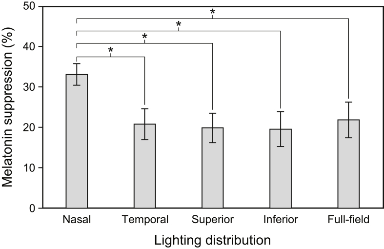
The magnitude of the stimulus to the biological clock will depend upon the distribution of circadian phototransduction circuits across the retinae and the spatial distribution of luminous stimuli in the environment. The present study compared nocturnal melatonin suppression for light exposures to the superior, inferior, nasal, and temporal retina in one eye independent of shading from the brow and the nose. The stimulus was a 40° diameter luminous disc, half of which was blue light (LED, λpeak = 470 nm) and the other amber light (LED, λpeak = 590 nm). Experimentally, the orientation of the bipartite disc was rotated to each of the four cardinal points of the visual field. A full, 40° blue disc was also employed by replacing the amber half-disc with another blue half-disc. The blue full- and half-discs always produced 100 photopic lx at the cornea. As hypothesized, nocturnal melatonin suppression was statistically greatest when the blue half-disc was delivered to the nasal hemi-field (35%); the other three hemi-fields were equally affected by the blue half-disc (≈20%). Melatonin suppression for the full-disc was 24%, which was not statistically different than the average suppression for the four hemi-fields of 27%.

Keywords: ANOVA, analysis of variance; Blue light; CLA, circadian light; CS, circadian stimulus; Circadian phototransduction; EML, equivalent melanopic lux; LED, light-emitting diode; Melatonin suppression; Monocular; Nasal retina; RGB, red, green, blue; α-opic, alpha-opic; λpeak, peak wavelength.


Conflict of interest statement

The author(s) have no potential conflicts of interest with respect to the research, authorship, and/or publication of this article.

Figures

Fig. 1
Fig. 1
The five experimental stimulus conditions and the dark control. All six experimental conditions were comprised of a 40° diameter circular field at the participant's eye. Half of each bipartite field was illuminated with a narrowband blue LED light source (λpeak = 470 nm) while the other half was illuminated with a phosphor converted amber LED light source (λpeak = 590 nm). Alone, the blue half-field delivered a CS = 0.60 at the cornea (100 lx), whereas the amber half-field delivered a CS < 0.01 (30 lx). Every bipartite field delivered a total photopic illuminance of 130 lx at the cornea. The full-field light condition was illuminated with the same blue LED, delivering the same CS = 0.60 (100 lx) at the cornea as each half-field. The temporal and nasal conditions were always set with respect to the participant's dominant eye. (For interpretation of the references to colour in this figure legend, the reader is referred to the Web version of this article.)
Fig. 2
Fig. 2
The apparatus used for all experimental conditions, including the dark control. Illustrated is one of the four bipartite fields with the septum (white bar) dividing the blue and amber half-fields and the RGB LED fixation light at its centre. The other three bipartite fields were created by rotating the bipartite field around its centre axis, represented by the fixation light. The blue full-field was created by replacing the amber half-field with a second blue half-field and the two blue half-fields were calibrated to provide a combined CS of 0.60. The lights illuminating both half-fields were turned off for the dark control. The height of the apparatus was adjusted for each participant with a variable-height stand to maintain a level gaze of the apparatus. (For interpretation of the references to colour in this figure legend, the reader is referred to the Web version of this article.)
Fig. 3
Fig. 3
Relative spectral power distributions for the blue and amber LED light sources. (For interpretation of the references to colour in this figure legend, the reader is referred to the Web version of this article.)
Fig. 4
Fig. 4
The experimental protocol for each of the six counterbalanced sessions. The grey bars correspond to the initial dim light exposure. The white bar designates the presentation of one of the five experimental lighting conditions or the dark control (black bar). S1–3 indicate when saliva samples were collected.
Fig. 5
Fig. 5
Main effect of lighting distribution on nocturnal melatonin suppression. The error bars represent SEM, * represents p < 0.05.

Similar articles

Cited by

References

    1. Baver S.B., Pickard G.E., Sollars P.J., Pickard G.E. Two types of melanopsin retinal ganglion cell differentially innervate the hypothalamic suprachiasmatic nucleus and the olivary pretectal nucleus. Eur. J. Neurosci. 2008;27(7):1763–1770. doi: 10.1111/j.1460-9568.2008.06149.x. - DOI - PubMed
    1. Blume C., Garbazza C., Spitschan M. Effects of light on human circadian rhythms, sleep and mood. Somnologie. 2019;23(3):147–156. doi: 10.1007/s11818-019-00215-x. - DOI - PMC - PubMed
    1. Bullough J.D., Brons J.A., Qi R., Rea M.S. Predicting discomfort glare from outdoor lighting installations. Light. Res. Technol. 2008;40(3):225–242. doi: 10.1177/1477153508094048. - DOI
    1. Burgess H.J., Fogg L.F. Individual differences in the amount and timing of salivary melatonin secretion. PloS One. 2008;3(8):e3055. doi: 10.1371/journal.pone.0003055. - DOI - PMC - PubMed
    1. Cogan D.G. A simplified entoptic pupillometer. Am. J. Ophthalmol. 1941;24(12):1431–1433. doi: 10.1016/S0002-9394(14)77456-2. - DOI

LinkOut - more resources