Review
. 2021 Apr 22;6(11):4110-4140.
doi: 10.1016/j.bioactmat.2021.03.043. eCollection 2021 Nov.

Bone physiological microenvironment and healing mechanism: Basis for future bone-tissue engineering scaffolds

Affiliations
Review

Bone physiological microenvironment and healing mechanism: Basis for future bone-tissue engineering scaffolds

Guanyin Zhu et al. Bioact Mater. .

Abstract

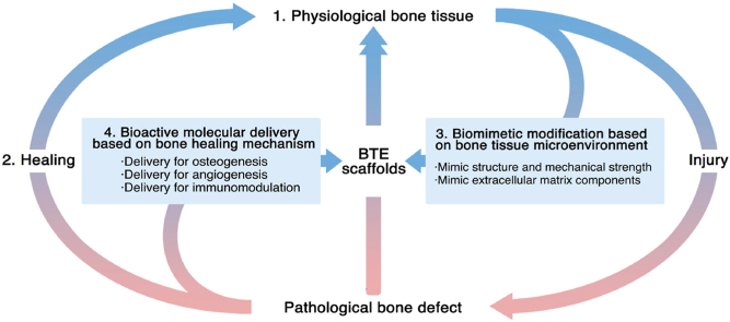
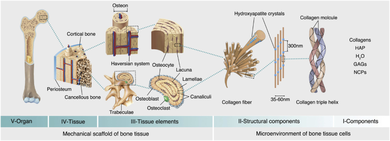
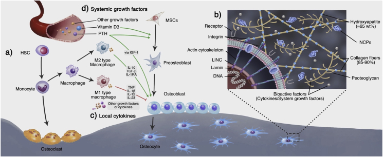
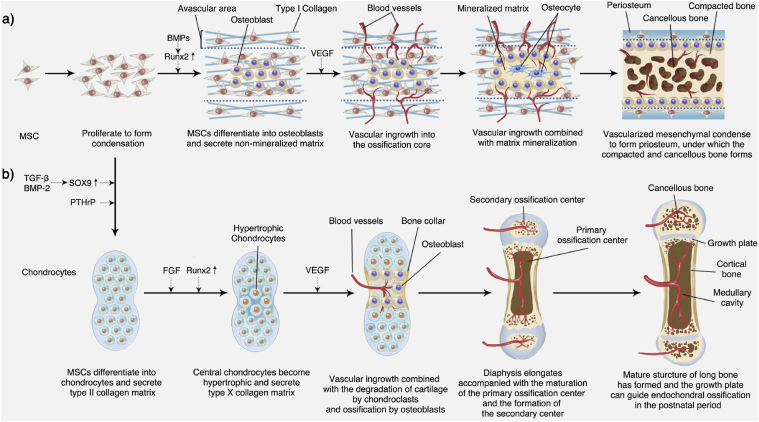
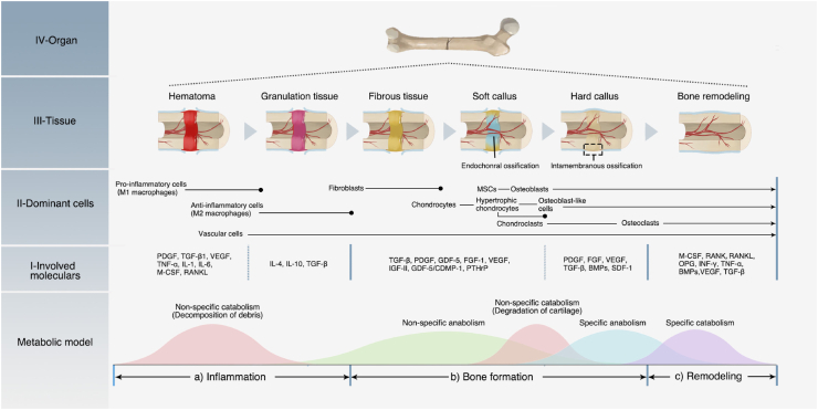
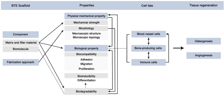
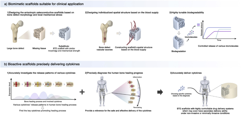
Bone-tissue defects affect millions of people worldwide. Despite being common treatment approaches, autologous and allogeneic bone grafting have not achieved the ideal therapeutic effect. This has prompted researchers to explore novel bone-regeneration methods. In recent decades, the development of bone tissue engineering (BTE) scaffolds has been leading the forefront of this field. As researchers have provided deep insights into bone physiology and the bone-healing mechanism, various biomimicking and bioinspired BTE scaffolds have been reported. Now it is necessary to review the progress of natural bone physiology and bone healing mechanism, which will provide more valuable enlightenments for researchers in this field. This work details the physiological microenvironment of the natural bone tissue, bone-healing process, and various biomolecules involved therein. Next, according to the bone physiological microenvironment and the delivery of bioactive factors based on the bone-healing mechanism, it elaborates the biomimetic design of a scaffold, highlighting the designing of BTE scaffolds according to bone biology and providing the rationale for designing next-generation BTE scaffolds that conform to natural bone healing and regeneration.

Keywords: Bone biology; Bone regeneration; Bone tissue engineering; Cytokine; Scaffold.


Conflict of interest statement

The authors declare that they have no known competing financial interests or personal relationships that could have appeared to influence the work reported in this paper.

Figures

Image 1
Graphical abstract
Fig. 1
Fig. 1
Bone hierarchical structure. The V, IV, and III levels construct the mechanical support structure for bone tissues, and the II and I levels construct the microenvironment for the bone tissue cells. These structures and components provide a biomimetic template for BTE scaffolds. HAP: hydroxyapatite; GAGs: glycosaminoglycans; NCPs: non-collagenous proteins. This Fig. is adapted from Refs. [[26], [27], [28], [29]].
Fig. 2
Fig. 2
Bone-tissue cells and extracellular matrix. a) In the bone marrow, HSCs and MSCs adjacent to the blood vessels can differentiate into osteoclasts and osteoblasts, which are directly involved in bone formation and resorption. b1) Osteoblasts can secrete the ECM and participate in its mineralization. They are gradually wrapped in new bone and differentiate into bone cells. In this process, they continuously receive biological signals from the components and biomolecules of the ECM. b2) The cells can also receive mechanical signals through sensors like integrins on the cell membrane. The integrin can adhere to the collagen of the ECM to form focal adhesions. The focal adhesions anchor the actin cytoskeleton, which links the LINC complex at the nuclear membrane. The LINC complexes interact with the lamins in the nuclear membrane, which finally transmits the mechanical signals to the nucleus and regulates the gene expression. c) In addition to osteoblasts and osteoclast cell lines that directly are involved in bone remodeling, the blood vessel growth brings endothelial cell lines. HSCs can also differentiate into the immune cell line. These cells communicate through cytokines and maintain the homeostasis of bone tissue. For example, as the most widely studied immune cells, macrophages can regulate osteoblast functions via inflammation-related cytokines. Generally, M1-type macrophages mainly inhibit osteogenesis, while M2-type macrophages mainly promote osteogenesis; d) Apart from the local regulation, bone homeostasis is also regulated by other organs through growth factors in the endocrine system.
Fig. 3
Fig. 3
Embryonic bone development. a) Intramembranous bone formation; b) Endochondral ossification.
Fig. 4
Fig. 4
Process and mechanism of bone healing. a) Inflammation phase; b) Bone formation phase; c) Remodeling phase. Bone healing is a dynamic and continuous process accompanied by an alternating metabolic model. In each phase, different cells and cytokines play the dominant roles.
Fig. 5
Fig. 5
Mimic bone's mechanical and morphological structure. a) Mimicking the mechanical strength of local bone tissues. Combining a CT-scan and a mechanical test of natural bone can help establish a relationship between imageological apparent density and bone modulus, which can be used to predict the local bone mechanical strength. After predicting the local bone's mechanical strength, a stiffness-matched scaffold with desirable bone conductivity can be constructed using AM. b) Underlying regulation mechanism of scaffolds' topology structures. The surface topology can cause cell membrane curvature. The curvature can activate curvature-sensing protein FBP17, which will further induce F-actin polymerization and actin reorganization in whole-cell. This cytoskeletal system change can activate the RhoA-ROCK, Akt/Erk, and YAP/TAZ effectors of the Hippo pathway, affecting the stem cells' differentiation. The ordered topology pattern and the stiff substrate can induce osteogenic differentiation, while actin depolymerization and the soft substrate can induce chondro/adipogenic differentiation. c) Mimicking the ECM structure by nanofiber scaffolds. Topology structure nanofibers can be constructed using electrospinning and can be used to develop porous scaffolds. Gao et al. modified nanofiber scaffolds with calcium phosphate (HA), to further mimic the bone ECM and release cytokines (BMP-7) and enhance the osteogenesis effects. This scaffold was proved to promote the in vitro osteogenic differentiation of hMSCs and enhance in vivo bone formation. This Fig. was adopted from Refs. [[223], [224], [225]].
Fig. 6
Fig. 6
Modify scaffolds with natural bone inorganic components. a) Chemical composition of HA in natural bone. M represents the cationic substitutions, while X and Y represent the anionic substitutions. These chemical groups are formed to meet the body's needs during growth and play essential roles in maintaining bone homeostasis and direct the bone cells' functions. b) Approaches for modifying crystallization on BTE scaffolds' surface. 1) Introduce nucleation sites on the surface followed by incubation in simulated body fluid; 2) Alternative exposure to Ca2+ and PO43− solutions; 3) Seeding osteogenic lineages on scaffold surface to secrete mineralized ECM followed by decellularization. This Fig. was adopted from Refs. [37,262].
Fig. 7
Fig. 7
Combine CaP with degradable polymer materials for bone tissue engineering. a) PEEK/β-TCP-PLLA scaffold via SLS. b) Degradation behaviors of the scaffolds with 0–50 wt% of PLLA content after PBS immersion, including Weight loss curve and SEM micrographs. c) Cell viability on 30 wt% PLLA scaffold after culture for different time intervals. d) Bone regeneration ability of the scaffolds. e) Histological analysis of new bone formation by H&E staining [272].
Fig. 8
Fig. 8
Construction of controlled release BTE scaffold. a) Construction scheme of an electroactive BTE scaffold with ability to control release and expression of hBMP-4 and its underlying mechanism. The addition of doxycycline could activate the expression of hBMP4, and the electrical stimulation could accelerate the release of hBMP4. b) SEM morphology of the scaffolds which maintained uniform porous structure during construction. c) Dox dose-dependent hBMP4 expression. (c1-c2) Detection of hBMP4 expression by RT-qPCR and ELISA methods. (c3-6) Dox regulation on expression of four osteogenesis-related genes. d) In vivo bone repair ability of the scaffolds [317].
Fig. 9
Fig. 9
Scaffolds for angiogenesis a) The category of angiogenesis scaffolds for BTE. 1) Scaffolds that can deliver osteoinductive and angioinductive molecules; 2) Scaffolds that deliver molecules with both osteoinductive and angioinductive abilities; 3) Scaffolds conducive for osteogenesis to deliver angioinductive molecules; 4) Scaffolds conductive for osteogenesis and angiogenesis without delivering inductive molecules. b) The approach for constructing scaffolds that support vascularization. The top-down approach represents the use of naturally derived polymer, which has inherent cell binding and degradation motif to construct scaffolds, while the bottom-up approach refers to the modification of synthetic polymer with growth factors, binding, and degrading protein sequences to form biodegradable angiogenesis scaffolds. c) Kolesky et al. used 3D bioprinting to construct a hydrogel matrix with a pre-designed vascular network. The procedure is as follows. 1) 3D-print a vascular network containing thermoreversible polymer pluronic and thrombin. Introduce cell components via printing cell-laden inks that contain gelatin, fibrinogen, and cells. 2) Cover ECM material containing gelatin, fibrinogen, cells, thrombin, and transglutaminase. Thrombin can cause the polymerization of fibrinogen into fibrin outside the vascular network and inside the ECM. Besides, transglutaminase further diffuses to crosslink gelatin and fibrin; 3) After casting, lower the temperature to liquefy pluronic and evacuate the vascular network. 4) Perfuse vascular ECs into the network to achieve vascularization. This Fig. was adopted from Refs. [252,320,321].
Fig. 10
Fig. 10
Mechanisms coupling angiogenesis and osteogenesis. a) Type-H vessels are mainly located at the metaphysis and endosteum, which are rich in osteoprogenitors and MSCs. Several factors have been proved to proliferate type-H ECs, including PDGF-BB secreted by preosteoclasts and TRAP+ macrophages (blue arrows), SLIT3 secreted by osteoblasts and osteoclasts (green arrows), and VEGF released by osteoblasts, osteoclasts and chondrocytes (pink arrows). The formation of type-H ECs will further facilitate osteogenesis by bone-producing cells (dotted arrow). b) The influence of type-H ECs on osteogenesis cells. Blood fluid stress can activate notch/Dll4 signaling, which can further induce the release of Noggin. Noggin promotes the proliferation and differentiation of osteoprogenitors and the maturation of chondrocytes. In a hypoxic environment, HIF-1α is upregulated in ECs and osteoprogenitors, which can trigger the expression of VEGF and promote vessel growth. Besides, the activation of HIF-1α facilitates the self-renewal and proliferation of osteoprogenitors. Compared to type L, type-H ECs have a strong expression of multiple cytokines, including FGF1, TGFβ, and PDGF, which play essential roles in the osteoprogenitors' survival, proliferation, and differentiation.
Fig. 11
Fig. 11
Crosstalk among immune cells and its effect on osteogenesis. During the bone homeostasis and healing process, immune cells can regulate the balance between osteoblastogenesis and osteoclastogenesis. They communicate and cooperate via cytokines [294,355], which can be used as biomolecules in BTE immunomodulatory scaffolds.
Fig. 12
Fig. 12
Considerations for constructing BTE scaffolds. Biomimetic and biomolecule delivery designs are not independent. An integral design consideration is needed. The biomaterials of scaffolds largely affect the mechanical strength, the basic biological property, and the biodegradability of scaffolds, while the fabrication approaches commonly determine the morphology of scaffolds. The physical–mechanical property (both mechanical strength and morphology) further influence the biological properties. The biomolecules inside the scaffolds are delivered via scaffold degradation, which could greatly affect the cell fate, particularly differentiation. This property of degradation is consistent with the biomolecules' delivery pattern, which could be affected by the scaffolds' matrix material and morphology. We should also beware that scaffold degradation must be accompanied by physical–mechanical and biological changes. The combined biological properties will influence the cell fate, including vascular cells, bone-producing cell lines, and immune cells. These cells will also interact with each other and determine the final tissue regeneration effects.
Fig. 13
Fig. 13
Outlook for next-generation BTE scaffold.

References

    1. Stevens M.M. Biomaterials for bone tissue engineering. Mater. Today. 2008;11(5):18–25.
    1. DiGirolamo D.J., Clemens T.L., Kousteni S. The skeleton as an endocrine organ. Nat. Rev. Rheumatol. 2012;8(11):674–683. - PubMed
    1. Loebel C., Burdick J.A. Engineering stem and stromal cell therapies for musculoskeletal tissue repair. Cell Stem Cell. 2018;22(3):325–339. - PMC - PubMed
    1. Defects B. 2011. Standard Guide for Pre-clinical in Vivo Evaluation in Critical Size Segmental Bone Defects.
    1. Hernlund E., Svedbom A., Ivergård M., Compston J., Cooper C., Stenmark J., McCloskey E.V., Jönsson B., Kanis J.A. Osteoporosis in the European union: medical management, epidemiology and economic burden. A report prepared in collaboration with the international osteoporosis foundation (IOF) and the European federation of pharmaceutical industry associations (EFPIA) Arch Osteoporos. 2013;8(1–2):136. - PMC - PubMed