. 2021 May 1;6(12):4402-4414.
doi: 10.1016/j.bioactmat.2021.04.027. eCollection 2021 Dec.

Carrier-free highly drug-loaded biomimetic nanosuspensions encapsulated by cancer cell membrane based on homology and active targeting for the treatment of glioma

Affiliations

Carrier-free highly drug-loaded biomimetic nanosuspensions encapsulated by cancer cell membrane based on homology and active targeting for the treatment of glioma

Yueyue Fan et al. Bioact Mater. .

Abstract

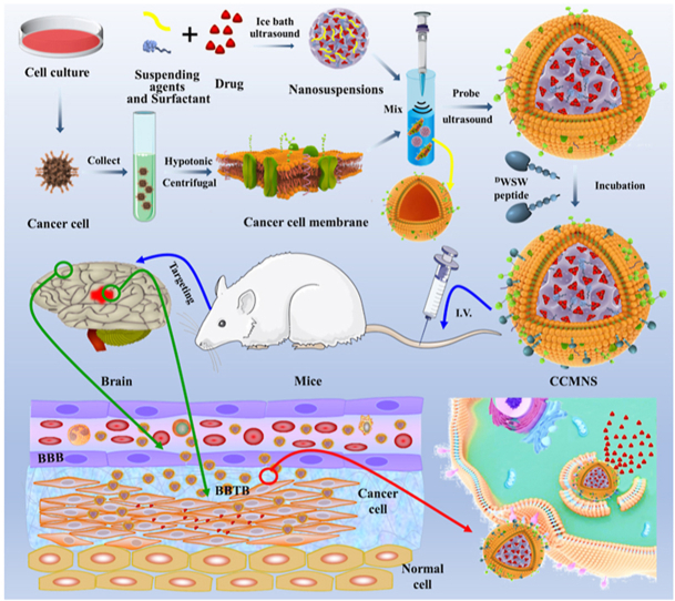
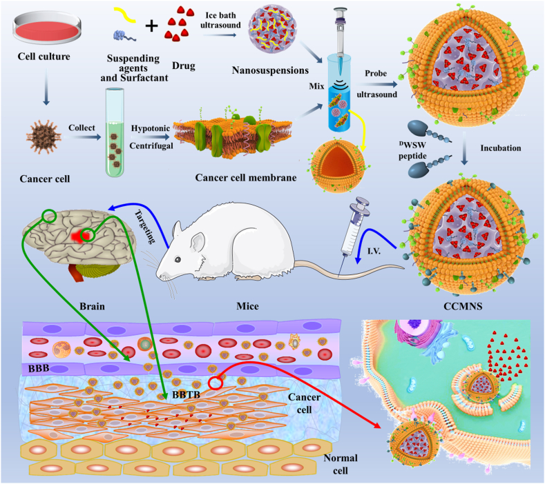
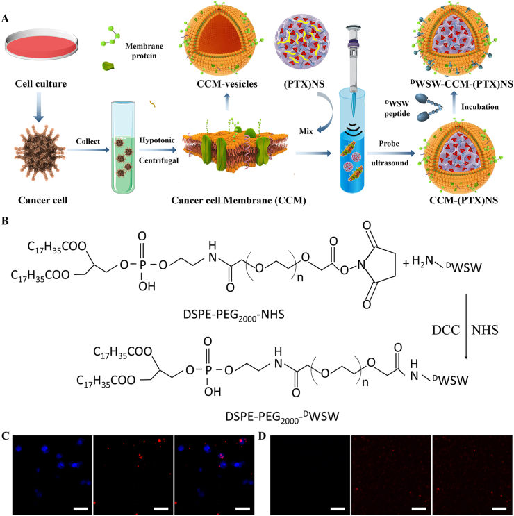
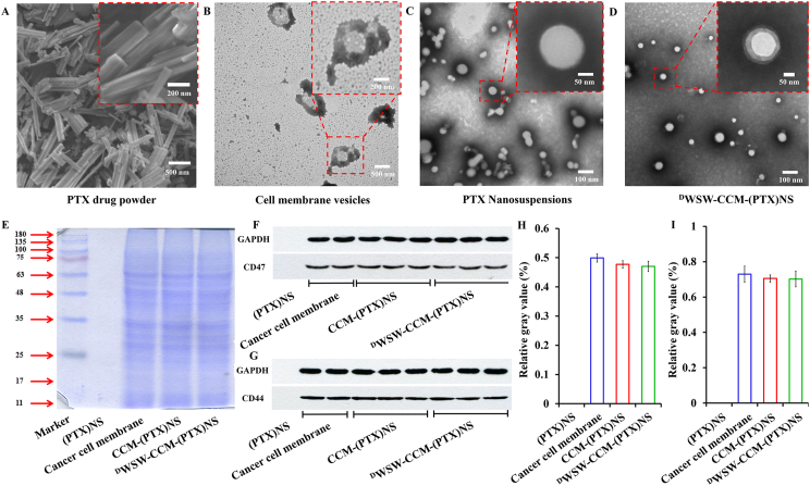
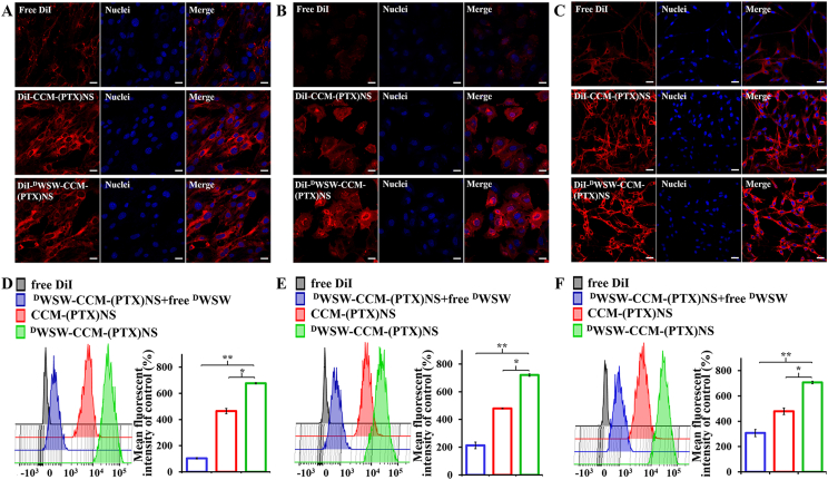
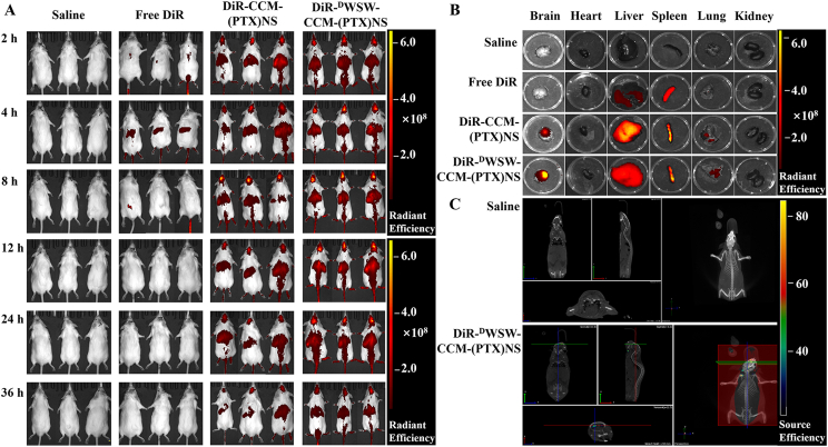
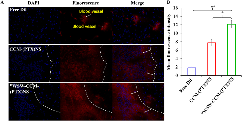
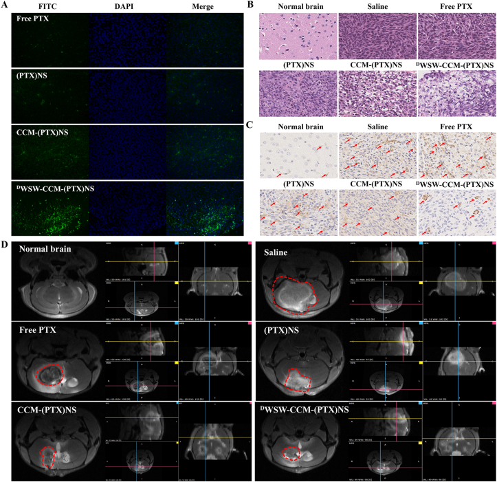
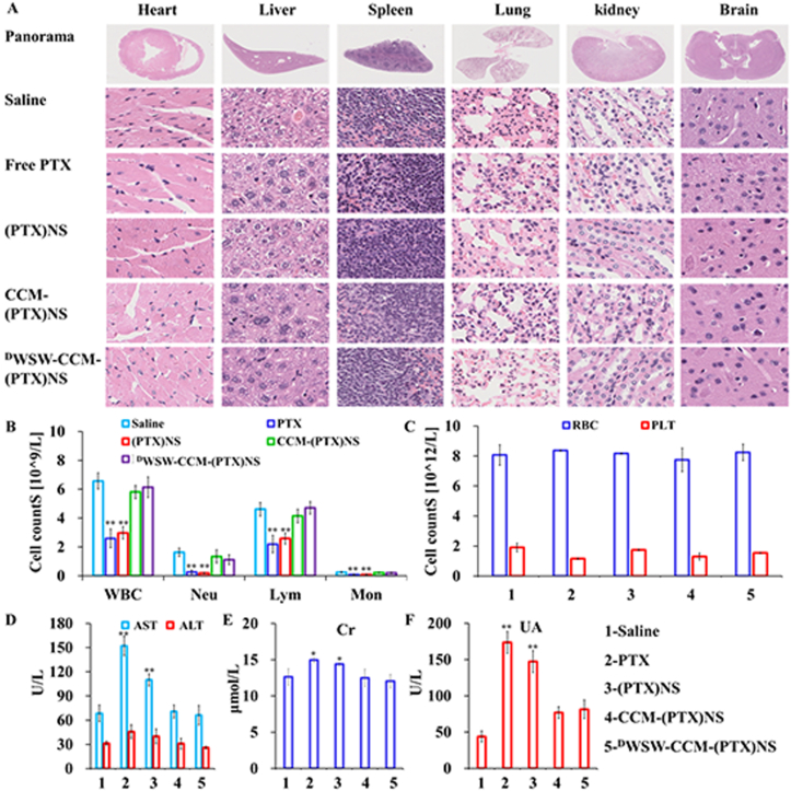
Nanosuspensions, as a new drug delivery system for insoluble drugs, are only composed of a drug and a small amount of stabilizer, which is dispersed in an aqueous solution with high drug-loading, small particle size, high dispersion, and large specific surface area. It can significantly improve the dissolution, bioavailability, and efficacy of insoluble drugs. In this study, paclitaxel nanosuspensions ((PTX)NS) were prepared by an ultrasonic precipitation method, with the characteristics of simple preparation and easy repetition. With the help of a homologous targeting mechanism, a kind of glioma C6 cancer cell membrane (CCM)-coated (PTX)NS was developed and modified with DWSW peptide to obtain DWSW-CCM-(PTX)NS with the functions of BBB penetration and tumor targeting. The results showed that the cancer cell membrane could effectively camouflage the nanosuspensions so that it was not cleared by the immune system and could cross the blood-brain-barrier (BBB) and selectively target tumor tissues. Cell uptake experiments and in vivo imaging confirmed that the uptake of DWSW-CCM-(PTX)NS by tumor cells and the distribution in intracranial gliomas increased. Cytotoxicity test and in vivo anti-glioma studies showed that DWSW-CCM-(PTX)NS could significantly inhibit the growth of glioma cells and significantly prolong the survival time of glioma-bearing mice. Finally, the cancer cell membrane coating endowed the nanosuspensions with the biological properties of homologous adhesion and immune escape. This study provides an integrated solution for improving the targeting of nanosuspensions and demonstrates the encouraging potential of biomimetic nanosuspensions applicable to tumor therapy.

Keywords: Biomimetic drug-delivery systems; Blood-brain barrier; Cancer cell membrane; Gliomas; Nanosuspensions; Paclitaxel.


Conflict of interest statement

We declare that we have no financial and personal relationships with other people or organizations that can inappropriately influence our work. There is no professional or other personal interest of any nature or kind in any product, service and/or company that could be construed as influencing the position presented in, or the review of the manuscript entitled “Carrier-free Highly Drug-loaded Biomimetic Nanosuspensions Encapsulated by Cancer Cell Membrane Based on Homology and Active Targeting for the Treatment of Glioma”.

Figures

Image 1
Graphical abstract
Fig. 1
Fig. 1
Graphic abstract of this study. Firstly, paclitaxel nanosuspensions were prepared by the ultrasonic-precipitation method. Secondly, the cancer cells were cultured and collected, and the cell membrane was separated from the plasma membrane of the source cells by hypotonic and differential centrifugation. Secondly, the cancer cells were cultured and collected, and the nuclei and membranes were separated by hypotonic and differential centrifugation. The obtained cancer cell membranes were combined with the nanosuspensions, and the mixture was fused by an ultrasonic probe to construct a biomimetic nanosuspension. The targeted ligand DWSW peptide was modified on the surface of the cell membranes by lipid insertion. Mice with in situ glioma were treated by intravenous injection, and the nanosuspensions penetrated the blood-brain barrier and the drug was delivered to the tumor to treat it.
Fig. 2
Fig. 2
Preparation and characterization of paclitaxel nanosuspension. (A) The suspending agent and the surfactant were dissolved in the water phase, the drug was dissolved in the oil phase, and the nanosuspension was prepared by the ultrasonic-precipitation method. (B) Nanosuspensions with different drug amounts where higher doses showed lower light transmission. (C) The particle size of the nanosuspensions with different drug amounts, showing the higher the dose, the larger the particle size. (D) X-ray diffraction patterns, (E) DSC scanning images, and (F) FTIR spectra of different components in the nanosuspensions.
Fig. 3
Fig. 3
(A) Preparation of PTX biomimetic nanosuspension. Cells were collected after culturing, and hypotonic treatment and centrifuging were used to obtain cell membranes. The membranes were combined with (PTX)NS using ultrasonic fusion to obtain biomimetic nanosuspensions and active targeting modification was carried out by the lipid insertion method. (B) Synthesis of targeting ligand DSPE-PEG2000-DWSW using the active ester method. In the presence of DCC and NHS, the –NH2 in the polypeptide reacted with the active ester in DSPE-PEG2000-NHS. (C) Cell nuclei obtained by low-speed centrifugation in the cell membrane preparation. (D) Cell membranes after purification. Hoechst 33258 (blue), DiI (red), (60x magnification).
Fig. 4
Fig. 4
Characterization of biomimetic nanosuspensions. (A) SEM of paclitaxel powder. The drug had a crystalline structure. Cell membrane vesicles (B), (PTX)NS(C) and DWSW-CCM-(PTX)NS(D) transmission electron microscopy images. The drug nanosuspension was approximately spherical and had a core-shell structure after being wrapped by the cancer cell membranes. (E) The membrane protein of the biomimetic nanosuspensions was determined by SDS-PAGE. WB measurement and relative gray value of membrane-specific proteins CD47 (F and H) and CD44 (G and I).
Fig. 5
Fig. 5
Cell uptake measured by CLSM and FCM. CLSM images and the corresponding flow cytometry analysis of bEnd.3 (A and D), HUVEC (B and E), and C6 (C and F) cells after incubation with DiI, DiI-CCM-(PTX)NS, or DiI-DWSW-CCM-(PTX)NS. The preparations showed different targeting capabilities. (20x magnification; red: DiI, blue: nuclei; *p < 0.05, **p < 0.01).
Fig. 6
Fig. 6
In vivo targeting evaluation. (A) In vivo real-time imaging of saline, DiR, CCM-(PTX)NS, and DWSW-CCM-(PTX)NS in glioma-bearing mice (n = 3). (B) Biodistribution of different DiR-encapsulated nanosuspensions in different organs. (C) 3D brain CT tumor localization scan. This indicated that the biomimetic nanosuspensions could effectively transport drugs to brain tissue.
Fig. 7
Fig. 7
(A) Distribution of brain tissue in vitro. DWSW-CCM-(PTX)NS had the highest fluorescence intensity and best targeting in tumor tissues. ImageJ was used for quantification (B). (DAPI: blue, DiI: red; 20x magnification; *p < 0.05, **p < 0.01).
Fig. 8
Fig. 8
In vivo anti-tumor effects in C6 glioma-bearing mice. (A) TUNEL staining of the tumors. (B) H&E staining of the tumor tissues. (C) Immunohistochemical staining of CD31 in the tumor tissues. (D) MRI of normal and glioma brains after treatment. The DWSW-CCM-(PTX)NS group showed the strongest anti-tumor effect. (DAPI: blue, FITC: green, CD31: brown; 40x magnification).
Fig. 9
Fig. 9
Preliminary safety evaluation. (A) Histological examination of the major organs derived from the mice after treatment. No major pathological changes were observed. Serum biochemical indicators of the mice after administration. (B) Immune system cells: WBC, Neu, Lym, and Mon counts. (C) Blood cells: RBC and PLT counts. (D) Liver and kidney function markers: AST, ALT, Cr, and UA. The data points represent the mean ± SD (n = 3). *p < 0.05, **p < 0.01; 40x magnification.

Similar articles

Cited by

References

    1. Wang N., Cheng X., Li N., Wang H., Chen H. Nanocarriers and their loading strategies. Adv Healthc Mater. 2019;8(6) e1801002. - PubMed
    1. Ahire E., Thakkar S., Darshanwad M., Misra M. Parenteral nanosuspensions: a brief review from solubility enhancement to more novel and specific applications. Acta Pharm. Sin. B. 2018;8(5):733–755. - PMC - PubMed
    1. Rabinow B.E. Nanosuspensions in drug delivery. Nat. Rev. Drug Discov. 2004;3(9):785–796. - PubMed
    1. Patel D., Zode S.S., Bansal A.K. Formulation aspects of intravenous nanosuspensions. Int. J. Pharm. 2020;586:119555. - PubMed
    1. Chai Z., Ran D., Lu L., Zhan C., Ruan H., Hu X., Xie C., Jiang K., Li J., Zhou J., Wang J., Zhang Y., Fang R.H., Zhang L., Lu W. Ligand-modified cell membrane enables the targeted delivery of drug nanocrystals to glioma. ACS Nano. 2019;13(5):5591–5601. - PubMed