

Endovascular and surgical approaches of iatrogenic vertebrovertebral arteriovenous fistula
- PMID: 33997554
- PMCID: PMC8093309
- DOI: 10.1016/j.jvscit.2020.12.019
Endovascular and surgical approaches of iatrogenic vertebrovertebral arteriovenous fistula
Abstract
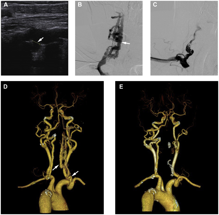
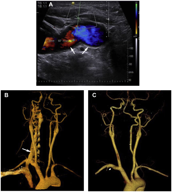
Vertebrovertebral arteriovenous fistula is an uncommon vascular disease with no clear management guidelines. It is most commonly caused by iatrogenic injury. We have presented the details of two iatrogenic cases and a review to discuss strategies for endovascular and surgical approaches. From the digital subtraction angiography findings, the vertebrovertebral arteriovenous fistulas were occluded by endovascular coil positioning (patient 1) and surgical ligation of the fistulas (patient 2). Although endovascular approaches are increasing in popularity and considered well-tolerated treatments, open surgical treatment is still reserved for the most complex cases and those not feasible for endovascular treatment.
Keywords: Endovascular surgery; Open surgery; Vertebrovertebral arteriovenous fistula.
© 2021 The Authors.
Figures
References
-
- Dodson T., Quindlen E., Crowell R., McEnany M.T. Vertebral arteriovenous fistulas following insertion of central monitoring catheters. Surgery. 1980;87:343–346. - PubMed
-
- Jamieson K.G. Vertebral arteriovenous fistula caused by angiography needle: report of a case. J Neurosurg. 1965;23:620–621. - PubMed
-
- Gao P., Chen Y., Zhang H., Zhang P., Ling F. Vertebral arteriovenous fistulae (AVF) in neurofibromatosis type 1: a report of two cases. Turk Neurosurg. 2013;23:289–293. - PubMed
Publication types
LinkOut - more resources
Full Text Sources
Other Literature Sources
