. 2021 Apr 21;2(2):100449.
doi: 10.1016/j.xpro.2021.100449. eCollection 2021 Jun 18.

Protocol for separation of the nuclear and the cytoplasmic fractions of Xenopus laevis embryonic cells for studying protein shuttling

Affiliations

Protocol for separation of the nuclear and the cytoplasmic fractions of Xenopus laevis embryonic cells for studying protein shuttling

Natalia Y Martynova et al. STAR Protoc. .

Abstract

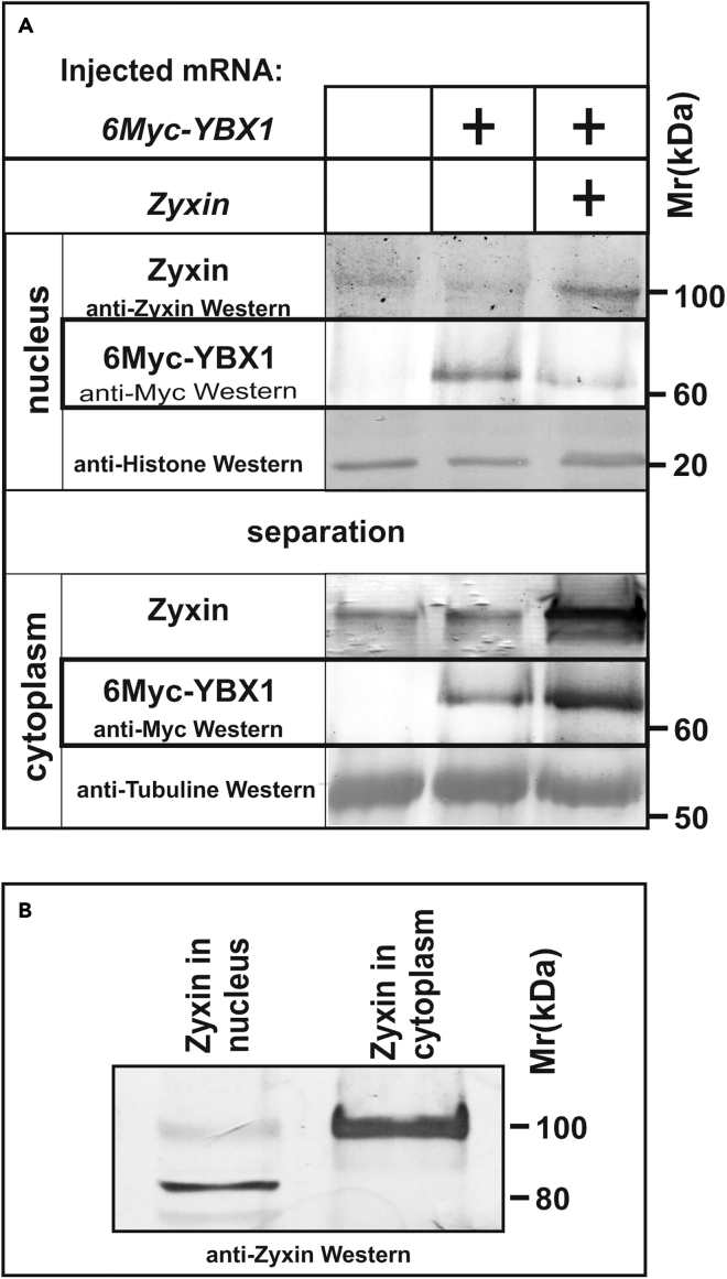
This protocol for the separation of nuclear and cytoplasmic fractions of cells of Xenopus laevis embryos was developed to study changes in the intracellular localization of the Zyxin and Ybx1 proteins, which are capable of changing localization in response to certain stimuli. Western blot analysis allows the quantification of changes in the distribution of these proteins between the cytoplasm and nucleus, whereas the posttranslational modifications specific to each compartment can be identified by changes in electrophoretic mobility. For complete details on the use and execution of this protocol, please refer to Parshina et al. (2020).

Keywords: Cell Biology; Cell separation/fractionation; Model Organisms; Molecular Biology; Protein Biochemistry.


Conflict of interest statement

The authors declare no competing interests.

Figures

None
Graphical abstract
Figure 1
Figure 1
The main steps of embryo preparation (A) Fertilized eggs before cleavage begins. (B) Embryos in Terasaki plates before manipulation. (C)Capillary preparation for microinjection. (D) Microinjection in embryos: general view of the workspace. (E) Two-cell stage embryo. (F) Embryo at the gastrula stage (stage 12), top (animal) view. (G) Embryo at the gastrula stage (stage 12), bottom (vegetal) view. (H) Alive (four on the left) and dead (two on the right, indicated by a red dashed line) embryos. Scale bars are 500 μm.
Figure 2
Figure 2
The main steps of extracts preparation (A) 60 embryos collected in Eppendorf tube to obtain crude extract (step 2) (B) Crude extract after centrifugation of embryos (step 3) (C) Crude extract, diluted 10 times with buffer N, before centrifugation (D) Separated fractions: left -cytoplasmic fraction, right - the pellet containing the cell nuclei (step 4) (E) Separated fractions before washing: on the left, the cytoplasmic fraction in buffer E (150 mM KCl), on the right, the nuclei in buffer with 0.8 M sucrose (step 5). (F) Fractions after extraction in buffer E and centrifugation: on the left is a cytoplasmic extract without any pellet, on the right is a nuclear extract containing a pellet of pigment and insoluble material.
Figure 3
Figure 3
Methanol/chloroform protein precipitation (A) The resulting nuclear (right) and cytoplasmic (left) extracts. (B) Methanol/chloroform precipitation. The protein layer appears on the interface between the methanol/water and chloroform phases (step 9, b). (C) The protein pellet is collected at the bottom of the tube (step 9,e).
Figure 4
Figure 4
Examples of using the protocol for separating the nuclear and cytoplasmic fractions to study changes in the localization and mobility of the cytoskeletal protein Zyxin and its effect on the localization of the transcription factor Ybx1 (A) While Zyxin reduces the nuclear concentration of 6Myc-Ybx1, it increases the cytoplasmic concentration of 6Myc-Ybx1. Xenopus laevis embryos at the 2–4 cell stage were injected with either 6 myc-ybx1 (200 pg per blastomere) and zyxin (300 pg per blastomere) or only 6 myc-ybx1 mRNA (200 pg per blastomere) and incubated at 18°C until the late gastrula stage. Nuclear and cytoplasmic fractions and precipitated samples were prepared as described in the protocol. The samples were analyzed by SDS-PAGE in a 10% gel according to the method of Laemmli and electroblotted onto PVDF membranes. Detection was performed with the following antibodies: monoclonal anti-Myc alkaline phosphatase conjugated antibodies (Sigma) for Myc-tagged protein Ybx1 and anti-Zyxin rabbit polyclonal monospecific antibodies for Zyxin (Figure reprinted with permission from Parshina et al. 2020). (B) Changes in the mobility of the band corresponding to Zyxin upon separation into nuclear and cytoplasmic fractions. An equal volume of 4× SDS Laemmli sample buffer was added to 50 μL of the obtained nuclear and cytoplasmic extracts, and then, the samples were boiled for 10 min at 80°C. The samples were analyzed by SDS-PAGE in 7.5% Laemmli gels and electroblotted onto a PVDF membrane. Detection was carried out using rabbit polyclonal monospecific antibodies against the C-terminal Lim domain of Zyxin.

References

    1. Lemaitre J.M., Bocquet S., Buckle R., Mechali M. Selective and rapid nuclear translocation of a c- Myc-containing complex after fertilization of Xenopus laevis eggs. Mol. Cell. Biol. 1995;15:5054–5062. - PMC - PubMed
    1. Martynova N.Y., Eroshkin F.M., Ermolina L.V., Ermakova G.V., Korotaeva A.L., Smurova K.M., Gyoeva F.K., Zaraisky A.G. The LIM-domain protein Zyxin binds the homeodomain factor Xanf1/Hesx1 and modulates its activity in the anterior neural plate of Xenopus laevis embryo. Dev. Dyn. 2008;237:736–749. - PubMed
    1. Martynova N.Y., Ermolina L.V., Ermakova G.V., Eroshkin F.M., Gyoeva F.K., Baturina N.S., Zaraisky A.G. The cytoskeletal protein Zyxin inhibits Shh signaling during the CNS patterning in Xenopus laevis through interaction with the transcription factor Gli1. Dev. Biol. 2013;380:37–48. - PubMed
    1. Nieuwkoop P.D., Faber J. Garland Science; 1994. Normal table of Xenopus laevis (Daudin): a systematical and chronological survey of the development from the fertilized egg till the end of metamorphosis. ISBN 9780815318965.
    1. Parshina E.A., Eroshkin F.M., Оrlov E.E., Gyoeva F.K., Shokhina A.G., Staroverov D.B., Belousov V.V., Zhigalova N.A., Prokhortchouk E.B., Zaraisky A.G., Martynova N.Y. Cytoskeletal protein Zyxin inhibits the activity of genes responsible for embryonic stem cell status. Cell Rep. 2020;33:108396. - PubMed

Publication types

LinkOut - more resources