

Decoupling of Global Brain Activity and Cerebrospinal Fluid Flow in Parkinson's Disease Cognitive Decline
- PMID: 33998068
- PMCID: PMC8453044
- DOI: 10.1002/mds.28643
Decoupling of Global Brain Activity and Cerebrospinal Fluid Flow in Parkinson's Disease Cognitive Decline
Abstract
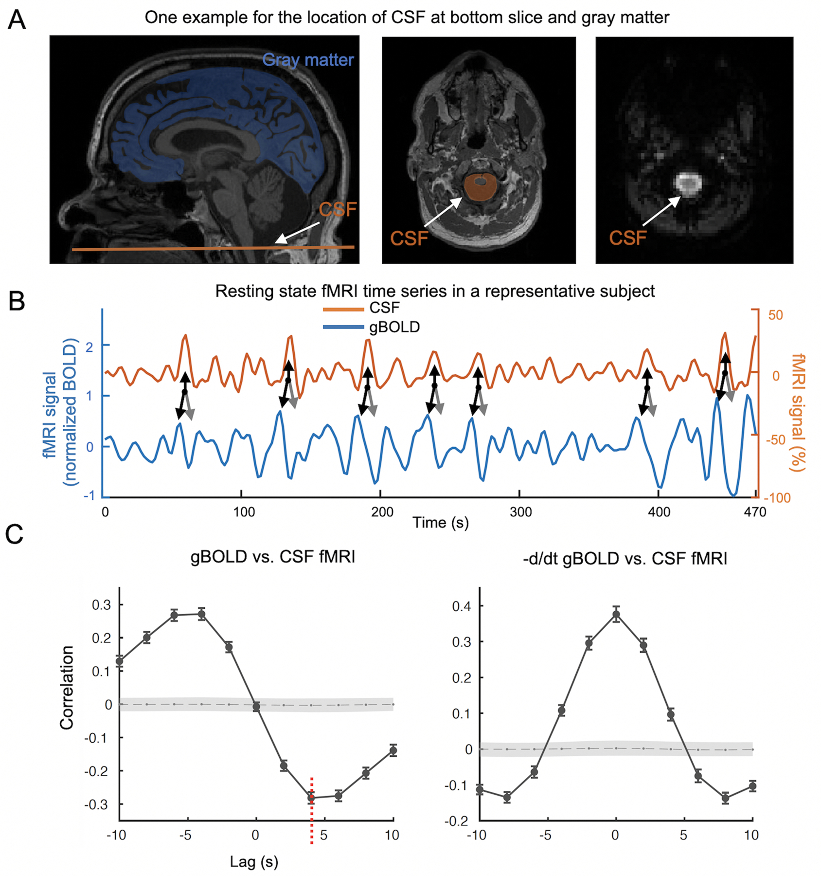
Background: Deposition and spreading of misfolded proteins (α-synuclein and tau) have been linked to Parkinson's disease cognitive dysfunction. The glymphatic system may play an important role in the clearance of these toxic proteins via cerebrospinal fluid (CSF) flow through perivascular and interstitial spaces. Recent studies discovered that sleep-dependent global brain activity is coupled to CSF flow, which may reflect glymphatic function.
Objective: The objective of this current study was to determine if the decoupling of brain activity-CSF flow is linked to Parkinson's disease cognitive dysfunction.
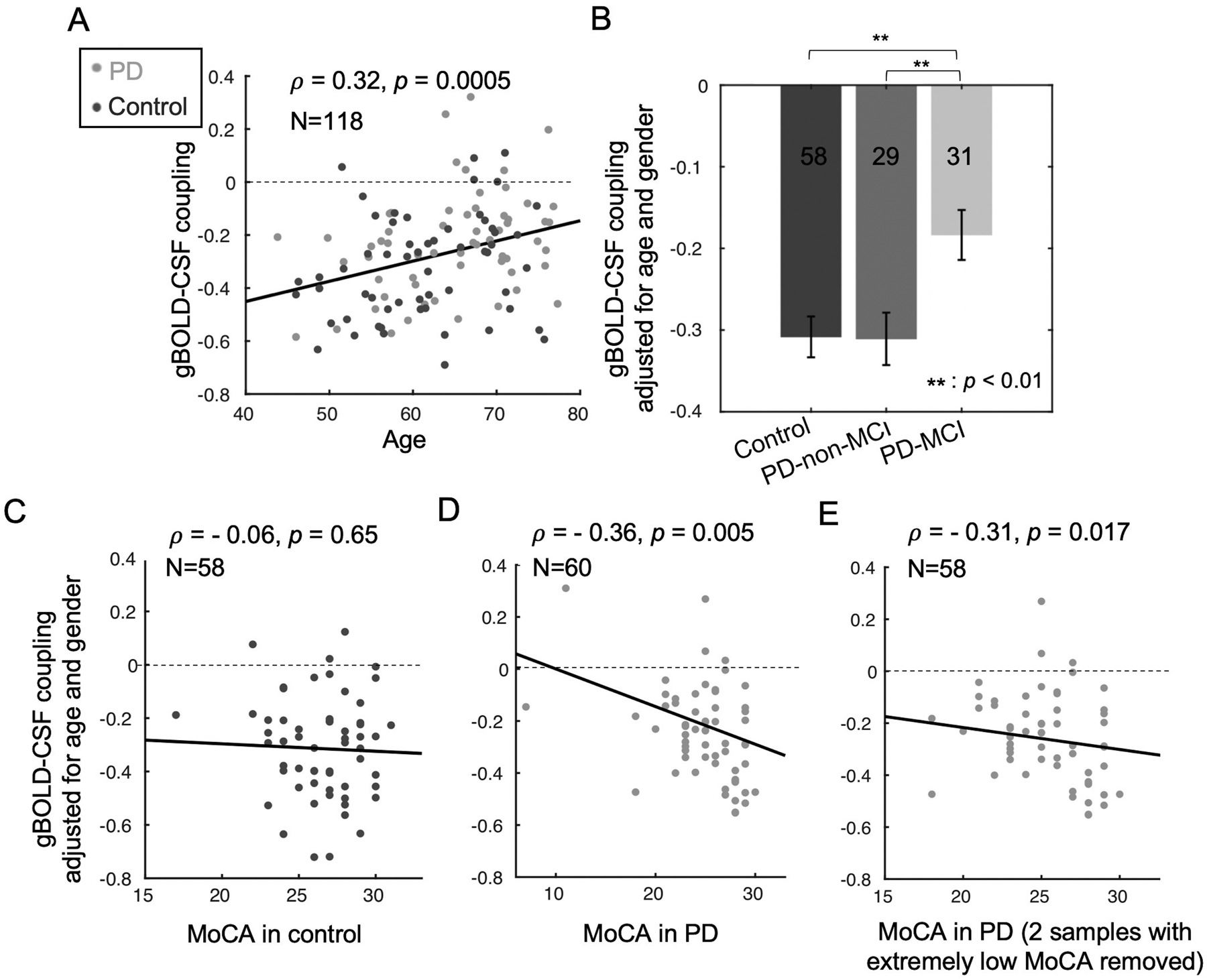
Methods: Functional and structural MRI data, clinical motor (Unified Parkinson's Disease Rating Scale), and cognitive (Montreal Cognitive Assessment [MoCA]) scores were collected from 60 Parkinson's disease and 58 control subjects. Parkinson's disease patients were subgrouped into those with mild cognitive impairment (MoCA < 26), n = 31, and those without mild cognitive impairment (MoCA ≥ 26), n = 29. The coupling strength between the resting-state global blood-oxygen-level-dependent signal and associated CSF flow was quantified, compared among groups, and associated with clinical and structural measurements.
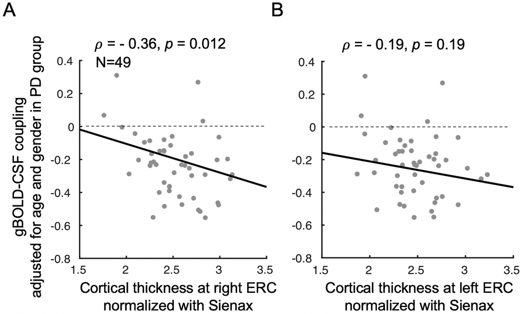
Results: Global blood-oxygen-level-dependent signal-CSF coupling decreased significantly (P < 0.006) in Parkinson's disease patients showing mild cognitive impairment, compared with those without mild cognitive impairment and controls. Reduced global blood-oxygen-level-dependent signal-CSF coupling was associated with decreased MoCA scores present in Parkinson's disease patients (P = 0.005) but not in controls (P = 0.65). Weaker global blood-oxygen-level-dependent signal-CSF coupling in Parkinson's disease patients also was associated with a thinner right entorhinal cortex (Spearman's correlation, -0.36; P = 0.012), an early structural change often seen in Alzheimer's disease.
Conclusions: The decoupling between global brain activity and associated CSF flow is related to Parkinson's disease cognitive impairment. © 2021 International Parkinson and Movement Disorder Society.
Keywords: Parkinson's disease; cerebrospinal fluid flow; cognitive impairment; global resting-state fMRI signal; glymphatic system.
© 2021 International Parkinson and Movement Disorder Society.
Figures
References
-
- Parkinson J. An essay on shaking palsy. 1817; 14: 223–236. - PubMed
-
- Aarsland D, Kurz MW. The epidemiology of dementia associated with Parkinson disease. J. Neurol. Sci 2010; 289: 18–22. - PubMed
-
- Mattila PM, Rinne JO, Helenius H, Dickson DW, Röyttä M. Alpha-synuclein-immunoreactive cortical Lewy bodies are associated with cognitive impairment in Parkinson’s disease. Acta Neuropathol. 2000; 100: 285–290. - PubMed
-
- Hurtig HI, Trojanowski JQ, Galvin J, et al. Alpha-synuclein cortical Lewy bodies correlate with dementia in Parkinson’s disease. Neurology. 2000; 54: 1916–1921. - PubMed
Publication types
MeSH terms
Substances
Grants and funding
LinkOut - more resources
Full Text Sources
Other Literature Sources
Medical
