. 2021;81(4):1749-1761.
doi: 10.3233/JAD-210095.

PINK1 Activation Attenuates Impaired Neuronal-Like Differentiation and Synaptogenesis and Mitochondrial Dysfunction in Alzheimer's Disease Trans-Mitochondrial Cybrid Cells

Affiliations

PINK1 Activation Attenuates Impaired Neuronal-Like Differentiation and Synaptogenesis and Mitochondrial Dysfunction in Alzheimer's Disease Trans-Mitochondrial Cybrid Cells

Fang Du et al. J Alzheimers Dis. 2021.

Abstract

Background: Mitochondrial dysfunction, bioenergetic deficit, and extensive oxidative stress underlie neuronal perturbation during the early stage of Alzheimer's disease (AD). Previously, we demonstrated that decreased PTEN-induced putative kinase 1 (PINK1) expression is associated with AD pathology in AD-affected human brains and AD mice.

Objective: In the present study, we highlight the essential role of PINK1 in AD-relevant mitochondrial perturbation and neuronal malfunction.

Methods: Using trans-mitochondrial "cybrid" (cytoplasmic hybrid) neuronal cells, whose mitochondria are transferred from platelets of patients with sporadic AD, we observed the effect of PINK1 in neuronal-like differentiation and synaptogenesis and mitochondrial functions.

Results: In AD cybrid cells, the downregulation of PINK1 is correlated to the alterations in mitochondrial morphology and function and deficit in neuronal-like differentiation. Restoring/increasing PINK1 by lentivirus transduction of PINK1 robustly attenuates mitochondrial defects and rescues neurite-like outgrowth. Importantly, defective PINK1 kinase activity fails to reverse these detrimental effects. Mechanistically, AD cybrid cells reveal a significant decrease in PINK1-dependent phosphorylated mitofusin (Mfn) 2, a key mitochondrial membrane protein that participates in mitochondrial fusion, and an insufficient autophagic activity for the clearance of dysfunctional mitochondria. Overexpression of PINK1, but not mutant PINK1 elevates phosphorylation of Mfn2 and autophagy signaling LC3-II. Accordingly, PINK1-overexpressed AD cybrids exhibit increases in mitochondrial length and density and suppressed reactive oxygen species. These results imply that activation of PINK1 protects against AD-affected mitochondrial dysfunction and impairment in neuronal maturation and differentiation.

Conclusion: PINK1-mediated mitophagy is important for maintaining mitochondrial health by clearance of dysfunctional mitochondria and therefore, improves energy homeostasis in AD.

Keywords: Alzheimer’s disease; PINK1; cybrid cells; mitochondrial dysfunction; mitophagy.


Figures

Fig. 1.
Fig. 1.
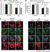
PINK1 expression in non-AD and AD cybrid cells during differentiation. A, B) Immunoblotting of protein extracts from non-AD and AD cybrid cells for PINK1 expression. β-actin was used for protein loading control. A) Quantification of immunoreactive bands for PINK1 relative to β-actin. B) Representative immunoblot for PINK1 and β-actin. (n = 5 per group). C) Representative fluorescent images of PINK1 (green), mitochondria (MitoRed, red), merge of PINK1 and mitochondria (yellow), and MAP2 (purple) from non-AD and AD cybrid cells under differentiated conditions induced by SAT (14 days after 10 nM staurosporine [SAT] treatment). Enlarged views of an individual non-AD or AD cybrid cell are shown in the right panels. Scale bars = 50 μm. D-F) Quantification of fluorescent intensity of PINK1 (D), MAP2 (E) and mitochondrial density (F, MitoRed staining) of cybrid cells using the image J program. G, H) Analysis of the relationship between PINK1 expression and MAP2 level (G) or mitochondrial content (H). (n = 9–11 cybrid cells of each group).
Fig. 2.
Fig. 2.
Effect of PINK1 on neuronal differentiation in AD cybrid cells. A) Representative morphological images from differentiated non-AD and AD cybrid cells with or without transducing PINK1 or mPINK1 lentivirus. Scale bar = 50 μm. B) Quantification of length of neuronal processes from indicated cybrid cells (12 neuronal processes for each group) using the image J program. C-F) Immunoblotting of protein extracts from non-AD and AD cybrid cells with or without PINK1/mPINK1 lentivirus transduction for MAP2 and synaptophysin (Syn, C and D) and PINK1 (E and F) expressions. β-actin was used for protein loading control. Quantification of immunoreactive bands for the corresponding protein relative to β-actin in C and E. Representative immunoblots for the indicated proteins in D and F. n = 3 per group.
Fig. 3.
Fig. 3.
Effect of PINK1 on AD-related mitochondrial dysfunction. A, B) Enzymatic activity of cytochrome c oxidase (CcO, A) and cellular ATP levels (B) were determined in cell lysates from non-AD and AD cybrid cells with or without PINK1 or mPINK1 lentivirus transfection. C, D) Generation of ROS in the indicated groups of cells was detected by electron paramagnetic resonance (EPR) spectra. Quantifications of the peak of EPR (C) of the indicated cybrid cells and Representative EPR images (D). n = 5 per group.
Fig. 4.
Fig. 4.
Effect of PINK1 on mitochondrial morphology in AD cybrid cells. A, B) Quantifications of mitochondrial length (A) and density (B). Mitochondria are visible by MitoRed staining from differentiated non-AD and AD cybrid cells with or without PINK1 or mPINK1 lentivirus transfection. For the analysis of mitochondrial length, 25 mitochondria in the growth cone of axon-like neurites from 5 cybrids per group were analyzed. 25 cybrid cells of each group were analyzed for mitochondrial density. C) Representative fluoresce images of mitochondria (MitoRed), GFP (green) and nucleus (blue) from non-AD and AD cybrid cells. Scale bars = 10 μm.
Fig. 5.
Fig. 5.
PINK1 phosphorylates Mitofusin 2 (Mfn2) at Ser442 and activates autophagy signaling in AD cybrid cells. A, B) Immunoblotting of protein extracts from non-AD and AD cybrid cells with or without PINK1/mPINK1 lentivirus transfection for detecting phosphorylated-Mfn2 (p-Mfn2, Ser442, A and B), phosphorylated Drp1 (p-Drp1, Ser616, A and C), and LC3II (D and E). β-actin was used for protein loading control. Quantifications of immunoreactive bands for the corresponding protein relative to β-actin in B, C and E. **p < 0.01 versus non-AD transduced by lentivirus-GFP. n = 3 per group.

Similar articles

Cited by

References

    1. Ryman DC, Acosta-Baena N, Aisen PS, Bird T, Danek A, Fox NC, Goate A, Frommelt P, Ghetti B, Langbaum JB, Lopera F, Martins R, Masters CL, Mayeux RP, McDade E, Moreno S, Reiman EM, Ringman JM, Salloway S, Schofield PR, Sperling R, Tariot PN, Xiong C, Morris JC, Bateman RJ, Dominantly Inherited Alzheimer Network (2014) Symptom onset in autosomal dominant Alzheimer disease: A systematic review and meta-analysis. Neurology 83, 253–260. - PMC - PubMed
    1. Ittner LM, Gotz J (2011) Amyloid-beta and tau–a toxic pas de deux in Alzheimer’s disease. Nat Rev Neurosci 12, 65–72. - PubMed
    1. Busche MA, Hyman BT (2020) Synergy between amyloid-beta and tau in Alzheimer’s disease. Nat Neurosci 23, 1183–1193. - PMC - PubMed
    1. Pascoal TA, Mathotaarachchi S, Mohades S, Benedet AL, Chung CO, Shin M, Wang S, Beaudry T, Kang MS, Soucy JP, Labbe A, Gauthier S, Rosa-Neto P (2017) Amyloid-beta and hyperphosphorylated tau synergy drives metabolic decline in preclinical Alzheimer’s disease. Mol Psychiatry 22, 306–311. - PMC - PubMed
    1. Cenini G, Voos W (2019) Mitochondria as potential targets in Alzheimer disease therapy: An update. Front Pharmacol 10, 902. - PMC - PubMed

Publication types

Substances