. 2021 Aug;6(4):315-326.
doi: 10.1089/can.2020.0159. Epub 2021 Mar 17.

Protective Effects of Cannabidivarin and Cannabigerol on Cells of the Blood-Brain Barrier Under Ischemic Conditions

Affiliations

Protective Effects of Cannabidivarin and Cannabigerol on Cells of the Blood-Brain Barrier Under Ischemic Conditions

Nicole L Stone et al. Cannabis Cannabinoid Res. 2021 Aug.

Abstract

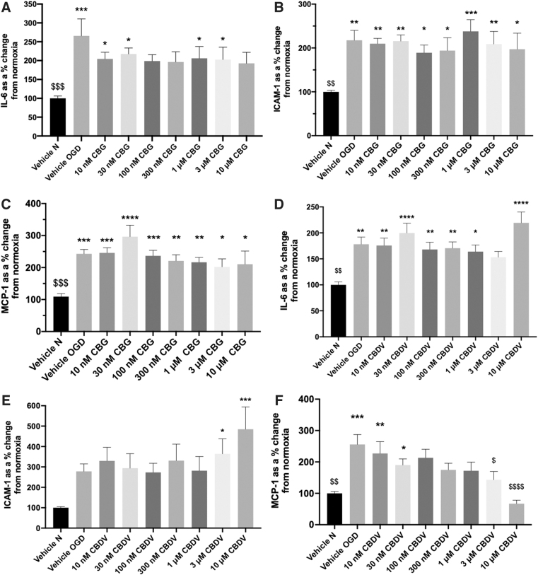
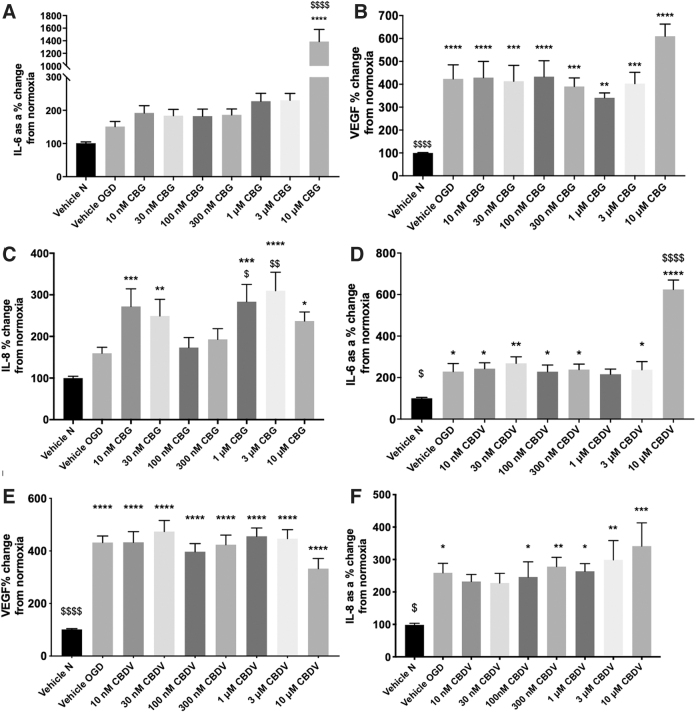
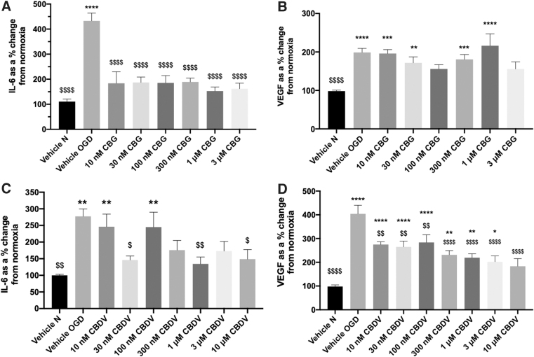
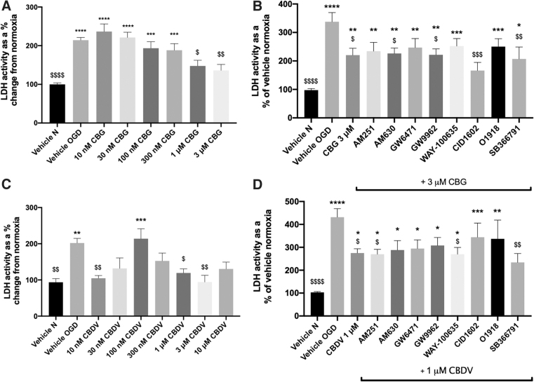
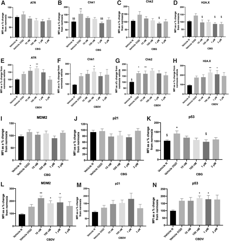
Background and Objectives: Preclinical studies have shown cannabidiol is protective in models of ischemic stroke. Based on results from our recent systematic review, we investigated the effects of two promising neuroprotective phytocannabinoids, cannabigerol (CBG) and cannabidivarin (CBDV), on cells of the blood-brain barrier (BBB), namely human brain microvascular endothelial cells (HBMECs), pericytes, and astrocytes. Experimental Approach: Cultures were subjected to oxygen-glucose deprivation (OGD) protocol to model ischemic stroke and cell culture medium was assessed for cytokines and adhesion molecules post-OGD. Astrocyte cell lysates were also analyzed for DNA damage markers. Antagonist studies were conducted where appropriate to study receptor mechanisms. Results: In astrocytes CBG and CBDV attenuated levels of interleukin-6 (IL-6) and lactate dehydrogenase (LDH), whereas CBDV (10 nM-10 μM) also decreased vascular endothelial growth factor (VEGF) secretion. CBDV (300 nM-10 μM) attenuated levels of monocyte chemoattractant protein (MCP)-1 in HBMECs. In astrocytes, CBG decreased levels of DNA damage proteins, including p53, whereas CBDV increased levels of DNA damage markers. Antagonists for CB1, CB2, PPAR-γ, PPAR-α, 5-HT1A, and TRPV1 had no effect on CBG (3 μM) or CBDV (1 μM)-mediated decreases in LDH in astrocytes. GPR55 and GPR18 were partially implicated in the effects of CBDV, but no molecular target was identified for CBG. Conclusions: We show that CBG and CBDV were protective against OG mediated injury in three different cells that constitute the BBB, modulating different hallmarks of ischemic stroke pathophysiology. These data enhance our understanding of the protective effects of CBG and CBDV and warrant further investigation into these compounds in ischemic stroke. Future studies should identify other possible neuroprotective effects of CBG and CBDV and their corresponding mechanisms of action.

Keywords: blood–brain barrier; cannabidivarin; cannabigerol; cannabinoids; ischemia; neuroprotection.


Conflict of interest statement

S.O.S. is director of CanPharmaConsulting, scientific advisor to pharmaceutical companies developing cannabinoid-based medicines. None of these companies were involved in this research.

Figures

FIG. 1.
FIG. 1.
The effects of CBG and CBDV on HBMEC monocultures. Medium was analyzed for IL-6 (A, D), ICAM-1 (B, E), and MCP-1 (C, F) 24 h after 4-h OGD. Data were normalized to total protein (calculated using a BCA assay) and are given as a % change from the normoxia vehicle presented as means with error bars representing SEM. n=6–9 from three experimental repeats. *, Significant difference compared with vehicle normoxia (vehicle N) (*p<0.05, **p<0.01, ***p<0.001, and ****p<0.0001). $p < 0.05, $$p < 0.01, $$$p < 0.001, and $$$$p < 0.0001) significant difference to vehicle OGD, one-way ANOVA with Dunnett's post hoc analysis. ANOVA, analysis of variance; BCA, bicinchoninic acid; CBDV, cannabidivarin; CBG, cannabigerol; HBMEC, human brain microvascular endothelial cell; ICAM-1, intracellular adhesion molecule-1; IL-6, interleukin-6; MCP-1, monocyte chemoattractant protein-1; OGD, oxygen-glucose deprivation; SEM, standard error of the mean.
FIG. 2.
FIG. 2.
The effects of CBDV and CBG on pericyte monocultures. Medium 24 h after 4-h OGD was analyzed for IL-6, VEGF, and IL-8 (A–F). Data were normalized to total protein and are given as a % change from the normoxia vehicle, presented as means with error bars representing SEM. n=6–9 from 3 experimental repeats. *, Significant difference compared with vehicle normoxia (vehicle N) (*p<0.05, **p<0.01, ***p<0.001, and ****p<0.0001). $p < 0.05, $$p < 0.01, $$$p < 0.001, and $$$$p < 0.0001) significant difference to vehicle OGD, one-way ANOVA with Dunnett's post hoc analysis. VEGF, vascular endothelial growth factor.
FIG. 3.
FIG. 3.
The effects of CBG and CBDV on astrocyte monocultures. (A-D) Medium 24 h after 8-h OGD were analyzed for IL-6 and VEGF. Data were normalized to total protein and are given as a % change from the normoxia vehicle, presented as means with error bars representing SEM. n=5–9 from 3 experimental repeats. *, Significant difference compared with vehicle normoxia (vehicle N) (*p<0.05, **p<0.01, ***p<0.001, and ****p<0.0001). $p < 0.05, $$p < 0.01, $$$p < 0.001, and $$$$p < 0.0001) significant difference to vehicle OGD, one-way ANOVA with Dunnett's post hoc analysis.
FIG. 4.
FIG. 4.
The effects of CBG and CBDV treatment alone (A, B) and with antagonists (C, D) on LDH release from astrocyte monocultures. Medium 24 h after 8-h OGD were analyzed LDH. Data were normalized to total protein and are given as a % change from the normoxia vehicle, presented as means with error bars representing SEM. n=5–6 from 3 experimental repeats. *, Significant difference compared with vehicle normoxia (vehicle N) (*p<0.05, **p<0.01, ***p<0.001, and ****p<0.0001). $p < 0.05, $$p < 0.01, $$$p < 0.001, and $$$$p < 0.0001) significant difference to vehicle OGD, one-way ANOVA with Dunnett's post hoc analysis. LDH, lactate dehydrogenase.
FIG. 5.
FIG. 5.
The effects of CBG and CBDV on DNA damage markers (ATR [A,E], Chk1 [B,F], Chk2 [C,G], H2A.X [D,H], MDM2 [I,L], p21 [J,M], and p53 [K,N] from astrocyte cell lysates, 24 h after 8-h OGD. Data were normalized to total protein and are given as MFI as a % change from the normoxia vehicle (vehicle N); means with error bars represent SEM. n=6–8 from 3 experimental repeats. *, Significant difference compared with vehicle normoxia (vehicle N) (*p<0.05, **p<0.01). $p<0.05, $$p<0.01 significant difference to vehicle OGD, one-way ANOVA with Dunnett's post hoc analysis. MFI, mean fluorescent intensity.

References

    1. Abbott NJ, Patabendige AAK, Dolman DEM, et al. . Structure and function of the blood-brain barrier. Neurobiol Dis. 2010;37:13–25 - PubMed
    1. McConnell HL, Kersch CN, Woltjer RL, et al. . The translational significance of the neurovascular unit. J Biol Chem. 2017;292:762–770 - PMC - PubMed
    1. Sosunov AA, Wu X, Tsankova NM, et al. . Phenotypic heterogeneity and plasticity of isocortical and hippocampal astrocytes in the human brain. J Neurosci. 2014;34:2285–2298 - PMC - PubMed
    1. Wang Q, Tang XN, Yenari MA. The inflammatory response in stroke. J Neuroimmunol. 2007;184:53–68 - PMC - PubMed
    1. Douglas DR, Luoma V, Reddy U. Acute management of ischaemic stroke. Anaesth Intensive Care Med. 2020;21:1–7

Publication types

MeSH terms

LinkOut - more resources