

A Convenient and Biosafe Replicon with Accessory Genes of SARS-CoV-2 and Its Potential Application in Antiviral Drug Discovery
- PMID: 33999369
- PMCID: PMC8127439
- DOI: 10.1007/s12250-021-00385-9
A Convenient and Biosafe Replicon with Accessory Genes of SARS-CoV-2 and Its Potential Application in Antiviral Drug Discovery
Abstract
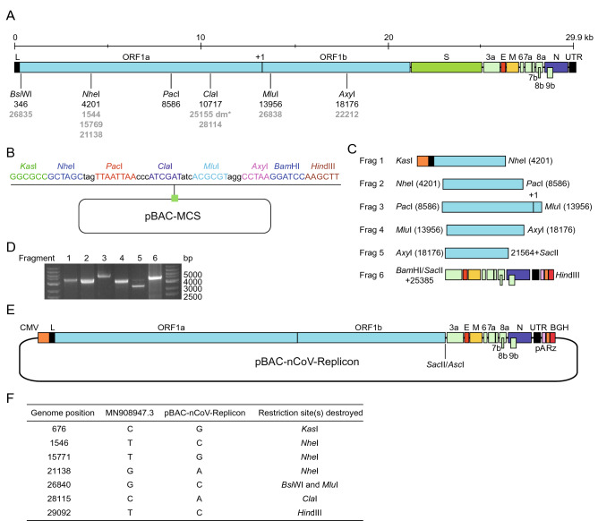
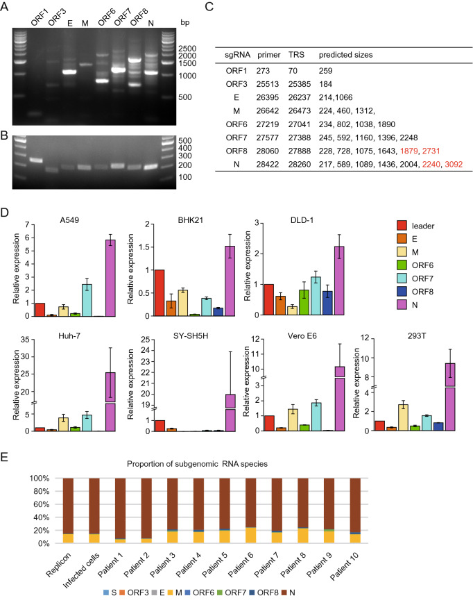
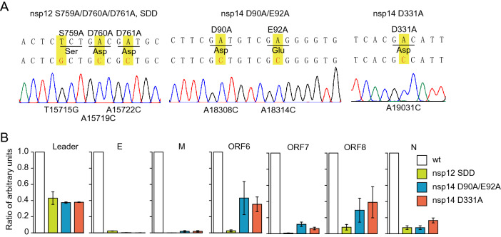
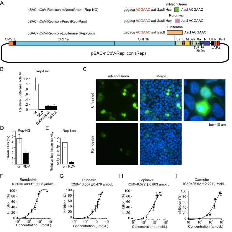
SARS-CoV-2 causes the pandemic of COVID-19 and no effective drugs for this disease are available thus far. Due to the high infectivity and pathogenicity of this virus, all studies on the live virus are strictly confined in the biosafety level 3 (BSL3) laboratory but this would hinder the basic research and antiviral drug development of SARS-CoV-2 because the BSL3 facility is not commonly available and the work in the containment is costly and laborious. In this study, we constructed a reverse genetics system of SARS-CoV-2 by assembling the viral cDNA in a bacterial artificial chromosome (BAC) vector with deletion of the spike (S) gene. Transfection of the cDNA into cells results in the production of an RNA replicon that keeps the capability of genome or subgenome replication but is deficient in virion assembly and infection due to the absence of S protein. Therefore, such a replicon system is not infectious and can be used in ordinary biological laboratories. We confirmed the efficient replication of the replicon by demonstrating the expression of the subgenomic RNAs which have similar profiles to the wild-type virus. By mutational analysis of nsp12 and nsp14, we showed that the RNA polymerase, exonuclease, and cap N7 methyltransferase play essential roles in genome replication and sgRNA production. We also created a SARS-CoV-2 replicon carrying a luciferase reporter gene and this system was validated by the inhibition assays with known anti-SARS-CoV-2 inhibitors. Thus, such a one-plasmid system is biosafe and convenient to use, which will benefit both fundamental research and development of antiviral drugs.
Keywords: Antiviral drug screening; Replicon; Reverse genetics; SARS-CoV-2.
© 2021. Wuhan Institute of Virology, CAS.
Conflict of interest statement
The authors declare that they have no conflict of interest.
Figures
Similar articles
-
Reverse genetic systems of SARS-CoV-2 for antiviral research.Antiviral Res. 2023 Feb;210:105486. doi: 10.1016/j.antiviral.2022.105486. Epub 2022 Dec 22. Antiviral Res. 2023. PMID: 36657881 Free PMC article. Review.
-
Construction of a Noninfectious SARS-CoV-2 Replicon for Antiviral-Drug Testing and Gene Function Studies.J Virol. 2021 Aug 25;95(18):e0068721. doi: 10.1128/JVI.00687-21. Epub 2021 Aug 25. J Virol. 2021. PMID: 34191580 Free PMC article.
-
Development of a Single-Cycle Infectious SARS-CoV-2 Virus Replicon Particle System for Use in Biosafety Level 2 Laboratories.J Virol. 2022 Feb 9;96(3):e0183721. doi: 10.1128/JVI.01837-21. Epub 2021 Dec 1. J Virol. 2022. PMID: 34851142 Free PMC article.
-
A BSL-2 compliant mouse model of SARS-CoV-2 infection for efficient and convenient antiviral evaluation.J Virol. 2024 Jul 23;98(7):e0050424. doi: 10.1128/jvi.00504-24. Epub 2024 Jun 20. J Virol. 2024. PMID: 38899934 Free PMC article.
-
Reverse genetics systems for SARS-CoV-2.J Med Virol. 2022 Jul;94(7):3017-3031. doi: 10.1002/jmv.27738. Epub 2022 Apr 5. J Med Virol. 2022. PMID: 35324008 Free PMC article. Review.
Cited by
-
Identification of potential SARS-CoV-2 inhibitors among well-tolerated drugs using drug repurposing and in vitro approaches.Sci Rep. 2025 Apr 22;15(1):13975. doi: 10.1038/s41598-025-88388-4. Sci Rep. 2025. PMID: 40263343 Free PMC article.
-
Reverse genetic systems of SARS-CoV-2 for antiviral research.Antiviral Res. 2023 Feb;210:105486. doi: 10.1016/j.antiviral.2022.105486. Epub 2022 Dec 22. Antiviral Res. 2023. PMID: 36657881 Free PMC article. Review.
-
The adenosine analog prodrug ATV006 is orally bioavailable and has preclinical efficacy against parental SARS-CoV-2 and variants.Sci Transl Med. 2022 Sep 7;14(661):eabm7621. doi: 10.1126/scitranslmed.abm7621. Epub 2022 Sep 7. Sci Transl Med. 2022. PMID: 35579533 Free PMC article.
-
Development of the coronavirus reverse genetic system: Core technology for pathogenesis mechanisms research and vaccine/drug development.Virulence. 2025 Dec;16(1):2525930. doi: 10.1080/21505594.2025.2525930. Epub 2025 Jun 28. Virulence. 2025. PMID: 40580492 Free PMC article. Review.
-
Reverse genetics systems for SARS-CoV-2: Development and applications.Virol Sin. 2023 Dec;38(6):837-850. doi: 10.1016/j.virs.2023.10.001. Epub 2023 Oct 11. Virol Sin. 2023. PMID: 37832720 Free PMC article. Review.
References
-
- Almazan F, Dediego ML, Galan C, Escors D, Alvarez E, Ortego J, Sola I, Zuniga S, Alonso S, Moreno JL, Nogales A, Capiscol C, Enjuanes L. Construction of a severe acute respiratory syndrome coronavirus infectious cDNA clone and a replicon to study coronavirus RNA synthesis. J Virol. 2006;80:10900–10906. doi: 10.1128/JVI.00385-06. - DOI - PMC - PubMed
-
- Carrillo A, Stewart KD, Sham HL, Norbeck DW, Kohlbrenner WE, Leonard JM, Kempf DJ, Molla A. In vitro selection and characterization of human immunodeficiency virus type 1 variants with increased resistance to ABT-378, a novel protease inhibitor. J Virol. 1998;72:7532–7541. doi: 10.1128/JVI.72.9.7532-7541.1998. - DOI - PMC - PubMed
MeSH terms
Substances
LinkOut - more resources
Full Text Sources
Other Literature Sources
Medical
Miscellaneous
