Review
. 2022 Jun;289(12):3360-3373.
doi: 10.1111/febs.16023. Epub 2021 May 30.

Bottom-up reconstitution of focal adhesion complexes

Affiliations
Review

Bottom-up reconstitution of focal adhesion complexes

Stephanie Schumacher et al. FEBS J. 2022 Jun.

Abstract

Focal adhesions (FA) are large macromolecular assemblies relevant for various cellular and pathological events such as migration, polarization, and metastatic cancer formation. At FA sites at the migrating periphery of a cell, hundreds of players gather and form a network to respond to extra cellular stimuli transmitted by the integrin receptor, the most upstream component within a cell, initiating the FA signaling pathway. Numerous cellular experiments have been performed to understand the FA architecture and functions; however, their intricate network formation hampers unraveling the precise molecular actions of individual players. Here, in vitro bottom-up reconstitution presents an advantageous approach for elucidating the FA machinery and the hierarchical crosstalk of involved cellular players.

Keywords: PIP2; actin; integrin; talin; vinculin.


Conflict of interest statement

The authors declare no conflicts of interest.

Figures

Fig. 1
Fig. 1
Simplified schematic of the FA machinery focusing on the activation of integrin. Components of the ECM (gray) bind to integrin receptors (α subunit in light green and β subunit in light blue), which reach through the plasma membrane (dark gray) into the cytosol. Intracellular proteins kindlin (pink) and talin (blue) bind to the cytoplasmic tail of β‐integrin together with additional signaling factors like FAK, Pax and Skelemin (gray). Activated talin extends through all FA layers from the integrin receptor to the actin cytoskeleton (red/pink) and vinculin (dark yellow) enforces the talin‐actin interaction. The FA machinery is tightly regulated and allows bidirectional signal transduction from outside‐in and from inside‐out.
Fig. 2
Fig. 2
Domain architecture of integrin receptors. (A) α‐integrins consist of a β‐propeller head, thigh, Genu, Calf‐1, calf‐2, TM helix, and cytoplasmic tail domains. β‐integrins comprise a βI head, hybrid, PSI, I‐EGF1‐4, β‐tail, TM helix, and cytoplasmic tail domains. Integrin receptors can adopt a bent (left), an extended‐closed (center), and an extended‐open (right) conformation. They bind ligands typically at the cleft between the α‐ and β‐subunit heads (depicted in orange). (B) The cytoplasmic tails of β‐integrins can contain two different linear binding motifs (NPxY or NxxY) for various interaction partners (in boxes on the right) depending on β‐integrin isotype.
Fig. 3
Fig. 3
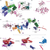
Domain architecture of talin, kindlin, and vinculin. (A) Talin consists of a globular FERM head (F0‐3) and a tail of 13 helical rod domains (R1–R13, in different colors) and a dimerization helix (DD, behind R13). In the autoinhibited form, all rod domains are entangled and the structure is secured by key interactions between F2–R12 and F3–R13 (insert). (B) Model of activated talin in an extended conformation with individual domains in different colors. Actin‐binding sites (ABS2 and ABS3) become accessible. Potential vinculin‐binding sites are highlighted with violet balls. (C) Kindlin consists of a globular FERM domain (F0–F3) and an additional PH domain. (D) Vinculin consists of a head domain comprising helical bundles D1–D4 and a helical tail that folds back in the autoinhibited state. The tail domain is released upon activation and opens binding sites for talin as well as for actin.
Fig. 4
Fig. 4
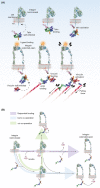
Schematic of integrin activation and FA initiation. (A) Autoinhibited talin can approach to the PIP2‐enriched membranes, resulting in the release of talin head and rod domains autoinhibition. Binding of talin to the cytoplasmic tails of β‐integrin primes the integrin receptor, which then binds to ligands in the extended‐open conformation. Opened talin can bind to actin, and this interaction is strengthened by crosslinking of talin and actin by activated vinculin. Further force‐dependent extension of talin (indicated by lightning bolts) uncovers additional vinculin binding sites. (B) Models of the cooperative activation of integrin by kindlin and talin. Integrin receptors can be activated by sequential binding of kindlin and talin (violet path), by simultaneous binding of both proteins to the same integrin receptor (cis cooperation, green path), or by synchronous binding of both proteins to different, clustered integrin receptors (trans cooperation, blue path).

References

    1. Geiger B, Bershadsky A, Pankov R & Yamada KM (2001) Transmembrane crosstalk between the extracellular matrix–cytoskeleton crosstalk. Nat Rev Mol Cell Biol 2, 793–805. - PubMed
    1. Winograd‐Katz SE, Fassler R, Geiger B & Legate KR (2014) The integrin adhesome: from genes and proteins to human disease. Nat Rev Mol Cell Biol 15, 273–288. - PubMed
    1. Wozniak MA, Modzelewska K, Kwong L & Keely PJ (2004) Focal adhesion regulation of cell behavior. Biochim Biophys Acta 1692, 103–119. - PubMed
    1. Legate KR, Wickstrom SA & Fassler R (2009) Genetic and cell biological analysis of integrin outside‐in signaling. Genes Dev 23, 397–418. - PubMed
    1. Hynes RO (2002) Integrins: bidirectional, allosteric signaling machines. Cell 110, 673–687. - PubMed

LinkOut - more resources