

1.7-Micron Optical Coherence Tomography Angiography for Characterization of Skin Lesions-A Feasibility Study
- PMID: 33999817
- PMCID: PMC8834583
- DOI: 10.1109/TMI.2021.3081066
1.7-Micron Optical Coherence Tomography Angiography for Characterization of Skin Lesions-A Feasibility Study
Abstract
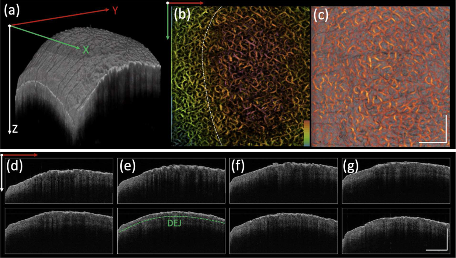
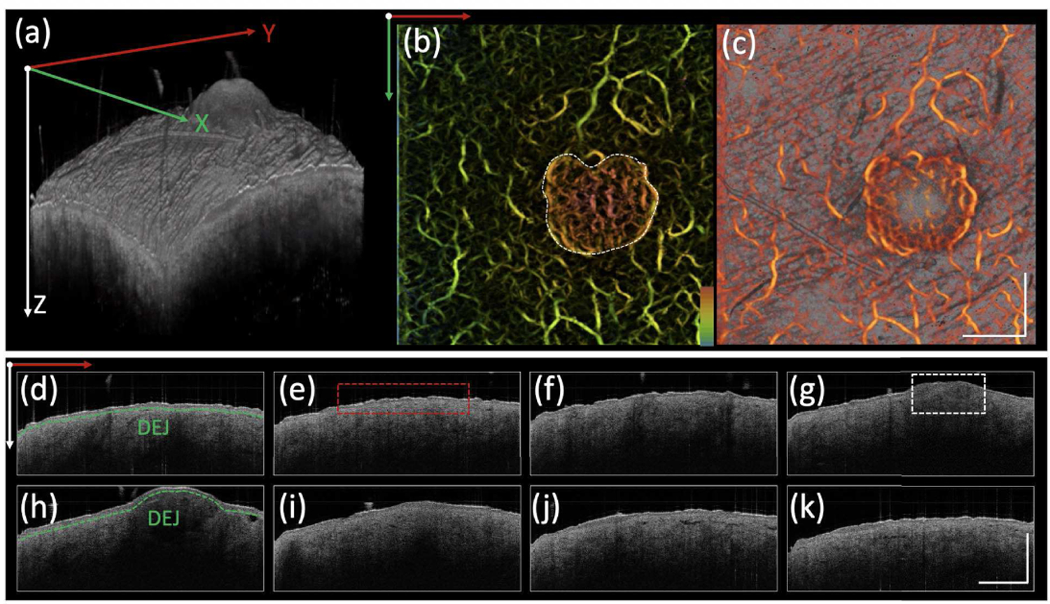
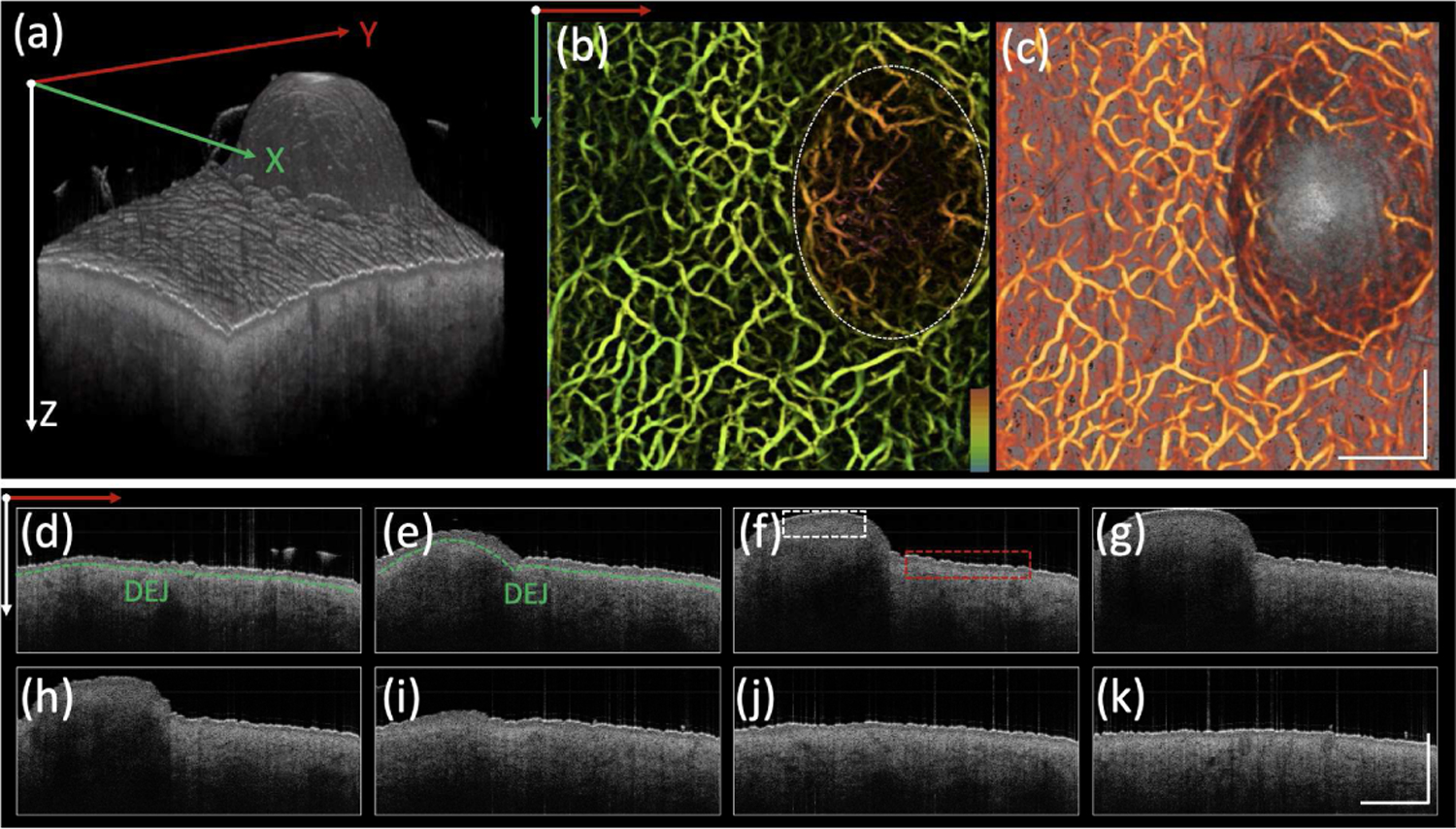
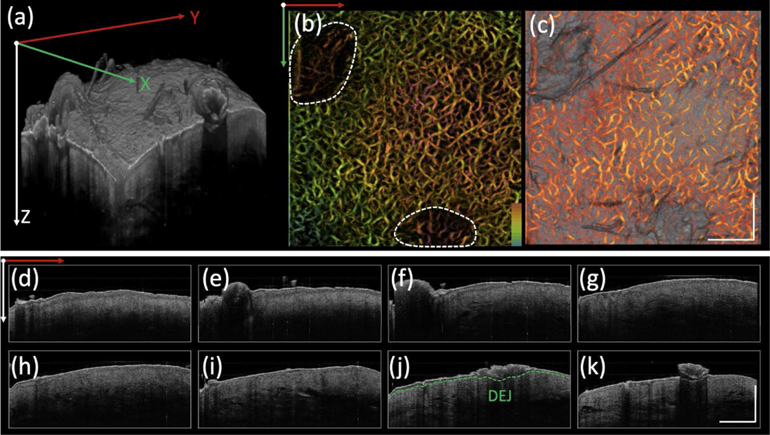
Optical coherence tomography (OCT) is a non-invasive diagnostic method that offers real-time visualization of the layered architecture of the skin in vivo. The 1.7-micron OCT system has been applied in cardiology, gynecology and dermatology, demonstrating an improved penetration depth in contrast to conventional 1.3-micron OCT. To further extend the capability, we developed a 1.7-micron OCT/OCT angiography (OCTA) system that allows for visualization of both morphology and microvasculature in the deeper layers of the skin. Using this imaging system, we imaged human skin with different benign lesions and described the corresponding features of both structure and vasculature. The significantly improved imaging depth and additional functional information suggest that the 1.7-micron OCTA system has great potential to advance both dermatological clinical and research settings for characterization of benign and cancerous skin lesions.
Figures
Similar articles
-
Assessment of chronic radiation proctopathy and radiofrequency ablation treatment follow-up with optical coherence tomography angiography: A pilot study.World J Gastroenterol. 2019 Apr 28;25(16):1997-2009. doi: 10.3748/wjg.v25.i16.1997. World J Gastroenterol. 2019. PMID: 31086467 Free PMC article.
-
Dynamic Optical Coherence Tomography in Dermatology.Dermatology. 2016;232(3):298-311. doi: 10.1159/000444706. Epub 2016 Apr 23. Dermatology. 2016. PMID: 27104356 Review.
-
Optical coherence tomography angiography of normal skin and inflammatory dermatologic conditions.Lasers Surg Med. 2018 Mar;50(3):183-193. doi: 10.1002/lsm.22788. Epub 2018 Jan 22. Lasers Surg Med. 2018. PMID: 29356051 Free PMC article.
-
Optical coherence tomography angiography and photoacoustic imaging in dermatology.Photochem Photobiol Sci. 2019 May 15;18(5):945-962. doi: 10.1039/c8pp00471d. Photochem Photobiol Sci. 2019. PMID: 30735220 Review.
-
Penetration-enhanced optical coherence tomography angiography with optical clearing agent for clinical evaluation of human skin.Photodiagnosis Photodyn Ther. 2020 Jun;30:101734. doi: 10.1016/j.pdpdt.2020.101734. Epub 2020 Apr 8. Photodiagnosis Photodyn Ther. 2020. PMID: 32171879
Cited by
-
Current trends in the characterization and monitoring of vascular response to cancer therapy.Cancer Imaging. 2024 Oct 23;24(1):143. doi: 10.1186/s40644-024-00767-8. Cancer Imaging. 2024. PMID: 39438891 Free PMC article. Review.
-
Computerized Texture Analysis of Optical Coherence Tomography Angiography of Choriocapillaris in Normal Eyes of Young and Healthy Subjects.Cells. 2022 Jun 15;11(12):1934. doi: 10.3390/cells11121934. Cells. 2022. PMID: 35741063 Free PMC article.
-
Imaging the ex-vivo human cochlea using 1.3-μm and 1.7-μm optical coherence tomography.J Biomed Opt. 2025 Apr;30(4):046007. doi: 10.1117/1.JBO.30.4.046007. Epub 2025 Apr 17. J Biomed Opt. 2025. PMID: 40248314 Free PMC article.
-
Pilot study of optical coherence tomography angiography-derived microvascular metrics in hands and feet of healthy and diabetic people.Sci Rep. 2023 Jan 20;13(1):1122. doi: 10.1038/s41598-022-26871-y. Sci Rep. 2023. PMID: 36670141 Free PMC article.
-
Measuring collagen injury depth for burn severity determination using polarization sensitive optical coherence tomography.Sci Rep. 2022 Jun 21;12(1):10479. doi: 10.1038/s41598-022-14326-3. Sci Rep. 2022. PMID: 35729262 Free PMC article.
References
-
- Rogers HW, Weinstock MA, Feldman SR, Coldiron BM, Incidence Estimate of Nonmelanoma Skin Cancer (Keratinocyte Carcinomas) in the U.S. Population, 2012, JAMA Dermatol 151(10) (2015) 1081–6. - PubMed
-
- Altman AM, Bankson J, Matthias N, Vykoukal JV, Song YH, Alt EU, Magnetic resonance imaging as a novel method of characterization of cutaneous photoaging in a murine model, Arch Dermatol Res 300(5) (2008) 263–7. - PubMed
-
- Borsari S, Pampena R, Lallas A, Kyrgidis A, Moscarella E, Benati E, Raucci M, Pellacani G, Zalaudek I, Argenziano G, Longo C, Clinical Indications for Use of Reflectance Confocal Microscopy for Skin Cancer Diagnosis, JAMA Dermatol 152(10) (2016) 1093–1098. - PubMed
Publication types
MeSH terms
Grants and funding
LinkOut - more resources
Full Text Sources
Other Literature Sources
Medical
