. 2021 May 17;17(5):e1009561.
doi: 10.1371/journal.pgen.1009561. eCollection 2021 May.

DEFECTIVE EMBRYO AND MERISTEMS genes are required for cell division and gamete viability in Arabidopsis

Affiliations

DEFECTIVE EMBRYO AND MERISTEMS genes are required for cell division and gamete viability in Arabidopsis

Chin Hong Lee et al. PLoS Genet. .

Abstract

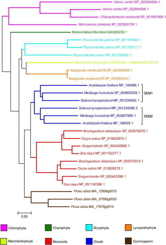
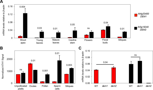
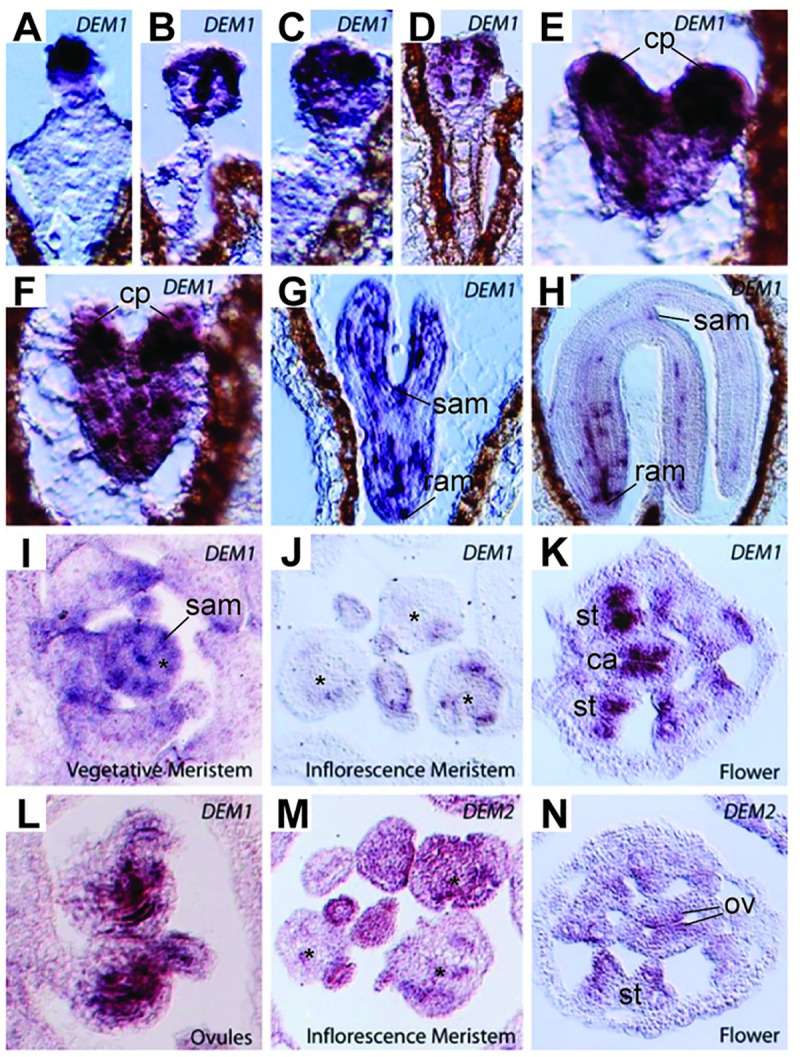
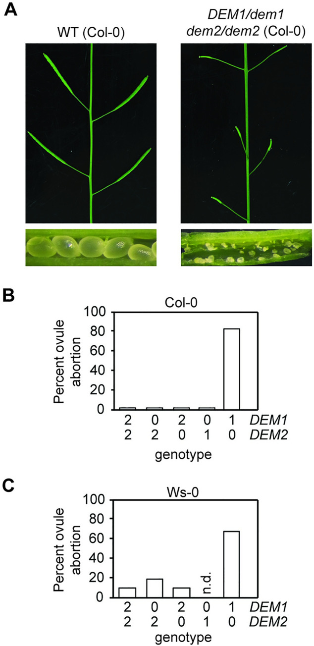
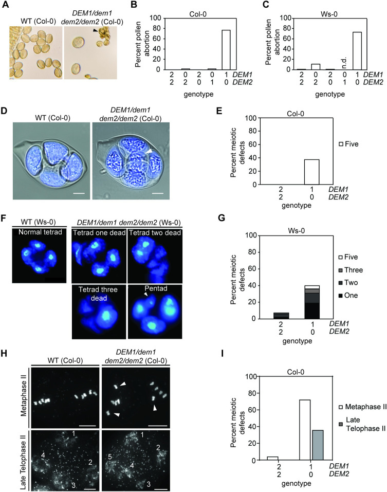
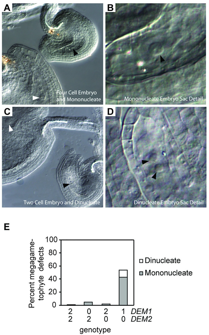
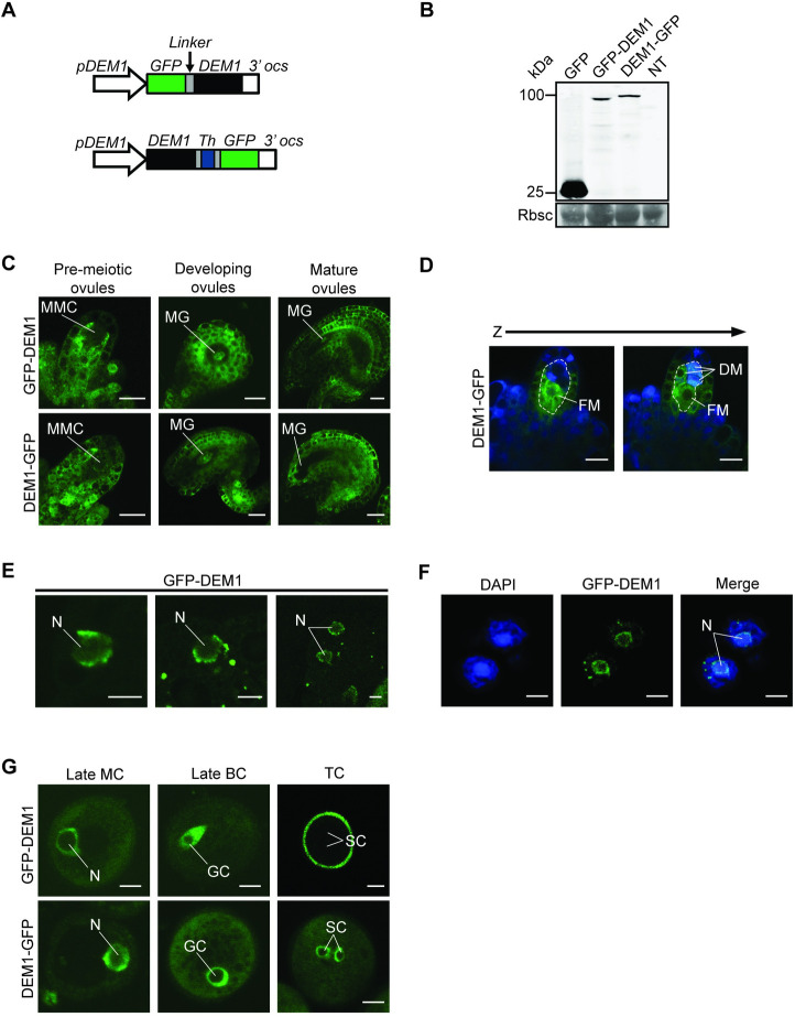
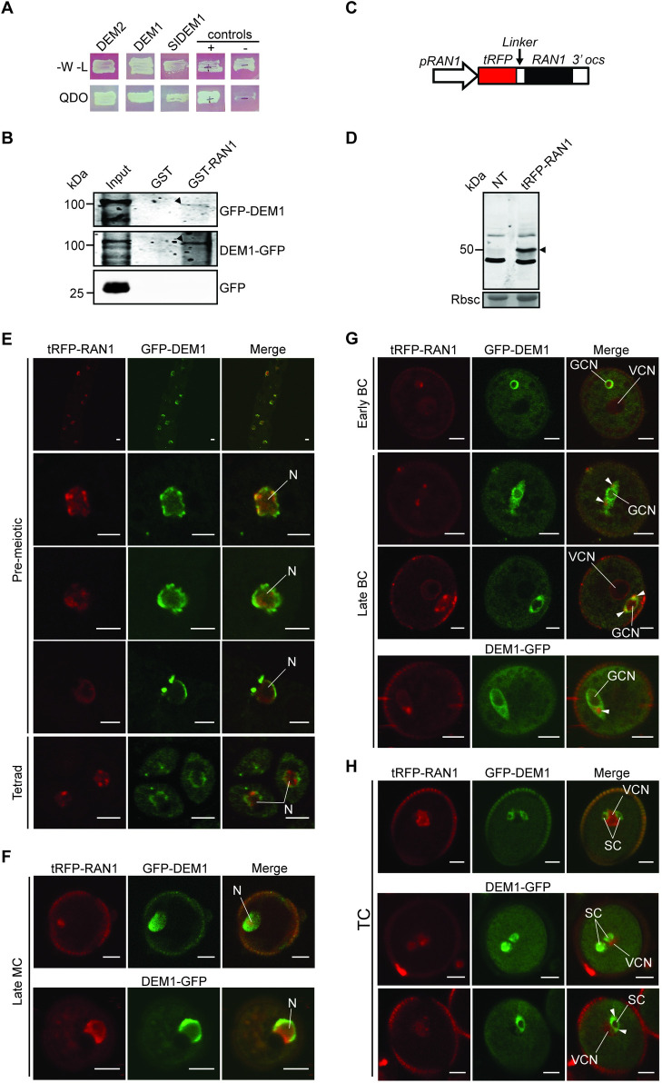
The DEFECTIVE EMBRYO AND MERISTEMS 1 (DEM1) gene encodes a protein of unknown biochemical function required for meristem formation and seedling development in tomato, but it was unclear whether DEM1's primary role was in cell division or alternatively, in defining the identity of meristematic cells. Genome sequence analysis indicates that flowering plants possess at least two DEM genes. Arabidopsis has two DEM genes, DEM1 and DEM2, which we show are expressed in developing embryos and meristems in a punctate pattern that is typical of genes involved in cell division. Homozygous dem1 dem2 double mutants were not recovered, and plants carrying a single functional DEM1 allele and no functional copies of DEM2, i.e. DEM1/dem1 dem2/dem2 plants, exhibit normal development through to the time of flowering but during male reproductive development, chromosomes fail to align on the metaphase plate at meiosis II and result in abnormal numbers of daughter cells following meiosis. Additionally, these plants show defects in both pollen and embryo sac development, and produce defective male and female gametes. In contrast, dem1/dem1 DEM2/dem2 plants showed normal levels of fertility, indicating that DEM2 plays a more important role than DEM1 in gamete viability. The increased importance of DEM2 in gamete viability correlated with higher mRNA levels of DEM2 compared to DEM1 in most tissues examined and particularly in the vegetative shoot apex, developing siliques, pollen and sperm. We also demonstrate that gamete viability depends not only on the number of functional DEM alleles inherited following meiosis, but also on the number of functional DEM alleles in the parent plant that undergoes meiosis. Furthermore, DEM1 interacts with RAS-RELATED NUCLEAR PROTEIN 1 (RAN1) in yeast two-hybrid and pull-down binding assays, and we show that fluorescent proteins fused to DEM1 and RAN1 co-localize transiently during male meiosis and pollen development. In eukaryotes, RAN is a highly conserved GTPase that plays key roles in cell cycle progression, spindle assembly during cell division, reformation of the nuclear envelope following cell division, and nucleocytoplasmic transport. Our results demonstrate that DEM proteins play an essential role in cell division in plants, most likely through an interaction with RAN1.


Conflict of interest statement

The authors have declared that no competing interests exist.

Figures

Fig 1
Fig 1. Phylogenetic tree of plant DEM proteins, rooted with the Chlorophyta sequences.
Software and parameters used in the analysis are described in Materials and Methods. The name of the organism is indicated on the tree, followed by the accession number or the Conifer Genome Integrative Explorer gene ID for Picea abies. Scale bar indicates 0.05 substitutions per site.
Fig 2
Fig 2. DEM1 and DEM2 mRNA levels in various tissues and sperm cells during vegetative and reproductive development.
(A) Quantitative real-time reverse transcriptase PCR (qRT-PCR) was used to examine DEM1 and DEM2 mRNA levels in total RNA extracted from a range of Arabidopsis tissues. Ratios of mRNA levels to that of β-actin mRNA are shown (average ± S.E. for three biological replicates). (B) Microarray-based expression analysis of DEM1 and DEM2 in Arabidopsis reproductive tissues and FACS-purified sperm cells. The figure is based on raw data published by Pina et al. [18], Borges et al. [19] and Boavida et al. [20], and shows mean signal intensities ± S.E. from ATH1 Genechips normalized by the invariant set method, as previously described [19]. Data were derived from two biological replicates for siliques [18], microdissected ovules and unpollinated pistils (UPs) [20], and three biological replicates for pollen and FACS-purified sperm cells [19]. (C) qRT-PCR analysis of DEM1 and DEM2 mRNA levels in total RNA extracted from Arabidopsis wild-type (WT), dem1-2 (dem1) and dem2-2 (dem2) floral buds. Ratios of mRNA levels to that of β-actin mRNA are shown (average ± S.E. for three to six biological replicates). P-values were calculated using one-way ANOVA to indicate significant differences between DEM1 and DEM2 expression in the different tissues and sperm cells of wild-type plants (A, B), or between floral buds of wild-type and dem plants (C).
Fig 3
Fig 3. DEM1 and DEM2 mRNA expression detected by in situ hybridization in developing embryos and post-embryonic shoot tissues of Arabidopsis ecotype Ws-0.
(A-H) Punctate expression of DEM1 in developing embryos. (A-D) Globular stage. (E-F) Heart stage. (G) Torpedo stage. (H) Maturing embryo. (I-J) DEM1 expression in shoot apices is not as clearly punctate as in developing embryos but is clearly expressed in vegetative shoot apical meristem and in some dividing cells. (I) Vegetative meristem. (J) Inflorescence meristem. (K-L) DEM1 expression can clearly be seen in sporophytic tissues of developing anthers and developing ovules. (K) Flower. (L) Ovules. (M-N) DEM2 is expressed in dividing cells of the inflorescence. (M) Inflorescence meristem. (N) Flower. cp, cotyledon primordia; ram, root apical meristem; sam, shoot apical meristem; *, central zone of meristem; st, stamen primordia; ca, carpel primordia; ov, developing ovules.
Fig 4
Fig 4. DEM1 and DEM2 are required for normal seed production in Arabidopsis.
(A-C) Plants harboring only one wild-type DEM1 allele (DEM1/dem1 dem2/dem2) in both the Col-0 and Ws-0 genetic backgrounds exhibited significant ovule abortion. (A) Comparison of wild-type (WT) and DEM1/dem1 dem2/dem2 siliques of Col-0 genetic background. Note the occurrence of degenerated ovules in DEM1/dem1 dem2/dem2 siliques compared to the appearance of normal seeds in WT siliques. (B-C) Percentage of aborted ovules in self-fertilized WT and dem genotypes for Col-0 (B) and Ws-0 (C) genetic backgrounds. At least 200 ovules were assayed, involving at least three separate plants for each genotype. X-axis of (B) and (C) represent the copy number of DEM1 and DEM2 wild-type alleles in each genotype. No dem1/dem1 DEM2/dem2 plants were recovered in the Ws-0 genetic background; n.d., not determined.
Fig 5
Fig 5. DEM1 and DEM2 are required for normal pollen development and male meiosis in Arabidopsis.
(A) Phenotype of pollen produced by wild-type (WT) and DEM1/dem1 dem2/dem2 plants in the Col-0 genetic background. Dead pollen grains are indicated by a black arrowhead. (B-C) Percentage of aborted mature pollen in WT and dem genotypes for the Col-0 (B) and Ws-0 (C) genetic backgrounds. At least 700 mature pollen in total were assayed, involving at least three separate plants for each genotype. n.d., not determined as dem1/dem1 DEM2/dem2 were not recovered in the Ws-0 genetic background. (D) Confocal image of DAPI-stained tetrads, combined with a DIC imaging to view membrane structures, shows an excess of meiotic products (i.e. a pentad) in a DEM1/dem1 dem2/dem2 plant (ecotype Col-0); an extra product of meiosis of diminished size is highlighted by the white arrowhead. Bar = 5 μm. (E) Percentage abnormal tetrads produced by WT and DEM1/dem1 dem2/dem2 plants in the Col-0 background. At least 80 meiotic events were scored, involving at least three separate plants for each genotype. (F) DAPI-stained defective male tetrads of DEM1/dem1 dem2/dem2 plants in ecotype Ws-0 showing one to three dead microspores, and an extra microspore of diminished size in a pentad (white arrowhead). (G) Percentage of defective tetrads in WT and DEM1/dem1 dem2/dem2 plants of ecotype Ws-0. At least 200 tetrads were assayed for each genotype. (H) DAPI-stained meiotic chromosome spreads revealed misaligned chromosomes (white arrowheads) away from the metaphase plate at metaphase II in DEM1/dem1 dem2/dem2 male meiocytes, and also shown is an example of five daughter cells produced at telophase II in DEM1/dem1 dem2/dem2 male meiocytes (ecotype Col-0). (I) Percentage of male meiocytes showing defects at metaphase II (misaligned chromosomes) and late telophase II (pentads) in wild-type and DEM1/dem1 dem2/dem2 plants in ecotype Col-0. At least 60 meiocytes were assayed, involving at least three separate plants for each genotype. X-axis of (B), (C), (E), (G), and (I) represent the copy number of DEM1 and DEM2 wild-type alleles in each genotype.
Fig 6
Fig 6. DEM1 and DEM2 are required for normal megagametophyte development in Arabidopsis.
(A-D) Defective female gametophytes produced in DEM1/dem1 dem2/dem2 plants (Ws-0 genetic background). Developing ovules were observed as cleared whole mounts. (A-B) Four cell embryo (white arrowhead) and mononucleate embryo sac (black arrowhead) in two adjacent ovules. (C-D) Two cell embryo (white arrowhead) and di-nucleate embryo sac (black arrowheads) in two adjacent ovules. (E) Percentage of mononucleate and di-nucleate embryo sacs in self-fertilized wild-type and DEM1/dem1 dem2/dem2 plants of ecotype Ws-0. At least 200 ovules were assayed, involving at least three separate plants for each genotype. X-axis represents the copy number of DEM1 and DEM2 wild-type alleles in each genotype.
Fig 7
Fig 7. GFP-tagged DEM1 is expressed in developing gametophytes and sporophytic tissues of Arabidopsis.
(A) Transgenes used to express DEM1 N-terminal or C-terminal GFP fusion proteins under the control of the DEM1 promoter (pDEM1). The coding sequence for six alanine residues (Linker) were inserted between GFP and DEM1 of pDEM1:GFP-DEM1. Similarly, the pDEM1:DEM1-GFP transgene contained the coding sequence for three alanine residues, followed by a thrombin cleavage site and three more alanine residues (Th). (B) Anti-GFP western blot on floral bud extracts of 35S:GFP (GFP), pDEM1:GFP-DEM1 (GFP-DEM1) and pDEM1:DEM1-GFP (DEM1-GFP) transgenic lines, and a non-transgenic control (NT). Molecular weight is indicated in kDa. The molecular masses of GFP-DEM1 and DEM1-GFP are approximately 100 kDa, and of GFP is 27 kDa. The Coomassie Brilliant Blue-stained (CBB) membrane shows Rubisco (Rbsc) as a loading control. (C) Ubiquitous and punctate GFP-tagged DEM1 expression in ovules prior to meiosis (pre-meiotic ovules), and in developing and mature post-meiotic ovules. In pre-meiotic ovules, very little GFP fluorescence could be seen in the megaspore mother cell (MMC), whereas high expression was observed in the sporophytic cells surrounding the MMC (bar = 10 μm). In developing ovules, high GFP expression is observed in the sporophytic tissue and in the megagametophyte (MG) (bar = 20 μm). In mature ovules, high GFP fluorescence was observed in the sporophytic tissues, but very little expression was seen in the megagametophyte (MG) (bar = 20 μm). (D) Two optical sections (Z-stack) showing high expression of DEM1-GFP in the functional megaspore (FM) of an ovule (bar = 10 μm). DM, dead megaspores. (E) Expression of GFP-DEM1 in male pre-meiotic cells; GFP-DEM1 localized around the nuclear envelope (bar = 5 μm). N, nucleus. (F) Expression of GFP-tagged DEM1 was also detected in meiotic products of male meiosis; two cells of a tetrad are shown in the figure. Note the subcellular localization of GFP-DEM1 around the nuclear envelope relative to the DAPI stained nucleus (N) (bar = 5 μm). (G) Expression of GFP-tagged DEM1 during pollen development (bar = 5 μm). N, nucleus of monocellular pollen; GC, generative cell; SC, sperm cells; MC, monocellular pollen; BC, bicellular pollen; TC, tricellular pollen.
Fig 8
Fig 8. DEM interacts with RAN1, and the proteins co-localize during male meiosis and pollen development.
(A) RAN1 interacts with Arabidopsis DEM1 and DEM2, and tomato DEM1 (SlDEM1) in the yeast two-hybrid system. Yeast were plated on SD media lacking tryptophan and leucine (-W-L) to select for the bait (DEM) and prey (RAN1) vectors and show equal viability of all strains. Yeast were also plated on quadruple dropout media (QDO) to test for interactions between the bait and the prey. Known interacting (+) and non-interacting (-) controls are also shown. (B) GST-RAN1, but not GST, precipitates 100 kDa GFP-DEM1 and DEM1-GFP from floral bud extracts (black arrowheads). GFP-expressing plants were included to rule out binding of RAN1 to GFP (lower panel). Input represents an anti-GFP western of floral bud extracts from each transgenic line. (C) Transgene used to express RAN1 N-terminal tagRFP (tRFP) fusion protein under the control of the RAN1 promoter (pRAN1). The coding sequence for six glycine residues (Linker) were inserted between tRFP and RAN1. (D) Anti-tRFP western blot of pRAN1:tRFP-RAN1 transgenic and non-transgenic (NT) floral bud extracts. The tRFP-RAN1 is approximately 50 kDa (black arrowhead). The CBB membrane shows Rubisco (Rbsc) as a loading control. (E-H) Expression and co-localization of GFP-tagged DEM1 and tagRFP-RAN1 (tRFP-RAN1) during male meiosis and pollen development in wild-type ecotype Col-0 plants. (E) GFP-DEM1 localized around the nuclear envelope with additional weak signal throughout the nucleus and cytoplasm of pre-meiotic cells and tetrads, whereas tRFP-RAN1 typically localized to the nucleus (N). We examined 18 pre-meiotic cells in total, all 18 cells expressed detectable tagRFP-RAN1 and GFP-DEM1 and showed a similar localization of the fusion proteins. Representative pre-meiotic cells are shown in the figure. (F) Monocellular microspores (MC) show localization of GFP-tagged DEM1 around the nuclear envelope and tRFP-RAN1 to the nucleus, including the nuclear periphery. (G) Bicellular pollen showing co-localization of tRFP-RAN1 and GFP-tagged DEM1 around the nuclear envelope and in extranuclear foci at opposite poles of the generative cell (white arrowheads). (H) In tricellular pollen, GFP-tagged DEM1 was expressed in sperm cells (SC) around the nuclear envelope and throughout the sperm cytoplasm, whereas RFP-tagged RAN1 was primarily expressed in the immediately adjacent vegetative nucleus. In some tricellular pollen, weak RAN1 fluorescence overlapped with strong DEM1 fluorescence in extranuclear foci at opposite poles of sperm cells (lower panel; white arrowheads). We examined 93 pollen in total and 74 (80%) expressed detectable tagRFP-RAN1, 45 (48%) expressed detectable GFP-DEM1 or DEM1-GFP, and 39 (42%) expressed both tagRFP-RAN1 and GFP-DEM1 or DEM1-GFP (E-H). Pollen at the same stage expressing both tagRFP-RAN1 and GFP-DEM1 or DEM1-GFP showed a similar localization of the fusion proteins, and representative pollen are shown in the figure (E-H). Bar = 5 μm. BC, bicellular pollen; TC, tricellular pollen; GCN, generative cell nucleus; VCN, vegetative cell nucleus.

References

    1. Qiu Y-L, Taylor AB, McManus HA. Evolution of the life cycle in land plants. Journal of Systematics and Evolution. 2012;50(3):171–94. 10.1111/j.1759-6831.2012.00188.x - DOI
    1. Andres F, Coupland G. The genetic basis of flowering responses to seasonal cues. Nature Reviews Genetics. 2012;13(9):627–39. Epub 2012/08/18. 10.1038/nrg3291 . - DOI - PubMed
    1. McCormick S. Control of male gametophyte development. The Plant Cell. 2004;16 Suppl:S142–53. Epub 2004/03/24. 10.1105/tpc.016659 - DOI - PMC - PubMed
    1. Yadegari R, Drews GN. Female gametophyte development. The Plant Cell. 2004;16 Suppl:S133–41. Epub 2004/04/13. 10.1105/tpc.018192 - DOI - PMC - PubMed
    1. Liu J, Qu LJ. Meiotic and mitotic cell cycle mutants involved in gametophyte development in Arabidopsis. Molecular Plant. 2008;1(4):564–74. Epub 2008/07/01. 10.1093/mp/ssn033 . - DOI - PubMed

Publication types

MeSH terms