. 2021 May 17;17(5):e1009544.
doi: 10.1371/journal.pgen.1009544. eCollection 2021 May.

Light-dependent N-end rule-mediated disruption of protein function in Saccharomyces cerevisiae and Drosophila melanogaster

Affiliations

Light-dependent N-end rule-mediated disruption of protein function in Saccharomyces cerevisiae and Drosophila melanogaster

Leslie M Stevens et al. PLoS Genet. .

Abstract

Here we describe the development and characterization of the photo-N-degron, a peptide tag that can be used in optogenetic studies of protein function in vivo. The photo-N-degron can be expressed as a genetic fusion to the amino termini of other proteins, where it undergoes a blue light-dependent conformational change that exposes a signal for the class of ubiquitin ligases, the N-recognins, which mediate the N-end rule mechanism of proteasomal degradation. We demonstrate that the photo-N-degron can be used to direct light-mediated degradation of proteins in Saccharomyces cerevisiae and Drosophila melanogaster with fine temporal control. In addition, we compare the effectiveness of the photo-N-degron with that of two other light-dependent degrons that have been developed in their abilities to mediate the loss of function of Cactus, a component of the dorsal-ventral patterning system in the Drosophila embryo. We find that like the photo-N-degron, the blue light-inducible degradation (B-LID) domain, a light-activated degron that must be placed at the carboxy terminus of targeted proteins, is also effective in eliciting light-dependent loss of Cactus function, as determined by embryonic dorsal-ventral patterning phenotypes. In contrast, another previously described photosensitive degron (psd), which also must be located at the carboxy terminus of associated proteins, has little effect on Cactus-dependent phenotypes in response to illumination of developing embryos. These and other observations indicate that care must be taken in the selection and application of light-dependent and other inducible degrons for use in studies of protein function in vivo, but importantly demonstrate that N- and C-terminal fusions to the photo-N-degron and the B-LID domain, respectively, support light-dependent degradation in vivo.


Conflict of interest statement

The authors have declared that no competing interests exist.

Figures

Fig 1
Fig 1. An amino terminal domain encoding ubiquitin, fused to the LOV2 domain from oat phototropin I mediates blue light/N-end rule-mediated loss of Ura3p function in yeast.
(A) A schematic diagram showing the organization of the construct encoding blue/light, N-end rule targeted Ura3p, under the transcriptional control of the copper-inducible CUP1 promoter (PCUP1). From 5’ to 3’, the transgene encodes a single copy of the ubiquitin open reading frame (UBI), the LOV2 domain from plant phototropin I (LOV), a single copy of the influenza hemagglutinin epitope (HA), and the open reading frame encoding the yeast Ura3p protein (URA3). Protein synthesis initiates at the ubiquitin initiation codon (M) and the pair of glycine residues at the C-terminus of the ubiquitin open reading frame (GG) are followed immediately by an arginine codon (R). The ubiquitin domain is removed co-translationally, leaving the arginine residue immediately preceding the LOV domain as the N-terminal residue of the mature protein. In the corresponding UBI-R-DHFR-HA-URA3 construct, the sequence encoding the LOV2 domain have been replaced by DHFR coding sequences bearing an N-terminal arginine residue. (B) A schematic diagram of UBI-R-HA-URA3, which lacks the sequences encoding the light-sensitive LOV domain. (C) The UBI-R-LOV-HA-URA3 and UBI-R-DHFR-HA-URA3 transgenes were introduced into UBR1 ura3 and ubr1Δ ura3 mutant cells (introduced transgenes and yeast genotypes shown at top of panel), which were seeded onto selective plates lacking uracil and incubated in either darkness, under blue-light, or under red-light illumination. Under blue light, the UBI-R-LOV-HA-URA3 construct failed to restore growth in the absence of uracil, indicating the sensitivity of the expressed R-phLOV2-HA-Ura3p protein to blue light. When incubated under blue light in the absence of the Ubr1p activity (ubr1Δ ura3), growth in the absence of uracil was restored. In contrast to R-phLOV2-HA-Ura3p, the R-DHFR-HA-Ura3p protein did not confer light sensitivity upon growth in the absence of uracil. (D-K) Yeast cells expressing either UBI-R-phLOV2-HA-URA3 (D-G) or UBI-R-HA-URA3 (H-K) (transgenes shown at bottom of panels) were expressed in either Ubr1p-expressing (UBR1 ura3) (D, E, H, I) or Ubr1p-lacking (ubr1Δ ura3) (F, G, J, K) genetic backgrounds and incubated on selective plates lacking uracil either in darkness (D, F, H, J) or under blue light illumination (E, G, I, K). In the presence of Ubr1p and incubated under blue light illumination (E), UBI-R-phLOV2-HA-Ura3p-expressing cells arrested mainly as single cells, arguing that the light/Ubr1p-mediated loss of Ura3p protein function was rapid. In contrast, when grown in the dark (D) or in a ubr1Δ mutant background (G), these cells proliferated normally. In contrast, cells bearing a wild-type UBR1 gene and expressing UBI-R-HA-URA3, arrested as single cells both in darkness and under illumination (H, I), indicating that the presence of an N-end rule targeted arginine, in the absence of the phLOV2 domain, rendered the encoded protein functionally inactive regardless of light conditions (G, H), while the absence of the Ubr1p ubiquitin ligase protein left the R-HA-Ura3p protein functional in darkness and under blue light illumination (J, K). (L) shows a schematic representation of the mechanism through which R-phLOV-HA-tagged protein is presumed to be synthesized and degraded in response to blue-light illumination.
Fig 2
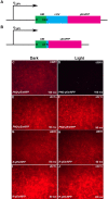
Fig 2. The PND mediates blue light/Ubr1-dependent loss of yEmRFP-associated fluorescence in yeast.
Two constructs encoding yEmRFP bearing either the PND (PND-yEmRFP) (A, C, D, E, F) or a single arginine (R) residue (R-yEmRFP) (B, G, H, I, J) at the amino terminus were introduced into UBR1 (C, D, G, H) and ubr1Δ (E, F, I, J) strains of yeast (labelled at top right of each panel) and seeded onto selective plates. Following 48 hours growth to confluence in darkness (C, E, G, I) or under blue light illumination (D, F, H, J) the surfaces of the patches were imaged for red fluorescence. Photographic imaging was carried out on the same day and under the same conditions, with the time of exposure (50 or 100 milliseconds [ms], noted at bottom right of each panel) the same for each of the light/dark pairings. This permitted a determination of relative levels of expression between light/dark pairings and between the strains and the constructs that they carried. Yeast expressing PND-yEmRFP in the presence of Ubr1p, exhibited a dramatic decrease in response to illumination (C, D). In the absence of Ubr1p, yeast bearing this construct expressed higher levels of fluorescence that were not affected by illumination (E, F). Yeast expressing R-yEmRFP in the presence of Ubr1p expressed levels of fluorescence that did not depend upon illumination and were greater than that expressed by PND-yEmRFP (compare G, H to C and note the difference in exposure times), while fluorescence levels were highest when this construct was expressed in yeast lacking Ubr1p, regardless of illumination (I, J). These results indicate that the PND leads to blue light/Ubr1p dependent loss of yEmRFP activity. Moreover, in the context of yEmRFP, the presence of the phLOV2 domain in the PND results in a less stable protein than yEmRFP bearing a simple N-end rule-targeted arginine.
Fig 3
Fig 3. Yeast cells in which the endogenous CDC28 gene has been replaced by PND-HA-CDC28 exhibit blue light/Ubr1-dependent cell cycle defects.
A schematic diagram of the site of chromosomal insertion of the PND-HA-CDC28 transgene-bearing plasmid is shown in (A). Homologous recombination results in the insertion of the entire plasmid at the genomic site of the restriction site (Msc I) that was used to linearize the plasmid. This results in the interruption of the endogenous CDC28 gene and its replacement by the PND-tagged form, under the transcriptional control of the copper-inducible CUP1 promoter. Cells were plated on selective medium and grown in the dark (B, D) and under blue-light illumination (C, E) in both UBR1 (B, C) and ubr1Δ (D, E) genetic backgrounds. Note that UBR1 cells expressing PND-HA-Cdc28p under illumination (C) arrest as large single cells exhibiting long outgrowths, similar to what has been described for TS mutants of cdc28 grown at non-permissive temperatures [65], and for cells expressing the dominant-negative Cdc28p [66].
Fig 4
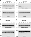
Fig 4. PND-HA-Cdc28p undergoes blue light/Ubr1-dependent degradation.
Cells expressing a chromosomal insertion of PND-HA-Cdc28p in either a UBR1 (A, B) or ubr1Δ (C, D) genetic background were grown in liquid culture in darkness to log phase, then divided and allowed to continue growth in darkness (A, C) or under blue-light illumination (B, D). Samples of culture medium were taken at 1-hour intervals and cells were processed for Western blot analysis. Western blots were divided into upper and lower sections with the upper sections probed using an antibody directed against the HA epitope in PND-HA-Cdc28p and the lower segments probed with an antibody directed against the endogenous protein Glyceraldehyde-3-phosphate dehydrogenase (GAPDH), which served as a loading control. Note that PND-HA-Cdc28p levels exhibited a dramatic decrease when grown in UBR1 cells under illumination (B). Growth in darkness (A) or under illumination in the absence of Ubr1p (D) resulted in constant levels of PND-HA-Cdc28p.
Fig 5
Fig 5. Mature PND-HA-Cdc28P undergoes rapid light dependent degradation.
Cells expressing PND-HA-Cdc28p in a UBR1 genetic background were grown in liquid culture in darkness to log phase, then divided and allowed to continue growth in darkness (A, C) or under blue-light illumination (B, D) in either the absence (A, B) or presence (C, D) of the translational inhibitor cycloheximide. Samples of culture medium were taken at 15-minute intervals and cells were processed for Western blot analysis, with an upper portion of each blot probed with an antibody against the HA epitope in PND-HA-Cdc28p and a lower portion probed with an antibody directed against endogenous GADPH, which served as a protein loading control. In the presence of Ubr1p, light-dependent loss of PND-HA-Cdc28p was very rapid, likely occurring within a single cell cycle, regardless of the absence (B) or presence (D) of cycloheximide.
Fig 6
Fig 6. Schematic diagrams of the transgene constructs encoding PND-, psd- and B-LID domain-tagged versions of the CactDN open reading frame.
Constructs encoding the three degron-tagged versions of CactDN were introduced into the Drosophila genome on the P-element transposon-based expression vector, pUASp [110], downstream of upstream activator sequences for the yeast Gal4 transcription factor (UASGAL4) and the promoter from the P-element transposase gene (PP-Transposase). Expression of the transgenes was accomplished by co-expression of a germline-specific source of the Gal4 transcription factor. (A) PND-HA-CactDN. (B) CactDN-psd. (C) CactDN-B-LID. Labels are as follows: UBI, a single copy of the ubiquitin open reading frame. LOV, encoding the LOV2 domain of plant phototropin I. HA, encoding a single copy of the influenza hemagglutinin (HA) epitope [61]. 3xMyc, sequences encoding three tandem copies of the 9E10 epitope from human c-myc [113]. ODC, an element encoding 23 amino acids from the synthetic ODC-like degron [83]. The single letters A, G, M. and R, represent codons encoding individual alanine, glycine, methionine and arginine. Specifically, M’s denote the initiation codons of the open reading frames of the three constructs. The three A’s present in the CactDN segments represent 3 serine-encoding codons that were mutated to alanines, rendering the encoded protein insensitive to Toll pathway signal-dependent proteolysis. GGR in PND-HA-CactDN represents codons encoding the two glycine residues at the C-terminus of ubiquitin and the subsequent arginine residue at the N-terminus of the LOV element. Finally, RRRG represents the codons encoding the critical C-terminal residues of the B-LID domain, which are likely to support degradation by the DesCEND mechanism [100,101].
Fig 7
Fig 7. Representative cuticular phenotypes of embryos expressing maternally provided degron-tagged cactDN constructs.
Embryos produced by females expressing the degron-tagged versions of CactDN described herein were collected and allowed to complete embryonic development in darkness, then subjected to cuticle preparation [112]. Levels of dorsalization denoted below are indicated at top right of each panel. (A) A completely dorsalized (DO) embryo produced by a female expressing cactDN-psd. (B) A strongly dorsalized (D1) embryo produced by a female expressing cactDN-B-LID. Note the presence of Filzkörper (Fk) structures (= tracheal spiracles). (C) A moderately dorsalized (D2) embryo from a female expressing PND-HA-CactDN. Note the presence of Filzkörper material and narrow ventral denticle (vd) bands. (D) A weakly dorsalized (D3) embryo from a female expressing PND-HA-CactDN, exhibiting the “twisted” phenotype. Note the asterisk marking the twist in the body axis. (E) A weakly dorsalized (D3) embryo, from a PND-HA-CactDN-expressing female, exhibiting the “U-shaped” or “tail-up” phenotype. (F) An apparently normal, unhatched (UH) embryo produced by a female expressing PND-HA-cactDN. In all panels, arrowheads mark the position of Filzkörper (Fk), arrows mark the position of ventral denticles (vd), and a left pointing angle mark (<) denotes the position of head skeletal (hs) elements. In all panels, anterior is to the left and the dorsal side of the egg is at top.
Fig 8
Fig 8. PND-CactDN and CactDN-B-LID undergo light-dependent loss in Drosophila embryos.
Embryos from females expressing a transgene encoding PND-HA-CactDN (A) or from females expressing two independent transgenic insertions encoding CactDN-B-LID (B), were collected and allowed to develop in either darkness (-) or under blue light illumination (+). Embryonic extracts were prepared from 2–4 hour-old embryos and Western blots of those extracts were probed with antibodies directed against the HA epitope (A) and against Cactus protein (B) are shown. The positions of bands corresponding to PND-HA-CactDN, CactDN-B-LID, and endogenous Cactus (Cact) are shown.
Fig 9
Fig 9. Laser illumination of live embryos expressing PND-HA-CactDN or CactDN-B-LID induces nuclear accumulation of Dorsal-GFP.
(A) Schematic showing the imaging setup that was used to visualize Dorsal-GFP in Drosophila embryos over a period of ~75 min spanning their development from nuclear cycle (nc) 12 up to gastrulation (st.6) under conditions that inactivate Cactus-degron fusions. Imaging was initiated at time = 0 (t0) and continued for a period of ~10 minutes during nc12 under low power 488 nm laser illumination. Immediately after this treatment (at t1) and extending into nc13 (a period of 15 minutes), embryos were illuminated for 20min under high power 488nm laser to initiate degron-mediated loss of CactusDN. After a 30–35’ rest in the dark at which point embryos had initiated gastrulation (t2), they were again illuminated for 5 min under low power 488nm laser to monitor the Dorsal-GFP gradient and the developmental state of the embryos (t2 + 5min). The dotted box represents the illuminated area. The remainder of the panels show four snapshots each, taken from movies of embryos containing Dorsal-GFP [85], either expressed alone (B-B“‘, control; see also S1 Movie) or together with the PND- and B-LID-tagged Cactus variants expressed under the control of the mat-α4-tub-Gal4:VP16 driver element [108]. The PND-HA-CactDN (C-C“‘, D-D“‘; see also S2 and S3 Movies) and the CactDN-B-LID (E-E“‘, F-F“‘; see also S4 and S5 Movies) fusion proteins were imaged using conditions outlined in panel A (C-C“‘, E-E“‘, S2 and S4 movies) or under low power 488nm laser illumination (light blue bar)(D-D“‘, F-F“‘, S3 and S5 Movies). Scale bars represent 65μm or 50μm, as noted; in the absence of Dorsal-GFP nuclear translocation, we used a slightly higher digital magnification (i.e. 50μm), in those cases to increase visibility of empty nuclei.
Fig 10
Fig 10. Laser illumination of live embryos expressing CactDN-psd induces transient cyclical nuclear accumulation of Dorsal-GFP.
Images shown are four snapshots taken from movies of embryos expressing Dorsal-GFP, together with the photosensitive degron-tagged CactusDN (CactDN-psd) expressed under the control of the mat-α4-tub-Gal4:VP16 driver element, imaged under different conditions. Panels represent snapshots from respective S6–S8 Movies. (A-A’”, B-B’”, C-C“‘) Imaging was initiated at time = 0 (t0) and continued for 10 minutes during nc12 under low power 488 nm, using the scheme diagrammed in Fig 9A. Just after this treatment (i.e. t1) and extending into nc13 (t1+15min) and nc14a (t1+20min), embryos were illuminated for 20min at 488nm high power to initiate degron-mediated loss of CactDN-psd (A’-A“‘; see also S6 Movie). As a control, embryos were also imaged under low power 488nm only (B-B’”; see also S7 Movie). Scale bars represent 65μm or 50μm, as noted; in the absence of Dorsal-GFP nuclear translocation, we used a slightly higher digital magnification (i.e. 50μm), to increase visibility of empty nuclei. (C-C“‘) Embryos were exposed to blue light earlier for 20 min, initiating at nc12 and into nc13 (blue bar, 488nm), and subsequently imaged under low power 488nm laser illumination. These images show that Dorsal-GFP enters nuclei in a transient manner, entering just before division but relocalize to the cytoplasm after nuclear division; see also S8 Movie.

Similar articles

Cited by

References

    1. Dang DT, Perrimon N. Use of a yeast site-specific recombinase to generate embryonic mosaics in Drosophila. Dev Genet. 1992;13: 367–375. 10.1002/dvg.1020130507 - DOI - PubMed
    1. Chou TB, Perrimon N. Use of a yeast site-specific recombinase to produce female germline chimeras in Drosophila. Genetics. 1992;131: 643–653. - PMC - PubMed
    1. Gu H, Marth J, Orban P, Mossmann H, Rajewsky K. Deletion of a DNA polymerase beta gene segment in T cells using cell type-specific gene targeting. Science. 1994. pp. 103–106. 10.1126/science.8016642 - DOI - PubMed
    1. Xu T, Rubin GM. Analysis of genetic mosaics in developing and adult Drosophila tissues. Development. 1993;117: 1223–1237. - PubMed
    1. Dietzl G, Chen D, Schnorrer F, Su K-C, Barinova Y, Fellner M, et al.. A genome-wide transgenic RNAi library for conditional gene inactivation in Drosophila. Nature. 2007;448: 151–156. 10.1038/nature05954 - DOI - PubMed

Publication types

MeSH terms