. 2021 Oct:230:12-47.
doi: 10.1016/j.ajo.2021.04.013. Epub 2021 May 14.

Assessing Photoreceptor Status in Retinal Dystrophies: From High-Resolution Imaging to Functional Vision

Affiliations

Assessing Photoreceptor Status in Retinal Dystrophies: From High-Resolution Imaging to Functional Vision

José-Alain Sahel et al. Am J Ophthalmol. 2021 Oct.

Abstract

Purpose: To describe the value of integrating phenotype/genotype data, disease staging, and evaluation of functional vision in patient-centered management of retinal dystrophies.

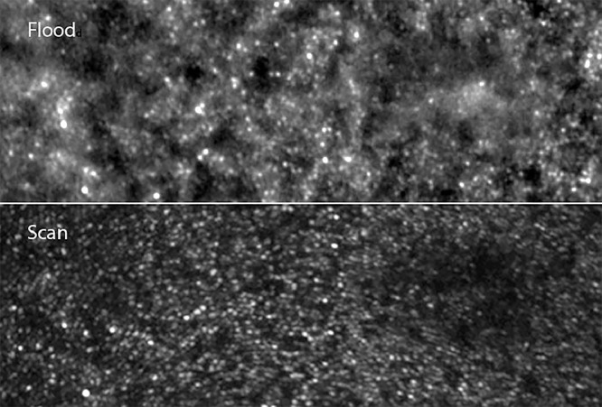
Methods: (1) Cross-sectional structure-function and retrospective longitudinal studies to assess the correlations between standard fundus autofluorescence (FAF), optical coherence tomography, visual acuity (VA), and perimetry (visual field [VF]) examinations to evaluate photoreceptor functional loss in a cohort of patients with rod-cone dystrophy (RCD); (2) flood-illumination adaptive optics (FIAO) imaging focusing on photoreceptor misalignment and orientation of outer segments; and (3) evaluation of the impact of visual impairment in daily life activities, based on functional (visual and mobility) vision assessment in a naturalistic environment in visually impaired subjects with RCD and subjects treated with Luxturna for RPE65-related Leber congenital amaurosis before and after therapy.

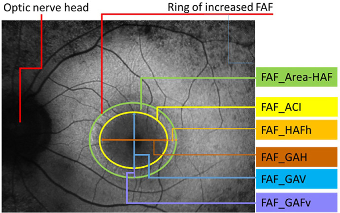
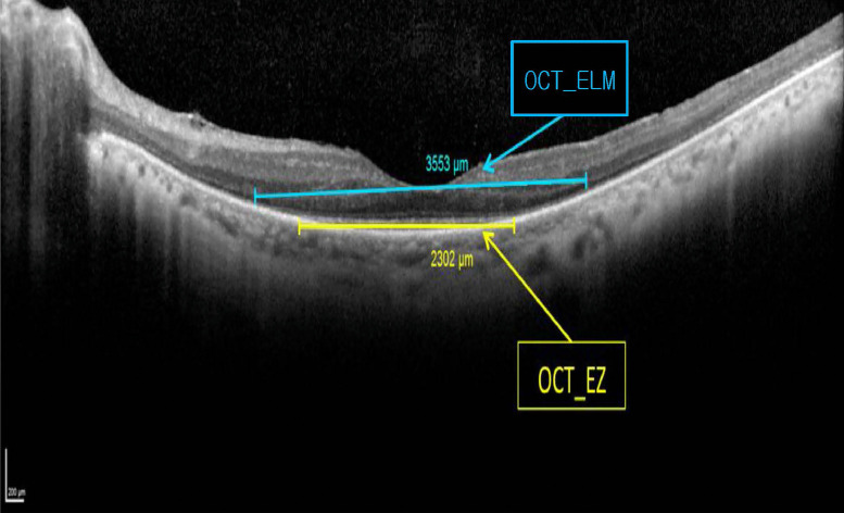
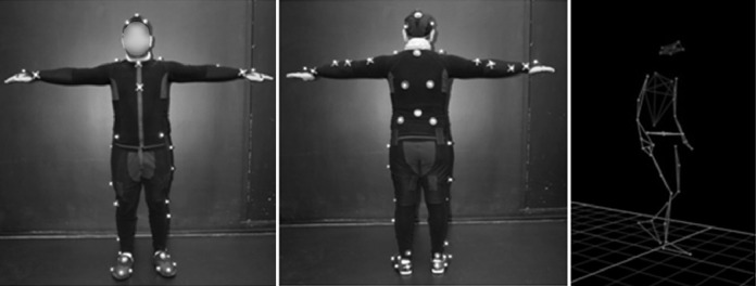
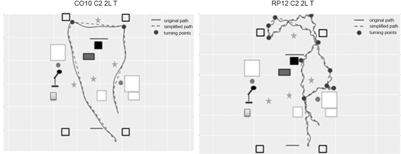
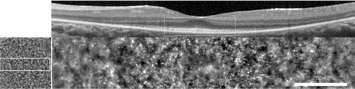
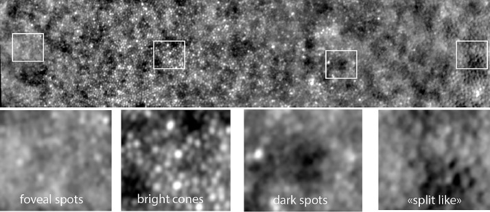
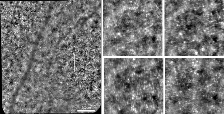
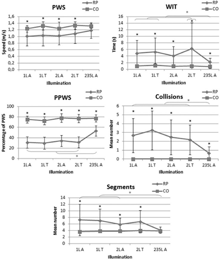
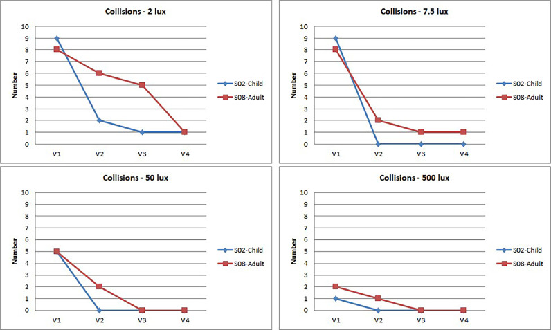
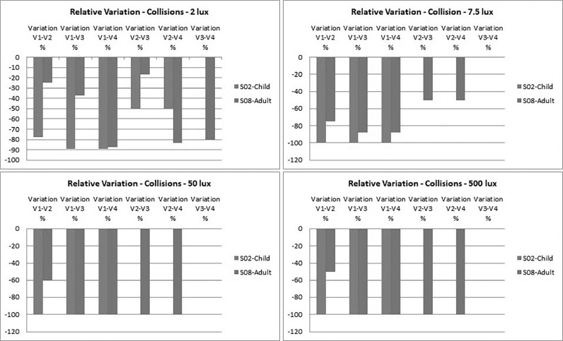
Results: The results of the cross-sectional transversal study showed that (1) VA and macular sensitivity were weakly correlated with the structural variables; and (2) functional impairment (VF) was correlated with reduction of anatomical markers of photoreceptor structure and increased width of autofluorescent ring. The dimensions of the ring of increased FAF evolved faster. Other criteria that differed among groups were the lengths of the ellipsoid zone, the external limiting membrane, and the foveal thickness. FIAO revealed a variety of phenotypes: paradoxical visibility of foveal cones; heterogeneous brightness of cones; dim, inner segment-like, and RPE-like mosaic. Directional illumination by varying orientation of incident light (Stiles-Crawford effect) and the amount of side illumination (gaze-dependent imaging) affected photoreceptor visibility. Mobility assessment under different lighting conditions showed correlation with VF, VA, contrast sensitivity (CS), and dark adaptation, with different predictive values depending on mobility study paradigms and illumination level. At high illumination level (235 lux), VF was a predictor for all mobility performance models. Under low illumination (1 and 2 lux), VF was the most significant predictor of mobility performance variables, while CS best explained the number of collisions and segments. In subjects treated with Luxturna, a very favorable impact on travel speed and reduction in the number of collisions, especially at low luminance, was observable 6 months following injection, in both children and adults.

Conclusions: Our results suggest the benefit of development and implementation of quantitative and reproducible tools to evaluate the status of photoreceptors and the impact of both visual impairment and novel therapies in real-life conditions. NOTE: Publication of this article is sponsored by the American Ophthalmological Society.


Figures

Figure 1
Figure 1
Gene replacement therapy is appropriate during the early stages of disease, when retinal photoreceptor cells are still intact. Early intervention with gene replacement, gene editing, or antisense oligonucleotides can potentially reverse vision loss and lead to close to normal visual outcomes. Neuroprotective strategies, particularly those targeted to preserve cones, are the best approaches to treat the disease with ongoing photoreceptor cell degeneration. Cone neuroprotection can stave off loss of high-acuity vision by protecting foveal cones. Stem cell therapy, optogenetic therapy, and retinal prostheses are used to restore vision during the later stages of retinal degeneration, after the loss of cone outer segments. These approaches can be applied independently from the causal mutation and are expected to restore low vision in blind patients. [From Sci Transl Med 2019;11(494):eaax2324. doi: 10.1126/scitranslmed.aax2324. (Courtesy of Dalkara D).]
Figure 2
Figure 2
View of the “Streetlab” artificial street.
Figure 3
Figure 3
View of the “Streetlab” control room.
Figure 4
Figure 4
Fundus autofluorescence picture of a retinitis pigmentosa (RP) patient with a ring of increased autofluorescence and the respective measured parameters.
Figure 5
Figure 5
Ellipsoid zone and external limiting membrane measured on the horizontal scan of the left eye of a retinitis pigmentosa patient.
Figure 6
Figure 6
Optical coherence tomography parameters analyzed in retinitis pigmentosa and control groups.
Figure 7
Figure 7
Example of a course configuration (course A). The platform was converted into an open space with office equipment (eg, tables, boxes, chairs, coat rack, lamps, plants) spread around the room. The course was designed as a triangle with a large white polystyrene block (height: 55 cm; length: 35 cm; width: 30 cm) at each angle, and the same starting and arrival points. To vary the difficulty, 15 obstacles of different sizes and contrast (high and low) were set on the course. Eleven had a fixed location and 4 (obstacles 1, 5, 8, and 9) changed location according to the course (A, B, C, and D), to avoid an adjustment bias. The ceiling of the platform is composed of 9 LED panels that produce a homogeneous atmosphere at any point of the space and according to the 3 selected light settings.
Figure 8
Figure 8
Vicon motion capture system description. The participants were equipped with a fitted velcro jumpsuit incorporating reflective markers on anatomical points based on the Plug-In-Gait (PIG) model. The latter reflected infrared emitted by Vicon cameras (T40), to collect walking parameters and head movements (not detailed).
Figure 9
Figure 9
Examples of trajectory segmentation.
Figure 10
Figure 10
Example of a mobility course presented to RPE65-related Leber congenital amaurosis subjects treated with Luxturna.
Figure 11
Figure 11
Experimental conditions for Streetlab mobility test undertaken by RPE65-related Leber congenital amaurosis patients treated with Luxturna.
Figure 12
Figure 12
Central patch of cone mosaic seen by optical coherence tomography (Top) and by flood adaptive optics–enhanced ophthalmoscopy (Bottom). Female patient affected with simplex rod-cone dystrophy of unknown genotype, since the subject denied testing.
Figure 13
Figure 13
Illustration of different cone phenotypes.
Figure 14
Figure 14
Illustration of the effect of directional imaging on the visualization of the cone mosaic. Left, reference (on-axis) image; Right, images of the area located in the square seen at 4 different off-axis light incidences. Note the variation in the appearance of the cone mosaic. Case of a 28-year-old man with rod-cone dystrophy associated with a likely pathogenic homozygous variant in USH2A (NM_206933.2; c.4628-2A>T).
Figure 15
Figure 15
Adaptive optics–enhanced ophthalmoscopy images illustrating the effect of gaze positioning on cone imaging. Both images show the same retinal area; in the top image this area was located on the image margin, while in the bottom the same area was placed centrally. Note the better visualization of the cone mosaic when placed in the center of the field of view (that is, the bottom image). Case of a 33-year-old male patient with rod-cone dystrophy of unknown genetic defect after screening on a 254 targeted gene next-generation sequencing panel (Audo et al, ref. 30).
Figure 16
Figure 16
Mobility performance in retinitis pigmentosa (RP) and control (CO) groups under different lighting conditions. PPWS = percentage of preferred walking speed; PWS = preferred walking speed; WIT = walking initiation time.
Figure 17
Figure 17
Manual Goldmann kinetic perimetry in the young patient. The surface the III4e isopter of the binocular visual central island had less than 20 degrees of diameter before treatment, and enlarged to more than 140 degrees with a thin ring scotoma.
Figure 18
Figure 18
Evolution of the percentage of preferred walking speed (PPWS) according to lighting conditions and visits. V = visit.
Figure 19
Figure 19
Relative variation for the percentage of preferred walking speed (PPWS) according to lighting conditions and visits. V = visit.
Figure 20
Figure 20
Evolution of collisions according to lighting conditions and visits. V = visit.
Figure 21
Figure 21
Relative variation for the percentage of preferred walking speed (PPWS) according to lighting conditions and visits. V = visit.
Figure 22
Figure 22
Comparison of flood adaptive optics–enhanced ophthalmoscopy (AOO) (Top) and scan AOO (Bottom). The fovea is on the right. Note the more regular aspect of the cone mosaic using scan AOO, but also the identification of additional disease features by flood AOO, such as brilliant central cones.

References

    1. Dryja TP, McGee TL, Reichel E, et al. A point mutation of the rhodopsin gene in one form of retinitis pigmentosa. Nature. 1990;343:364–366. - PubMed
    1. Russell S, Bennett J, Wellman JA, et al. Efficacy and safety of voretigene neparvovec (AAV2-hRPE65v2) in patients with RPE65-mediated inherited retinal dystrophy: A randomised, controlled, open-label, phase 3 trial. Lancet. 2017;390:849–860. - PMC - PubMed
    1. Sahel JA, Marazova K, Audo I. Clinical characteristics and current therapies for inherited retinal degenerations. Cold Spring Harb Perspect Med. 2014;5 - PMC - PubMed
    1. Busskamp V, Duebel J, Balya D, et al. Genetic reactivation of cone photoreceptors restores visual responses in retinitis pigmentosa. Science. 2010;329:413–417. - PubMed
    1. Sahel JA, Bennett J, Roska B. Depicting brighter possibilities for treating blindness. Sci Transl Med. 2019;11 - PubMed

Publication types

MeSH terms

LinkOut - more resources