Review
. 2022 Feb;28(1):9-15.
doi: 10.1177/15910199211018581. Epub 2021 May 18.

Chronic fusiform extracranial vertebral artery aneurysm with recurrent posterior circulation emboli: Case report and review of the literature

Affiliations
Review

Chronic fusiform extracranial vertebral artery aneurysm with recurrent posterior circulation emboli: Case report and review of the literature

Katherine Evans et al. Interv Neuroradiol. 2022 Feb.

Abstract

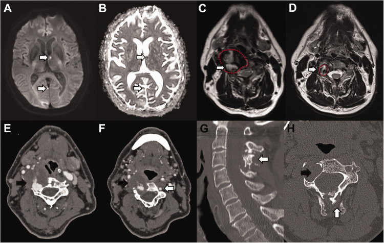
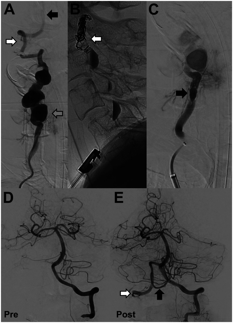
We report a case of a 64-year-old man with a fusiform right extracranial vertebral artery aneurysm, spanning over half the extra-cranial V2 (foraminal) segment, presenting with recurrent multi-focal posterior circulation embolic ischaemic stroke. The patient was treated with endovascular embolisation of the right vertebral artery to prevent further thrombo-embolic events. Distal and proximal occlusion of the aneurysmal vertebral artery was performed with a micro-vascular plug with partial aneurysm sack embolisation to aid thrombosis and reduce the risk of recanalisation. Two months post procedure MR angiography confirmed successful aneurysm occlusion with no post-procedural complication. The patient returned to his normal independent life. Endovascular treatment with vessel sacrifice is an effective treatment with low morbidity and we believe the MVP device to be a efficacious option in the vertebral artery.

Keywords: Vertebral artery; aneurysm; dissection; embolisation; embolism; stroke.


Conflict of interest statement

Declaration of conflicting interests: The author(s) declared no potential conflicts of interest with respect to the research, authorship, and/or publication of this article.

Figures

Figure 1.
Figure 1.
A and B: Multiple foci of restricted diffusion (white arrow), with corresponding low ADC value in the posterior circulation involving the right occipital lobe, left thalamus and cerebellum (not shown), in keeping with acute embolic infarction. Older non-restricting foci of embolic infarction were also evident on T2 sequences (not shown). C: T2 weighted sequence of the neck at the C4 vertebral level demonstrating an abnormal right vertebral artery (red broken line) with a central flow void and abnormal circumferential thick mixed T2 signal material (white arrow). Maximal diameter of the fusiform aneurysm was up to 35mm. D: T2 weighted sequence of the neck at the C4 vertebral level five years prior to presentation confirming significant growth (white arrow) of the right vertebral artery (red broken line). E and F: CT angiography demonstrated a grossly abnormal right vertebral artery measuring up to 5cm in diameter. The aneurysm demonstrated thick thrombus lining the lumen (black arrow) with areas of fissuring and crenulation. The contralateral left vertebral artery was of normal calibre and appearances (white arrow). G: Sagittal bone window reconstruction of CT demonstrates abnormal appearance to the spinous processes (white arrow) of C2 to C4 consistent with historic trauma. H: Axial CT at C3 level shows a grossly expanded right foramen transversarium consistent with a chronic process of fusiform aneurysm formation (black arrow).
Figure 2.
Figure 2.
A: Digital subtraction angiography (DSA)(anterior-posterior view) confirmed a chronic fusiform aneurysmal right vertebral artery with areas of saccular dilatation (grey arrow). The distal V3 segment of the vertebral artery (white arrow) and intra-cranial basilar artery were normal (black arrow). B: DSA (lateral view) endovascular embolization was performed with distal micro-vascular plug and platinum coils (white arrow). Contrast stagnation can be seen in the areas of saccular dilatation. C: Proximal cervical vertebral artery embolization was performed with a single micro-vascular plug (black arrow). D: Pre-embolization angiogram with injection in the left vertebral artery demonstrates normal appearances with no opacification of the right posterior inferior cerebellar artery territory (PICA). E: Post-embolization retrograde flow of contrast from the left vertebral artery fills the intra-dural right vertebral artery (white arrow) and perfuses the right PICA (black arrow).

References

    1. Matas R. Traumatisms and traumatic aneurisms of the vertebral artery and their surgical treatment with the report of a cured case. Ann Surg 1893; 18: 477–521. - PMC - PubMed
    1. Krueger BR, Okazaki H. Vertebral-basilar distribution infarction following chiropractic cervical manipulation. Mayo Clin Proc 1980; 55: 322–332. - PubMed
    1. Do KH, Leggit JC, Galifianakis A. Extracranial vertebral artery dissecting aneurysm with snowboarding: a case report. Curr Sports Med Rep 2018; 17: 16–19. - PubMed
    1. Egnor MR, Page LK, David C. Vertebral artery aneurysm – a unique hazard of head B banging by heavy metal rockers. Pediatr Neurosurg 1991; 17: 135–138. - PubMed
    1. Schievink WI, Piepgras DG. Cervical vertebral artery aneurysms and arteriovenous fistulae in neurofibromatosis type 1: case reports. Neurosurgery 1991; 29: 760–765. - PubMed

LinkOut - more resources