Review
. 2021 Nov 1;13(11):a040949.
doi: 10.1101/cshperspect.a040949.

Transcription Factor Dynamics

Affiliations
Review

Transcription Factor Dynamics

Feiyue Lu et al. Cold Spring Harb Perspect Biol. .

Abstract

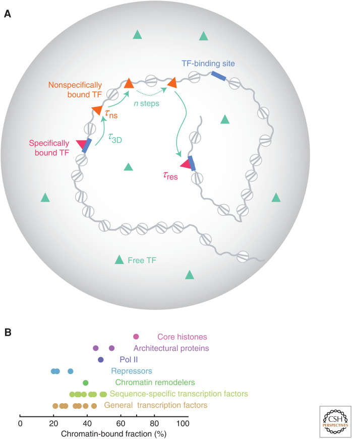
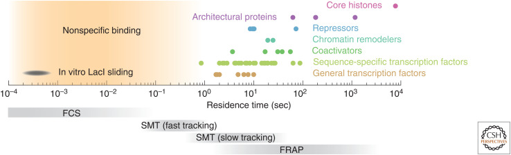
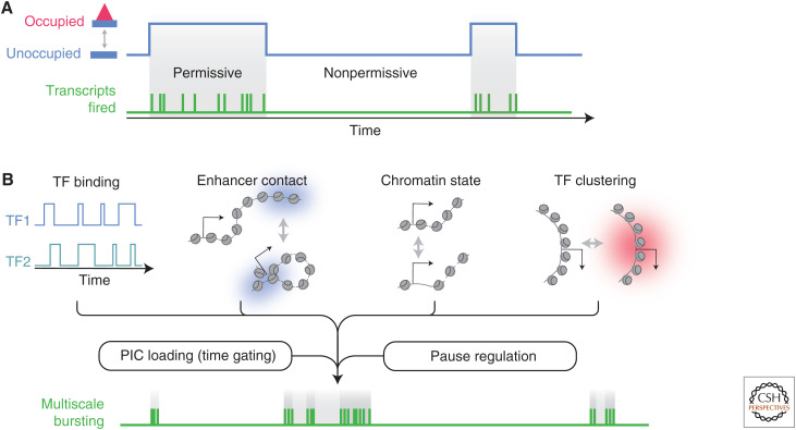
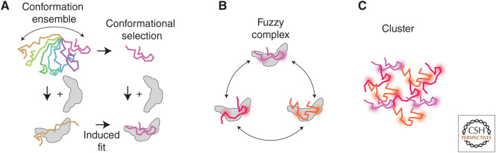
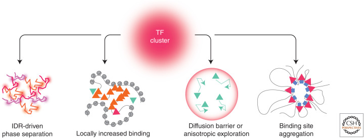
To predict transcription, one needs a mechanistic understanding of how the numerous required transcription factors (TFs) explore the nuclear space to find their target genes, assemble, cooperate, and compete with one another. Advances in fluorescence microscopy have made it possible to visualize real-time TF dynamics in living cells, leading to two intriguing observations: first, most TFs contact chromatin only transiently; and second, TFs can assemble into clusters through their intrinsically disordered regions. These findings suggest that highly dynamic events and spatially structured nuclear microenvironments might play key roles in transcription regulation that are not yet fully understood. The emerging model is that while some promoters directly convert TF-binding events into on/off cycles of transcription, many others apply complex regulatory layers that ultimately lead to diverse phenotypic outputs. Cracking this kinetic code is an ongoing and challenging task that is made possible by combining innovative imaging approaches with biophysical models.


Figures

Figure 1.
Figure 1.
Occupancy versus kinetics. ChIP-seq (top) measures the average occupancy of a given transcription factor (TF) at its binding site. Increased occupancy may result from higher TF-binding frequency (higher kon), or longer residence times (1/koff, bottom). Kinetic profiles can be decoded into distinct transcriptional outputs by promoters.
Figure 2.
Figure 2.
Transcription factor (TF) mobility in the nucleus. (A) Facilitated 3D diffusion model. While searching for its target sites (blue), a TF makes multiple, brief, and nonspecific contacts (τns < 1 sec; orange) with open chromatin before landing on its cognate site where it dwells longer (τres ∼ 1–100 sec; magenta). The average time between two specific binding events is defined as the search time τsearch = (n−1)*(τ3D + τns) + τ3D, where n ∼ 10–100 is the number of trials and τ3D is the averaged diffusion time between two trials. (Panel A is based on data in Chen et al. 2014.) (B) Experimentally measured chromatin-bound fraction (circles) for various TFs compiled from the community resource developed by Mir and colleagues (www.mir-lab.com/dynamics-database) and the recent literature. Only factors expressed as knockins or rescuing a knockout background are featured here.
Figure 3.
Figure 3.
Chromatin association timescales in vivo. Experimentally measured residence times (circles) compiled from the community resource developed by Mir and colleagues (www.mir-lab.com/dynamics-database) and the recent literature, relative to the temporal resolution of different imaging techniques (bottom, gray). (FCS) Fluorescence correlation spectroscopy, (SMT) single-molecule tracking, (FRAP) fluorescence recovery after photobleaching.
Figure 4.
Figure 4.
Decoding of transcription factor (TF) kinetics by promoters. (A) In simple systems, TF binding directly leads to permissive periods (gray) during which many Pol II are rapidly fired. In this one-to-one model, the TF residence time equals the burst duration. (B) Promoters often integrate complex regulation from multiple TFs and enhancers, as well as the state of chromatin at the promoter, and the kinetics of cluster formation. These interdependent inputs are processed by the transcription machinery, which applies further control layers, leading to multistate bursting dynamics. (PIC) Preinitiation complex.
Figure 5.
Figure 5.
Intrinsically disordered regions (IDRs) mediate different types of complexes. (A) Binding-coupled folding: free IDRs explore a vast conformation space (colors), but some IDRs adopt a fixed conformation when bound to a partner (gray). (B) In a fuzzy complex, the IDR is dynamic yet remains bound to its partner. (C) Nonstoichiometric complexes (clusters) can form via networked interactions between multivalent IDRs.
Figure 6.
Figure 6.
Clustering mechanisms. Transcription factor (TF) clustering can occur via phase separation, thanks to multivalent interactions between intrinsically disordered regions (IDRs) (left). Other mechanisms also exist; locally enhanced TF binding on highly accessible chromatin, locally anisotropic diffusion, and collapse of a TF-binding site. Triangles represent TFs; green, orange, and magenta denote, respectively, free, nonspecifically bound, and specifically bound species.

References

    1. Alexander JM, Guan J, Li B, Maliskova L, Song M, Shen Y, Huang B, Lomvardas S, Weiner OD. 2019. Live-cell imaging reveals enhancer-dependent Sox2 transcription in the absence of enhancer proximity. eLife 8: e41769. 10.7554/eLife.41769 - DOI - PMC - PubMed
    1. Bancaud A, Huet S, Daigle N, Mozziconacci J, Beaudouin J, Ellenberg J. 2009. Molecular crowding affects diffusion and binding of nuclear proteins in heterochromatin and reveals the fractal organization of chromatin. EMBO J 28: 3785–3798. 10.1038/emboj.2009.340 - DOI - PMC - PubMed
    1. Bartman CR, Hsu SC, Hsiung CCS, Raj A, Blobel GA. 2016. Enhancer regulation of transcriptional bursting parameters revealed by forced chromatin looping. Mol Cell 62: 237–247. 10.1016/j.molcel.2016.03.007 - DOI - PMC - PubMed
    1. Basu S, Mackowiak SD, Niskanen H, Knezevic D, Asimi V, Grosswendt S, Geertsema H, Ali S, Jerković I, Ewers H, et al. 2020. Unblending of transcriptional condensates in human repeat expansion disease. Cell 181: 1062–1079.e30. 10.1016/j.cell.2020.04.018 - DOI - PMC - PubMed
    1. Benabdallah NS, Williamson I, Illingworth RS, Kane L, Boyle S, Sengupta D, Grimes GR, Therizols P, Bickmore WA. 2019. Decreased enhancer–promoter proximity accompanying enhancer activation. Mol Cell 76: 473–484.e7. 10.1016/j.molcel.2019.07.038 - DOI - PMC - PubMed

Publication types

MeSH terms

LinkOut - more resources