. 2021 May 25;118(21):e2020389118.
doi: 10.1073/pnas.2020389118.

Short hydrogen bonds enhance nonaromatic protein-related fluorescence

Affiliations

Short hydrogen bonds enhance nonaromatic protein-related fluorescence

Amberley D Stephens et al. Proc Natl Acad Sci U S A. .

Abstract

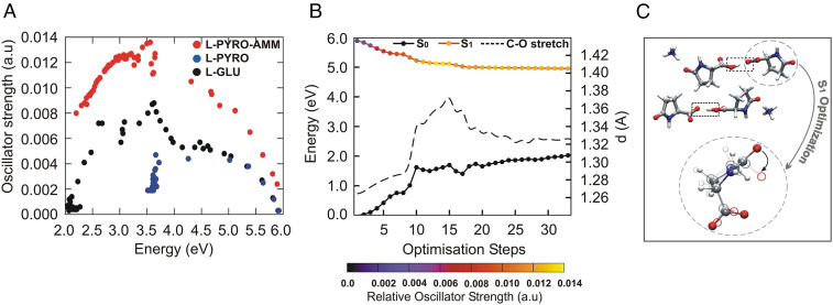
Fluorescence in biological systems is usually associated with the presence of aromatic groups. Here, by employing a combined experimental and computational approach, we show that specific hydrogen bond networks can significantly affect fluorescence. In particular, we reveal that the single amino acid L-glutamine, by undergoing a chemical transformation leading to the formation of a short hydrogen bond, displays optical properties that are significantly enhanced compared with L-glutamine itself. Ab initio molecular dynamics simulations highlight that these short hydrogen bonds prevent the appearance of a conical intersection between the excited and the ground states and thereby significantly decrease nonradiative transition probabilities. Our findings open the door to the design of new photoactive materials with biophotonic applications.

Keywords: intrinsic fluorescence; short hydrogen bond; ultraviolet fluorescence.


Conflict of interest statement

The authors declare no competing interest.

Figures

Fig. 1.
Fig. 1.
L-glu forms L-pyro-amm upon heating. (A) SEM image of crystals of L-pyro-amm dried. (B) XRD analysis of heated L-glu sample show the newly formed structure, L-pyro-amm. Geometry optimizations show that eight pyroglutamine groups in pairs, and four ammonium ions (144 atoms) for each dimer of pyroglutamine pairs, are complexed in the crystal. An SHB of 2.45 Å (within red dashed lines) is present near the ammonium ion (white: hydrogen, red: oxygen, blue: nitrogen, and gray: carbon). (C) Experimental (blue line) and theoretical (gray line) THz-TDS of the L-pyro-amm sample are in agreement and confirm the presence of the new L-pyro-amm structure.
Fig. 2.
Fig. 2.
Optical properties of L-pyro-amm are distinct from L-glu and L-pyro. (A) Absorption spectra of 0.3 M L-glu (black), L-pyro (blue), and L-pyro-amm (red) (L-glu incubated for 8 d at 65 °C) in water taken between 200 to 500 nm shows primarily features of L-pyro-amm. (B) Absorption spectra of L-glu, L-pyro, and L-pyro-amm obtained from periodic DFT calculations with the B3LYP functional. L-pyro-amm features the lowest lying excited states, which are characterized by the largest oscillator strengths. (C) Absorption spectra for L-glu and L-pyro-amm obtained from periodic simulations at room temperature. The spectra were computed by averaging over 25 frames randomly sampled from the AIMD simulations. (D) The excited-state electron density computed for L-pyro-amm from the optimized structure computed at the first peak (arrow in B). The lowest excited-state density shows a response from various parts of the crystal structure including the pyroglutamic acid ring and the SHB region (see dashed circle). The orange and green surfaces correspond to regions involving a decrease and increase in electron density, respectively, shown at an iso-value of 1 × 10⁻⁵. (E) A total of 1 M L-glu was incubated at 65 °C, and the excitation and emission spectra were measured over time. Excitation spectra were measured between 250 to 400 nm with the emission set at 420 nm, and emission spectra were measured between 380 to 560 nm with the excitation set at 360 nm.
Fig. 3.
Fig. 3.
Free energy profiles along the proton transfer coordinate show only L-pyro-amm displays a double-well potential. Free energy profiles along the proton transfer coordinate are displayed for L-glu (black), L-pyro (blue), and L-pyro-amm (red) at RT. The L-glu and L-pyro display single-well potentials, while the L-pyro-amm system is the only one the exhibits a double-well potential, implying that there is proton transfer from one side to the other.
Fig. 4.
Fig. 4.
Comparison of the HOMO and LUMO orbitals on two L-pyro-amm models. L-pyro-amm structures are presented where the ammonium cation is located directly near the HB (A) and where the ammonium cation is located away from the HB (B). While both models predicted charge-transfer HOMO–LUMO states, only in the case of A is the transition predicted to be in the vicinity of the experimentally observed peak, 304 nm compared with 669 nm for B.
Fig. 5.
Fig. 5.
L-pyro-amm optical properties as seen through excited state optimizations. (A) Scatter plot of the oscillator strengths versus the emission energy (defined as the difference between the first excited state and the ground state) during the excited optimizations of L-glu (black), L-pyro (blue), and L-pyro-amm (red). (B) Ground- and excited-state energies are plotted as a function of the excited-state optimization of the system shown in C. The curves on the excited state are color coded with the oscillator strengths. S0 and S1 refer to the ground- and excited-state energies, respectively. (C) A snapshot of the optimized excited-state cluster also showing the lengthening of the carbonyl bond and the deplanarization of the ring.
Fig. 6.
Fig. 6.
Excited-state AIMD highlights how the presence of an SHB and the ammonium ion is enhancing fluorescence. Excited-state AIMD performed for the (A) L-pyro dimer, (B) the L-pyro dimer constraining the SHB distance, and (C) L-pyro-amm is shown. (D) Accumulated nonradiative S1 → S0 decay probability, ANRP(t), for the L-pyro dimer (dimer-SHB) and the constrained SHB L-pyro (dimer-), where the HB distance was fixed at 3.0, 3.5, and 4.5 Å, and for the L-pyro-amm dimer (dimer-amm-SHB). (E) Accumulated nonradiative decay probability for L-pyro (red discontinuous line) and L-pyro-amm (black dashed line). (F) Carboxyl C = O distance histogram for the L-pyro-amm dimer at the SHB distance (red curve) or constraining the HB to 4.5 Å (blue curve). (G) Ring dihedral angle histogram for the L-pyro-amm dimer at the SHB distance (red curve) or constraining the HB to 4.5 Å (blue curve).

References

    1. Shukla A., et al. ., A novel UV laser-induced visible blue radiation from protein crystals and aggregates: Scattering artifacts or fluorescence transitions of peptide electrons delocalized through hydrogen bonding? Arch. Biochem. Biophys. 428, 144–153 (2004). - PubMed
    1. Amdursky N., et al. ., Blue luminescence based on quantum confinement at peptide nanotubes. Nano Lett. 9, 3111–3115 (2009). - PubMed
    1. Chan F. T., Pinotsi D., Schierle G. S. K., Kaminski C. F., Bio-nanoimaging: Protein Misfolding and Aggregation (Elsevier, 2013), pp. 147–155.
    1. Chan F. T. S., et al. ., Protein amyloids develop an intrinsic fluorescence signature during aggregation. Analyst (Lond.) 138, 2156–2162 (2013). - PMC - PubMed
    1. Pinotsi D., et al. ., Proton transfer and structure-specific fluorescence in hydrogen bond-rich protein structures. J. Am. Chem. Soc. 138, 3046–3057 (2016). - PubMed

Publication types

LinkOut - more resources