. 2021 May 17;7(1):110.
doi: 10.1038/s41420-021-00497-x.

mascRNA and its parent lncRNA MALAT1 promote proliferation and metastasis of hepatocellular carcinoma cells by activating ERK/MAPK signaling pathway

Affiliations

mascRNA and its parent lncRNA MALAT1 promote proliferation and metastasis of hepatocellular carcinoma cells by activating ERK/MAPK signaling pathway

Shu-Juan Xie et al. Cell Death Discov. .

Abstract

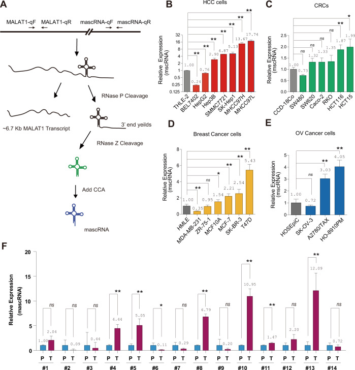
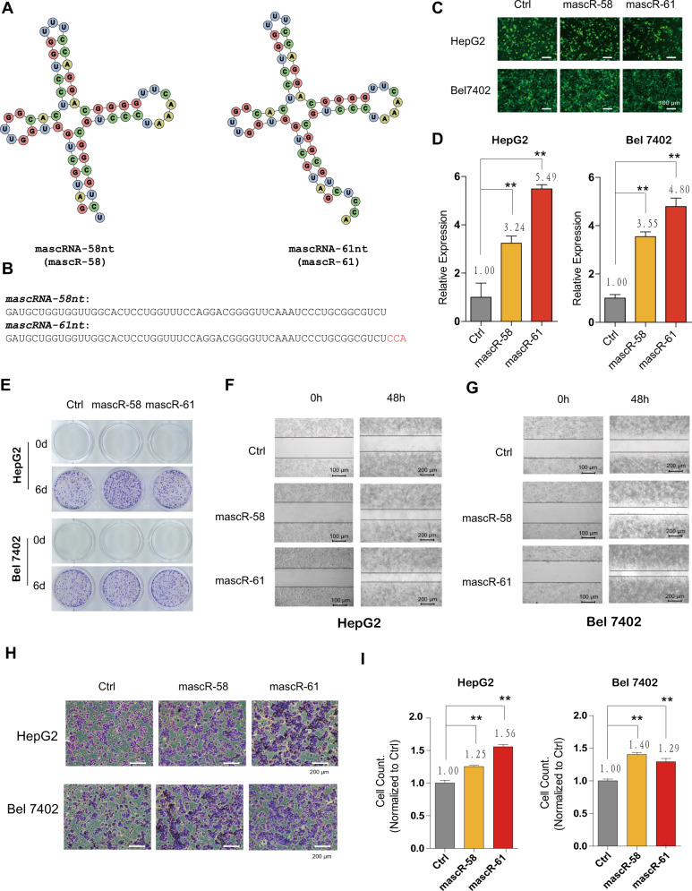
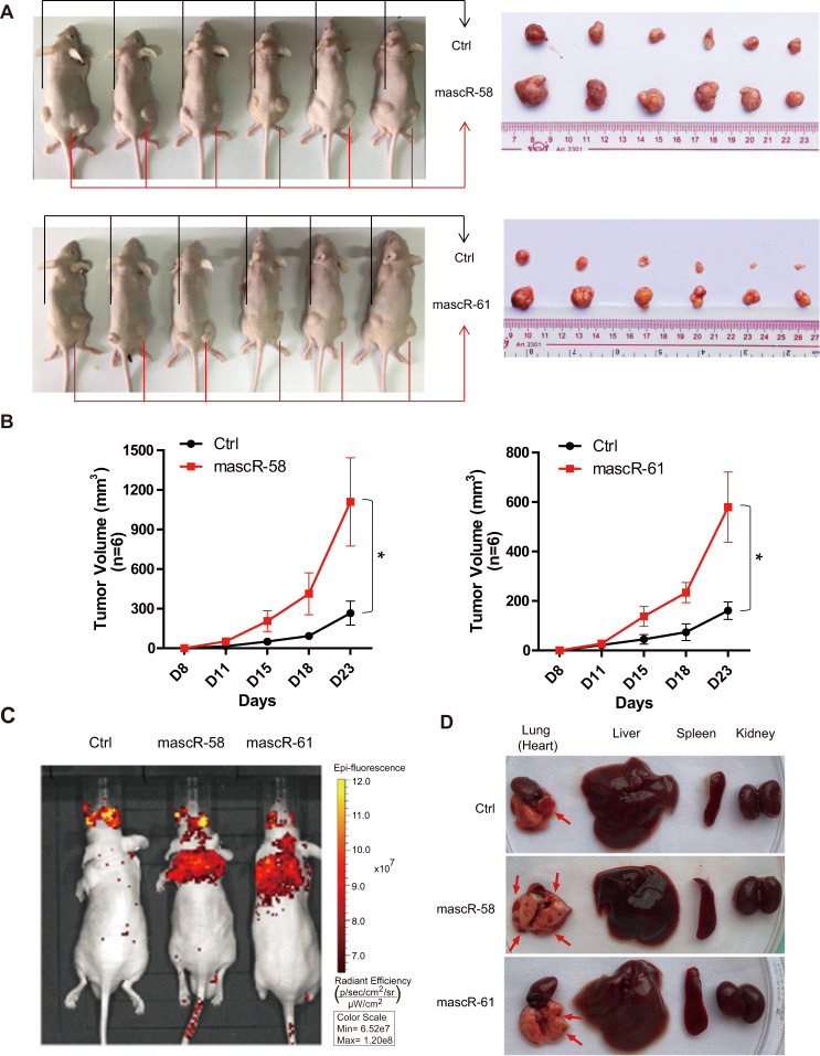
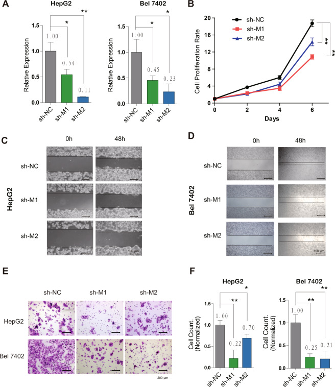
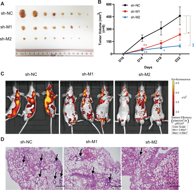
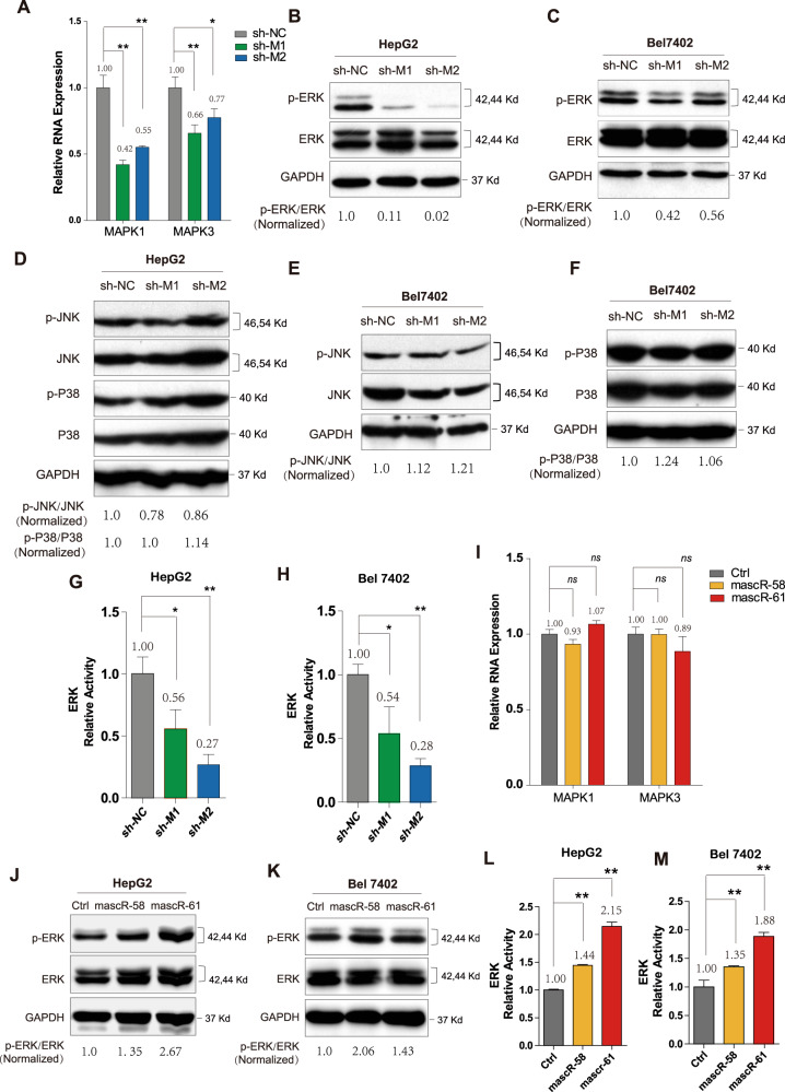
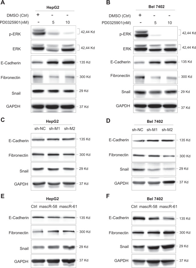
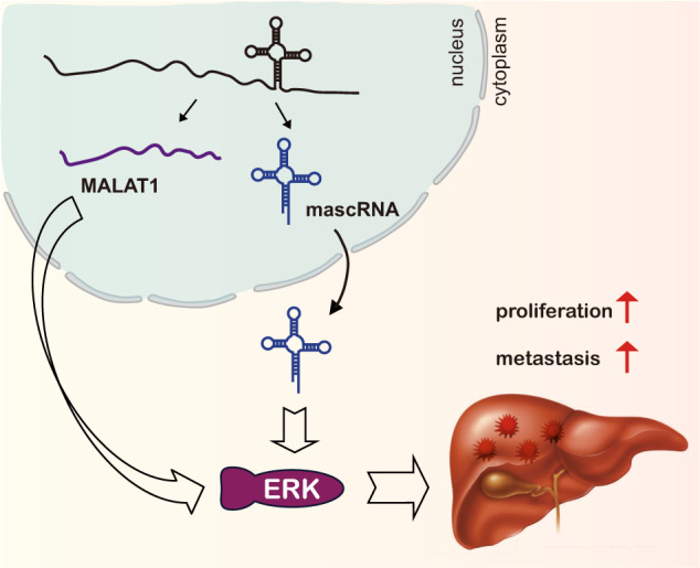
MALAT1-associated small cytoplasmic RNA (mascRNA) is a cytoplasmic tRNA-like small RNA derived from nucleus-located long noncoding RNA (lncRNA) metastasis-associated lung adenocarcinoma transcript 1 (MALAT1). While MALAT1 was extensively studied and was found to function in multiple cellular processes, including tumorigenesis and tumor progression, the role of mascRNA was largely unknown. Here we show that mascRNA is upregulated in multiple cancer cell lines and hepatocellular carcinoma (HCC) clinical samples. Using HCC cells as model, we found that mascRNA and its parent lncRNA MALAT1 can both promote cell proliferation, migration, and invasion in vitro. Correspondingly, both of them can enhance the tumor growth in mice subcutaneous tumor model and can promote metastasis by tail intravenous injection of HCC cells. Furthermore, we revealed that mascRNA and MALAT1 can both activate ERK/MAPK signaling pathway, which regulates metastasis-related genes and may contribute to the aggressive phenotype of HCC cells. Our results indicate a coordination in function and mechanism of mascRNA and MALAT1 during development and progress of HCC, and provide a paradigm for deciphering tRNA-like structures and their parent transcripts in mammalian cells.


Conflict of interest statement

The authors declare no competing interests.

Figures

Fig. 1
Fig. 1. mascRNA is enriched in multiple cancer cell lines and HCC clinical samples.
A Schematic diagram of mascRNA biogenesis. The position of primers for real-time RT-PCR of mature mascRNA or MALAT1 were marked with arrows on the upper diagram. mascRNA expression levels were measured by real-time RT-PCR in (B) normal hepatocyte THLE-2 and seven liver cancer cell lines (BEL7402, HepG2, Hep3B, SMMC-7721, SK-Hep1, MHCC97H, and MHCC97L), C normal colorectal cell line CCD-18Co and six colorectal cancer cell lines (SW480, SW620, Caco-2, RKO, HCT116, and HCT15), D HMLE and six breast cancer cell lines (MDA-MB-231, ZR-75-1, MCF10A, MCF-7, SK-BR-3, and T47D), E HOSEpiC and three ovarian cancer cell lines (SK-OV-3, A2780/TAX, and HO-8910PM), respectively. F mascRNA expression levels in 14 human primary HCC tissue (T) and paracancer tissue (P). Quantitative data are represented as mean ± SD. *P < 0.05, **P < 0.01.
Fig. 2
Fig. 2. mascRNA promotes HCC cell proliferation and metastasis in vitro.
A Secondary structure diagram of mascRNA without the terminal CCA (mascR-58, 58nt) and mature mascRNA containing CCA (mascR-61, 61nt). B Sequences of mascRNA-58nt and mascRNA-61nt. C GFP fluorescent signals of HepG2 and Bel7402 cells with control/oe-mascR-58nt/oe-mascR-61nt lentivirus infection. Scale bar represents 100 μm. D The expressions of mascRNA in HepG2/Bel7402 + control/oe-mascR-58nt/oe-mascR-61nt stable cells were measured by real-time RT-PCR. E The effect of mascRNA overexpression on cell proliferation was detected by colony formation assay. 1000 HepG2 or Bel7402 stable cells were seeded for each well in 12-well plate. Colonies were stained with 0.1% crystal violet. Wound healing assays were performed in (F) HepG2 and (G) Bel7402 stable cell lines. Movement of cells into wound was shown at 0 h (Scale bar represents 100 μm and 48 h (Scale bar represents 200 μm post scratch. Cell migration was assessed by recover of the scratch. H Transwell matrigel invasion assay showing the effect of mascRNA overexpression on the invasive activity of HepG2 or Bel7402 stable cells. Scale bar represents 200 μm. I The graphics presents the number of invaded cells, normalized to the control group. Quantitative data were represented as mean ± SD. *P < 0.05, **P < 0.01.
Fig. 3
Fig. 3. mascRNA promotes HCC tumor growth and metastasis in vivo.
A Subcutaneous tumor formation in male BALB/c-nu mice. mascR-58nt or mascR-61nt overexpressed Bel7402 stable cells were injected subcutaneous on the right side of mice, while the same number of corresponding control cells were injected on the left side of the same mice. After 4 weeks, the mice were dissected, and the tumors grown under the skin were removed, measured, and photographed. B The tumor volumes were measured on day 8, 11, 15, 18, and 23 after subcutaneous injection. n = 6/group. C In vivo metastasis assay of Bel7402 stable cells was performed by tail intravenous injection. Representative images of mice with the scramble control (Ctrl), mascR-58nt and mascR-61nt overexpression constructs were taken by the in vivo animal imaging system. The bioluminescence signals indicated the location of cancer cells. D Mice after the bioluminescence signal detection were sacrificed and dissected. Lung (heart), liver, spleen, and kidney were excised and the surface changes of organs were observed. Lesions in lung are indicated by red arrows. Quantitative data were represented as mean ± SD. *P < 0.05.
Fig. 4
Fig. 4. MALAT1 promotes HCC cell proliferation and metastasis in vitro.
A The expression of MALAT1 in HepG2/Bel7402 + shNC/shMALAT1-1/shMALAT1-2 stable cell lines were measured by real-time RT-PCR. B CCK8 assay was performed to detect the effect of MALAT1 on cell proliferation at day 0, 2, 4, and 6. Wound healing assays were performed in (C) HepG2 and (D) Bel7402 NC/MALAT1 knockdown stable cell lines. Movement of cells into wound was shown at 0 and 48 h post scratch. Scale bar represents 100 μm. E Transwell matrigel invasion assay showing the effect of MALAT1 knockdown on the invasive activity of HepG2 or Bel7402 stable cells. Scale bar represents 200 μm. F The graphics presents the number of invaded cells, normalized to NC group. Quantitative data were represented as mean ± SD. *P < 0.05, **P < 0.01.
Fig. 5
Fig. 5. MALAT1 promotes HCC tumor growth and metastasis in vivo.
A Subcutaneous tumor formation in male BALB/c-nu mice. shMALAT1-1 or shMALAT1-2 Bel7402 stable cells were injected subcutaneous on the right side of mice, while the same number of corresponding control cells were injected on the left side of the same mice. After 4 weeks, the mice were dissected, and the tumors grown under the skin were removed, measured, and photographed. B The tumor volumes were measured on day 10, 14, 18, and 22 after subcutaneous injection. n = 8/group. C In vivo metastasis assay of Bel7402 cells with MALAT1 downregulation was performed by tail intravenous injection. Representative images of mice with the scramble control (shNC), shMALAT1-1, or shMALAT1-2 knockdown constructs were taken by the in vivo animal imaging system. The bioluminescence signals indicated the location of cancer cells. D Mice after the bioluminescence signal detection were sacrificed and dissected. Organs were excised and the metastasis in lung was further investigated by immunohistochemistry analysis. Representative H&E staining images showing lung metastasis in control and shMALAT1 mice. Lesions in lung are indicated by black arrows. Scale bar represents 100 μm. Quantitative data were represented as mean ± SD. *P < 0.05, **P < 0.01.
Fig. 6
Fig. 6. mascRNA and MALAT1 active ERK/MAPK signaling pathway in HCC.
A The expressions of MAPK1 and MAPK3 were detected by real-time RT-PCR in control and shMALAT1 HepG2 stable cell lines. Western blotting for ERK and p-ERK in (B) HepG2 and (C) Bel7402 control and shMALAT1 stable cells. D Western blotting for JNK, p-JNK, P38, p-P38 in HepG2 control and shMALAT1 stable cells. Western blotting for (E) JNK, p-JNK, F P38, p-P38 in Bel7402 control and shMALAT1 stable cells. The activity of ERK signaling was examined by the dual-luciferase reporter assay in (G) HepG2 and (H) Bel7402 control and shMALAT1 stable cells. I The expressions of MAPK1 and MAPK3 were detected by real-time RT-PCR in control and mascRNA overexpressed HepG2 stable cell lines. Western blotting for ERK and p-ERK in (J) HepG2 and (K) Bel7402 control and mascRNA overexpressed stable cells. The activity of ERK signaling was examined by the dual-luciferase reporter assay in (L) HepG2 and (M) Bel7402 control and mascRNA overexpressed stable cells. All western blotting in this figure used GAPDH as a control. The band density of ERK, p-ERK, JNK, p-JNK, P38, and p-P38 were normalized to respective GAPDH, and the values of p-ERK/ERK, p-JNK/JNK, and p-P38/P38 were showed in the figure. Quantitative data were represented as mean ± SD. *P < 0.05, **P < 0.01.
Fig. 7
Fig. 7. mascRNA and MALAT1 regulate metastasis genes through ERK/MAPK signaling.
Western blotting for p-ERK, ERK, E-Cadherin, Fibronectin, Snail in (A) HepG2 and (B) Bel7402 cells treated with DMSO or 5/10 nM PD0325901. Western blotting for E-Cadherin, Fibronectin, Snail in (C) HepG2 and (D) Bel7402 control and shMALAT1 stable cells. Western blotting for E-Cadherin, Fibronectin, Snail in (E) HepG2 and (F) Bel7402 control and mascRNA overexpressed stable cells. GAPDH was used as control.
Fig. 8
Fig. 8. mascRNA and its parent lncRNA MALAT1 can both promote cell proliferation and metastasis of HCC cells by activating ERK/MAPK signaling.
Schematic overview of mascRNA and MALAT1 in HCC.

Similar articles

Cited by

References

    1. Bertone P, et al. Global identification of human transcribed sequences with genome tiling arrays. Science. 2004;306:2242–2246. doi: 10.1126/science.1103388. - DOI - PubMed
    1. Djebali S, et al. Landscape of transcription in human cells. Nature. 2012;489:101–108. doi: 10.1038/nature11233. - DOI - PMC - PubMed
    1. Kapranov P, et al. RNA maps reveal new RNA classes and a possible function for pervasive transcription. Science. 2007;316:1484–1488. doi: 10.1126/science.1138341. - DOI - PubMed
    1. Cai X, Cullen BR. The imprinted H19 noncoding RNA is a primary microRNA precursor. RNA. 2007;13:313–316. doi: 10.1261/rna.351707. - DOI - PMC - PubMed
    1. Su Z, Wilson B, Kumar P, Dutta A. Noncanonical roles of tRNAs: tRNA fragments and beyond. Annu. Rev. Genet. 2020;54:47–69. doi: 10.1146/annurev-genet-022620-101840. - DOI - PMC - PubMed

LinkOut - more resources