. 2021 May 17;11(1):10439.
doi: 10.1038/s41598-021-89836-7.

Radiation inducible MafB gene is required for thymic regeneration

Affiliations

Radiation inducible MafB gene is required for thymic regeneration

Daiki Hashimoto et al. Sci Rep. .

Abstract

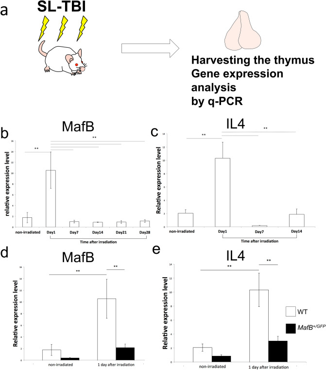
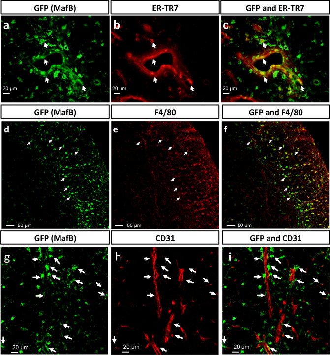
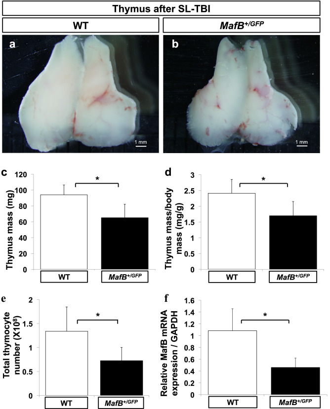
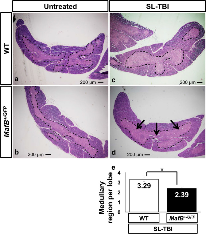
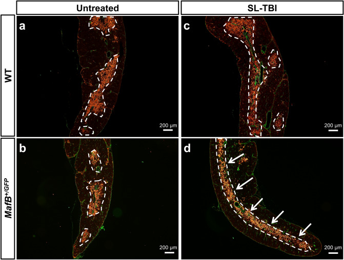
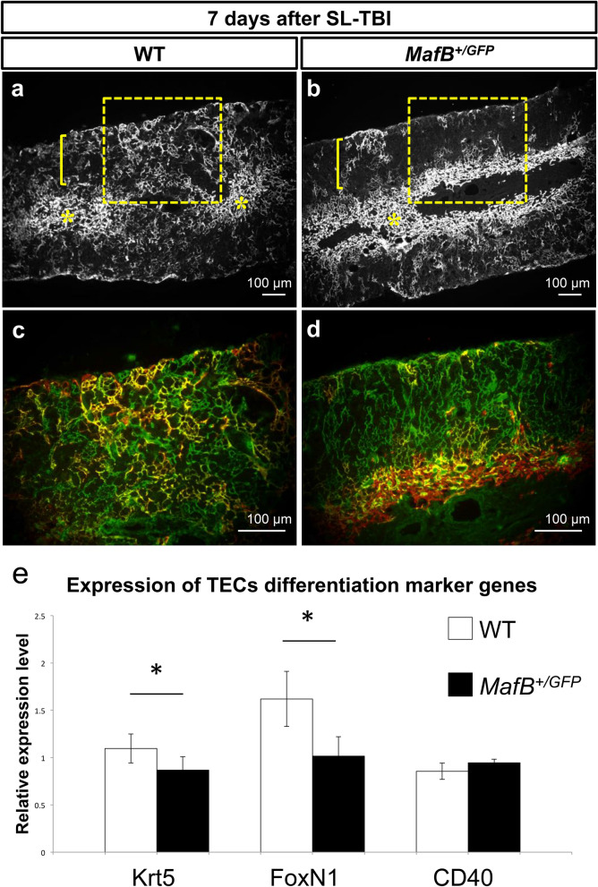
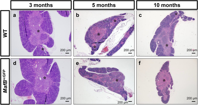
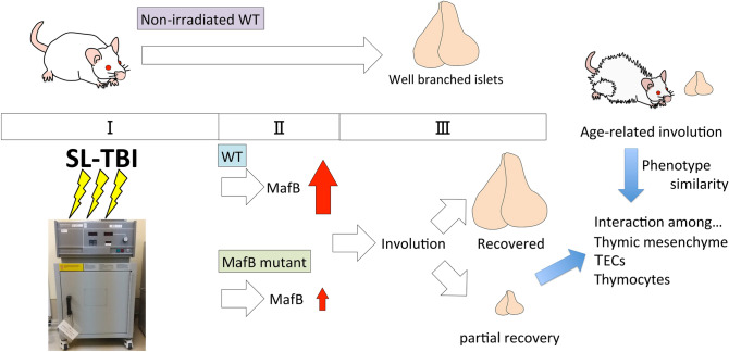
The thymus facilitates mature T cell production by providing a suitable stromal microenvironment. This microenvironment is impaired by radiation and aging which lead to immune system disturbances known as thymic involution. Young adult thymus shows thymic recovery after such involution. Although various genes have been reported for thymocytes and thymic epithelial cells in such processes, the roles of stromal transcription factors in these remain incompletely understood. MafB (v-maf musculoaponeurotic fibrosarcoma oncogene homolog B) is a transcription factor expressed in thymic stroma and its expression was induced a day after radiation exposure. Hence, the roles of mesenchymal MafB in the process of thymic regeneration offers an intriguing research topic also for radiation biology. The current study investigated whether MafB plays roles in the adult thymus. MafB/green fluorescent protein knock-in mutant (MafB+/GFP) mice showed impaired thymic regeneration after the sublethal irradiation, judged by reduced thymus size, total thymocyte number and medullary complexity. Furthermore, IL4 was induced after irradiation and such induction was reduced in mutant mice. The mutants also displayed signs of accelerated age-related thymic involution. Altogether, these results suggest possible functions of MafB in the processes of thymic recovery after irradiation, and maintenance during aging.


Conflict of interest statement

The authors declare no competing interests.

Figures

Figure 1
Figure 1
The induction of radiation induced genes, MafB and IL4, in the thymus. (a) The schematic illustration indicates the method to examine expression of radiation induced genes. Sublethal total body irradiation (SL-TBI) was performed. Thymic RNA was extracted and q-PCR was performed. (b) Prominent MafB gene induction after SL-TBI was detected at Day1 in WT. (c) The expression of IL4 after the SL-TBI. Prominent induction of IL4 was detected after 1 day SL-TBI. (d) The less prominent induction of MafB expression was reduced in the MafB+/GFP mutant mice. (e) The less prominent of IL4 induction after irradiation in the MafB+/GFP mutant mice. *P < 0.05, **P < 0.01 (Tukey Kramer method).
Figure 2
Figure 2
MafB-expressing cells in the adult thymus were predominantly F4/80 + macrophages/monocytes and ER-TR7 + perivascular fibroblasts. (a,b) GFP expression (green color, representing MafB-expressing cells) and ER-TR7 expression (red color, representing fibroblasts) were shown by immunofluorescence staining of MafB+/GFP thymus frozen sections (transverse). (c) White arrows indicate co-localization of GFP and ER-TR7 expression. Scale bar: 20 µm. (de) GFP expression (green color, MafB-expressing cells) and F4/80 expression (red color, representing macrophages/monocytes) were demonstrated by immunofluorescence staining of MafB+/GFP thymus frozen sections (transverse). (f) White arrows indicate co-localization of GFP and F4/80 expression. Scale bar: 50 µm. (gh) GFP expression (green color, representing MafB-expressing cells) and CD31 expression (red color, representing vascular endothelium) shown by immunofluorescence staining of MafB+/GFP thymus frozen sections (transverse). (i) White arrows indicate the localization of GFP positive cells adjacent to endothelial cells. Scale bar: 20 μm. All data shown are representative results of 3 independent experiments using adult specimens from different litters (n = 3).
Figure 3
Figure 3
MafB+/GFP mice showed impaired thymic recovery after sublethal total body irradiation (SL-TBI). (ae) Thymi from 9-week-old wild-type (WT) (white bars, n = 6) and MafB+/GFP (black bars, n = 6) mice were analyzed 28 days after SL-TBI. (a,b) Gross morphology of thymus. Scale bar: 1 mm. (c) Absolute thymus mass (mg; milligrams). (d) Normalized mass calculated as thymic mass divided by body mass (mg/g). (e) Total thymocyte number, measured using a haemocytometer. All data are shown as the means ± standard deviation (SD). *P < 0.05 (Student’s t test). (f) The thymi of MafB+/GFP mice showed reduced MafB mRNA expression compared to those of WT counterparts. Total RNA was extracted from whole thymi of untreated 5-week-old WT (white bar, n = 6) and MafB+/GFP (black bar, n = 6) mice. MafB mRNA expression was subsequently measured by qRT-PCR, and normalized to GAPDH mRNA expression. Data presented as the means ± SD. *P < 0.05 (Student’s t test). All data shown are representative results of at least 3 independent experiments using adult specimens from different litters (n ≥ 3).
Figure 4
Figure 4
MafB+/GFP mice showed impaired restoration of thymic architecture after SL-TBI. (ad) Hematoxylin and Eosin (H.E.) staining of 9-week-old MafB+/GFP and WT thymi paraffin serial sections (transverse), 28 days after SL-TBI. The borders of medullary region are indicated by black dotted lines. Scale bar: 200 µm. Multiple medullary region were observed in (a) untreated WT thymi, (b) untreated MafB+/GFP thymi, and (c) SL-TBI-treated WT thymi. (d) SL-TBI-treated MafB+/GFP thymi showed reduced number of medullary region compared to those of SL-TBI-treated WT littermates. Transverse sections of SL-TBI-treated MafB+/GFP thymi frequently exhibited a single large medulla region (indicated by black arrows), which was rarely observed in SL-TBI-treated WT counterparts. (e) Quantification of medullary region per thymic lobe was performed based on H.E.-stained serial sections (approximately 800 sections) from SL-TBI-treated WT and MafB+/GFP thymi. Data in (e) are shown as the means ± SD. *P < 0.05 (Student’s t test). All data shown are representative results of 3 independent experiments using adult specimens from different litters (n = 3).
Figure 5
Figure 5
Thymi from MafB+/GFP mice displayed aberrant organization of medullary thymic epithelial cell (mTEC) clusters after SL-TBI. (ad) ER-TR7 expression (green color, representing fibroblasts and other mesenchymal cells of the thymic capsule) and Keratin 14 expression (red color, representing mTECs) shown by immunofluorescence staining of 9-week-old MafB+/GFP and WT thymi frozen sections (transverse), 28 days after SL-TBI. The borders of Keratin 14 positive mTEC clusters are indicated by white dotted lines. Scale bar: 200 µm. Similar organization of Keratin 14 positive mTEC clusters was observed in (a) untreated WT thymi, (b) untreated MafB+/GFP thymi, and (c) SL-TBI-treated WT thymi. (d) Aberrant organization of mTEC clusters was prominently observed in SL-TBI-treated MafB+/GFP thymi, which often exhibited a single large mTEC cluster (indicated by white arrows). All data shown are representative results of 3 independent experiments using adult specimens from different litters (n = 3).
Figure 6
Figure 6
Cortical keratin 5/8 double positive cells were less prominently detected after SL-TBI; abnormal immature TECs marker expression in MafB mutant mice thymi. (ad) Immunofluorescence staining of 6-week-old MafB+/GFP and WT thymi frozen sections (transverse), 7 days after SL-TBI. (a,b) Low magnification images showing Keratin 5 expression (white color, representing mTECs). Yellow asterisks indicate the medulla regions, whereas yellow brackets indicate cortex regions. Enclosed areas include parts of the cortex, cortico-medullary junction (CMJ) and medulla regions. Scale bar: 100 µm. (c,d) High magnification images showing both Keratin 8 expression (green color, representing cTECs) and Keratin 5 expression (red color, mTECs) of enclosed areas in (a,b). Co-localization of Keratin 5 and Keratin 8 expression is indicated by yellow color. Scale bar: 100 µm. (a,b) Keratin 5 positive cells were detected in the medulla regions of both MafB+/GFP and WT thymi. Such cells were also present in the cortex regions of WT thymi, but were less apparent in the cortex regions of MafB+/GFP thymi. (c) Cells expressing both Keratin 5 and Keratin 8 were prominently detected in the cortex of WT thymi. (d) Keratin 5/8 double positive cells were less prominently detected in the cortex of MafB+/GFP thymi. All data shown are representative results of 3 independent experiments using adult specimens from different litters (n = 3). (e) The expression of TECs differentiation marker genes after irradiation. Krt5 and FoxN1 expressions were reduced in MafB+/GFP mutant compared with those of WT. CD40 is expressed in mature TECs in controls and such expression was not altered in mutants. *P < 0.05 (Student’s t test).
Figure 7
Figure 7
MafB+/GFP mice showed signs of accelerated age-related thymic involution. (af) Hematoxylin and Eosin (H.E.) staining of 3, 5 and 10-month-old MafB+/GFP and WT thymi paraffin sections (transverse). Scale bar: 200 µm. (a,b) 3 and 5-month-old WT thymi showed normal thymic architecture, judged by the presence of medullary region with a clear distinction between cortex (purple-colored regions) and medulla (pink-colored regions). Black asterisks indicate medullary region. (c) 10-month-old WT thymi displayed signs of age-related thymic involution, with slightly reduced number of medullary region and slight loss of distinction between cortex and medulla (relatively small intermixed pink and purple regions). (a,d) The thymi of 3-month-old MafB+/GFP mice were histologically similar to those of WT littermates. (b,c,e,f) The thymi of 5-month-old and 10-month-old MafB+/GFP mice exhibited slightly decreased number of medullary region compared to those of WT littermates. In contrast to WT counterparts, mutant thymi also displayed a greater loss of distinction between cortex and medulla, wherein a larger portion of the tissue area is occupied by medulla regions (pink) interspersed with cortex regions (purple). These observations suggest that MafB+/GFP thymi showed signs of accelerated age-related thymic involution compared to WT thymi. All data shown are representative results of 3 independent experiments using adult specimens from different litters (n = 3).
Figure 8
Figure 8
Implications of radiation induced MafB gene for thymic regeneration and aging. Summary of radiation (SL-TBI) induced MafB gene and its possible functions for thymic regeneration and age-related thymic involution. MafB is drastically induced at 1 day after irradiation. Its induction was decreased in MafB+/GFP mutant. In the case of WT, thymus is recovered after 28 days irradiation. On the other hand, MafB+/GFP mutant showed partial recovery including medullary region and epithelial abnormalities. Aged MafB+/GFP mice thymi showed signs of accelerated thymic involution with altered islets structures. Possible effects of MafB mutation on thymic cellular interaction; Mesenchyme derived regulation for TECs population could lead to change of the immature TECs marker expression such as FoxN1 and Krt5 which is evident after thymic irradiation. Such cellular changes in TECs population may affect to the thymic involution and thymocyte development.

Similar articles

Cited by

References

    1. Miller JF. The thymus and the development of immunologic responsiveness. Science. 1964;144:1544–1551. doi: 10.1126/science.144.3626.1544. - DOI - PubMed
    1. Miller JF. The golden anniversary of the thymus. Nat. Rev. Immunol. 2011;11:489–495. doi: 10.1038/nri2993. - DOI - PubMed
    1. Haynes BF, Markert ML, Sempowski GD, Patel DD, Hale LP. The role of the thymus in immune reconstitution in aging, bone marrow transplantation, and HIV-1 infection. Annu. Rev. Immunol. 2000;18:529–560. doi: 10.1146/annurev.immunol.18.1.529. - DOI - PubMed
    1. Williams KM, et al. Single cell analysis of complex thymus stromal cell populations: Rapid thymic epithelia preparation characterizes radiation injury. Clin. Transl. Sci. 2009;2:279–285. doi: 10.1111/j.1752-8062.2009.00128.x. - DOI - PMC - PubMed
    1. Song Y, Su M, Panchatsharam P, Rood D, Lai L. c-Met signalling is required for efficient postnatal thymic regeneration and repair. Immunology. 2015;144:245–253. doi: 10.1111/imm.12365. - DOI - PMC - PubMed

Publication types

MeSH terms

Substances

LinkOut - more resources