. 2021 May 17;4(1):589.
doi: 10.1038/s42003-021-02115-z.

Proline codon pair selection determines ribosome pausing strength and translation efficiency in bacteria

Affiliations

Proline codon pair selection determines ribosome pausing strength and translation efficiency in bacteria

Ralph Krafczyk et al. Commun Biol. .

Abstract

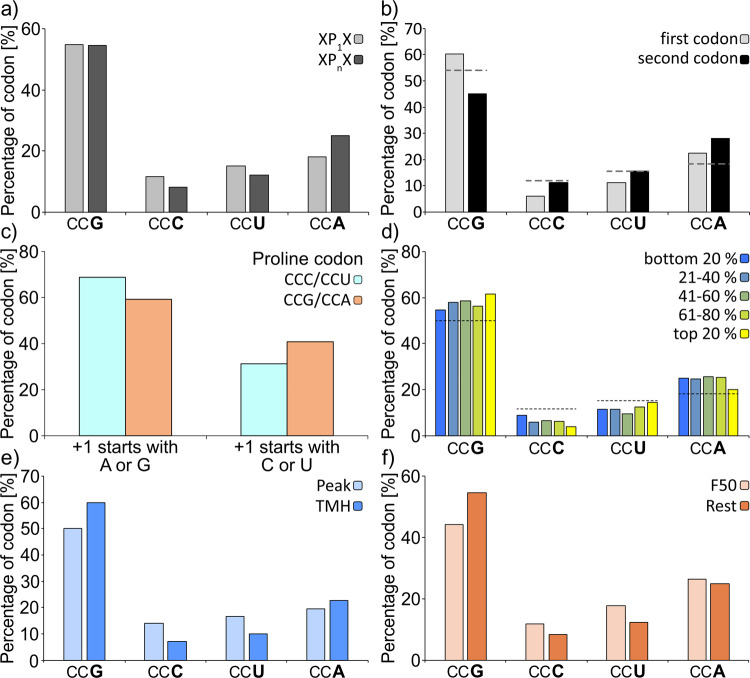
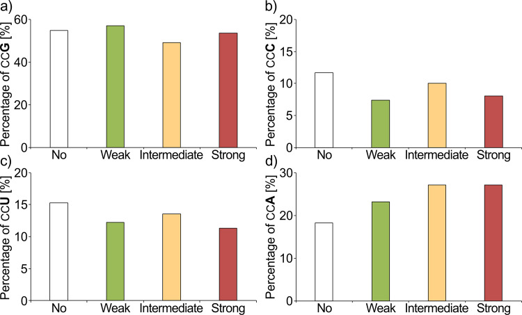
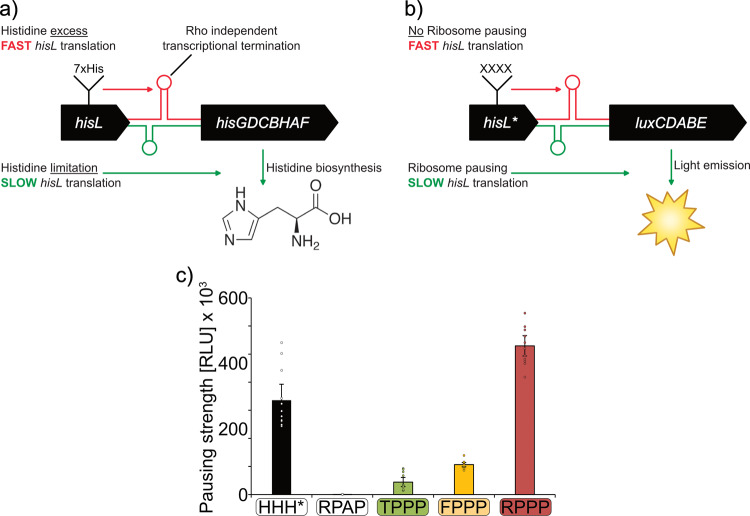
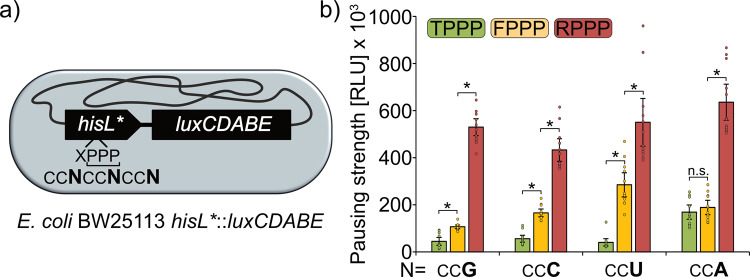
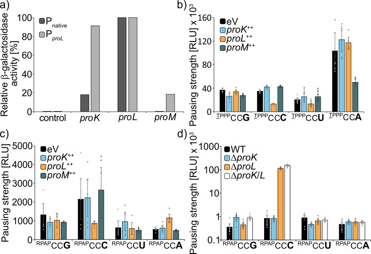
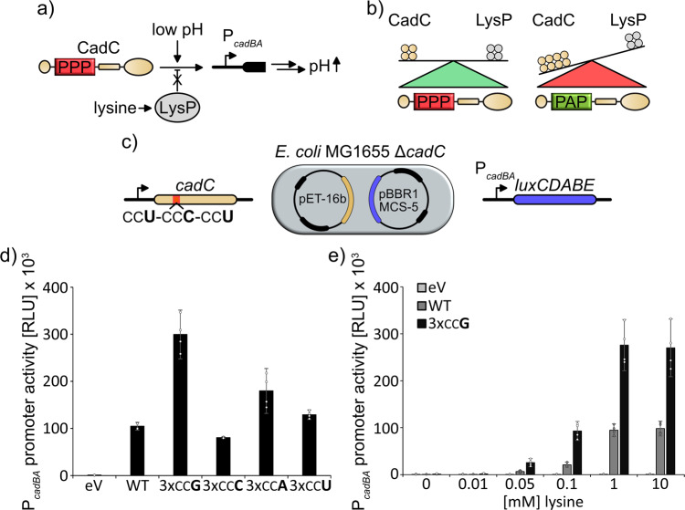
The speed of mRNA translation depends in part on the amino acid to be incorporated into the nascent chain. Peptide bond formation is especially slow with proline and two adjacent prolines can even cause ribosome stalling. While previous studies focused on how the amino acid context of a Pro-Pro motif determines the stalling strength, we extend this question to the mRNA level. Bioinformatics analysis of the Escherichia coli genome revealed significantly differing codon usage between single and consecutive prolines. We therefore developed a luminescence reporter to detect ribosome pausing in living cells, enabling us to dissect the roles of codon choice and tRNA selection as well as to explain the genome scale observations. Specifically, we found a strong selective pressure against CCC/U-C, a sequon causing ribosomal frameshifting even under wild-type conditions. On the other hand, translation efficiency as positive evolutionary driving force led to an overrepresentation of CCG. This codon is not only translated the fastest, but the corresponding prolyl-tRNA reaches almost saturating levels. By contrast, CCA, for which the cognate prolyl-tRNA amounts are limiting, is used to regulate pausing strength. Thus, codon selection both in discrete positions but especially in proline codon pairs can tune protein copy numbers.


Conflict of interest statement

The authors declare no competing interests.

Figures

Fig. 1
Fig. 1. Diversity of proline codons and their corresponding tRNAs.
a The genetic code contains four codons for proline: CCG, CCC, CCU, and CCA. b The three tRNAs ProK, ProL, and ProM recognize distinct sets of proline codons and exhibit different levels of abundance within the cell. All three prolyl-tRNAs are charged by the prolyl-tRNA synthetase ProS.
Fig. 2
Fig. 2. Bioinformatic analysis of proline codon bias in E. coli.
a Codon usage of either single (XP1X) or consecutive (XPnX) prolines (with X being any amino acid except proline and n > 1). p value = 1.7e−30, chi-squared test. b Codon usage of the first and second proline in PP-motifs. Only PP-motifs with two consecutive proline residues were included in this analysis. The dashed lines indicate the codon usage for single prolines. p value < 2.2e−16, chi-squared test. c Codon usage for amino acids in the +1-position downstream CCC/CCU (cyan) or CCG/CCA (orange) encoded single prolines. p value < 2.2e−16, chi-squared test. d Correlation between proline codon usage in PP-motifs and translation efficiency from least efficiently translated proteins (dark blue) to most efficiently translated proteins (yellow). The dashed lines indicate the codon usage for single prolines. e Difference between proline codon usage of PP-motifs in the peak region (light blue, amino acids 49–59 from the TMH start where PP-motifs are enriched to facilitate the efficient insertion of TMH into the membrane) and TMHs (blue; transmembrane helices in which PP-motifs are depleted for proper folding of transmembrane segments. p value = 0.13, chi-squared test. f Proline codon usage in PP-motifs in the first 50 codons (light orange) compared with the rest of proteins (orange). p value = 2.14e−7, chi-squared test.
Fig. 3
Fig. 3. Codon usage in PP-motifs of different pausing strength.
Pausing strength of PP-motifs depends on the upstream amino acid context,, resulting in weak, intermediate, and strong pausers. The pausing strength resulting from amino acid context is indicated by colored bars (no pausing—white; weak pausing—green; intermediate pausing—yellow; strong pausing—red). Codon usage in differently strong pausing motifs is shown for CCG (a), CCC (b), CCU (c), and CCA (d) codons. The difference is significant according to chi-squared test, p value = 4.2e−3.
Fig. 4
Fig. 4. The His-pausing system for in vivo measurement of pausing strength.
a Architecture of the histidine biosynthesis operon in E. coli. In its native state, the histidine biosynthesis gene cluster (hisGDCBHAF) is regulated by the His-leader peptide (hisL). This peptide contains seven consecutive histidines. At high histidine/histidyl-tRNA levels, translation efficiently proceeds through the His-leader peptide, resulting in the formation of an attenuator stem loop (red) that prevents transcription of the downstream genes. At low histidine and histidyl-tRNA levels translation is slowed down allowing for transcription and translation of the structural genes and synthesis of histidine (green). b Architecture of the His-pausing operon. An engineered His-leader peptide (hisL*) precedes the structural genes of the lux operon (luxCDABE). Here, His1 through His4 are exchanged by artificial sequence motifs (XXXX). In case of non-consecutive proline motifs (e.g., RPAP) there is no pausing, resulting in the formation of an attenuator stem loop (red) that prevents transcription of the downstream genes and low light emission. In the presence of motifs that contain consecutive prolines (e.g., RPPP) translation is slowed down allowing for transcription and translation of the structural genes and thus increased light emission (green). c Maximal luminescence emission at PP-motifs with increasing pausing strength. HisL*_Lux operons carrying a stop codon at the position corresponding to His4 (HHH*), non-consecutive (RPAP) or consecutive prolines of varying known pausing strength at the hisL* position (Weak: TPPP; green. Intermediate: FPPP; yellow. Strong: RPPP; red) were chromosomally integrated in E. coli BW25113 and tested for maximal luminescence emission. Threonine, phenylalanine, and arginine were encoded by ACC, TTT, and CGC, respectively. CCG was used as proline codon in all constructs. n = 12, Error bars indicate 95% confidence intervals.
Fig. 5
Fig. 5. Codon-dependent pausing strength at weak, intermediate, and strong PP-motifs.
a Genomic organization of the HisL*_Lux reporter. Synthetic His-Leader peptides (HisL*) preceding the lux genes (luxCDABE) were genomically integrated at the his-locus. In hisL*, His1 one was replaced by a variable amino acid (X) to modulate pausing strength. His2 through His4 were replaced by proline. In this regard several reporter strains (Supplementary data file S3) were generated with hisL* varying in the proline codon usage and are denoted as X_PPPCCN where the underlined X designates the preceding amino acid and the bold N designates the wobble base used for encoding the proline residues. b HisL*_Lux carrying PP-motifs of varying pausing strength (weak—TPPP: green; intermediate—FPPP: yellow; strong—RPPP: red) with different proline codon usage were chromosomally integrated in E. coli BW25113 and tested for maximal luminescence emission. n = 12, Error bars indicate 95% confidence intervals. Data for CCG codons are duplicated from Fig. 5 for better overview. Statistically significant differences according to unpaired two-sided t-tests (p value < 0.05) are indicated by asterisks.
Fig. 6
Fig. 6. Influence of prolyl-tRNA copy number on the codon-dependent pausing strength at PP-motifs.
a Approximation of E. coli BW25113 cells carrying the weak HisL*_Lux operon (TPPP) with different proline codon usage were transformed with pBBR1 MCS4-lacZ plasmids encoding ProK, ProL, or ProM under the control of their corresponding native promoters. n = 4. b E. coli BW25113 cells carrying the weak HisL*_Lux operon (TPPP) were transformed with pBBR1 MCS4-lacZ plasmids encoding for ProK, ProL, or ProM under control of PproL and tested for bioluminescence emission. n = 6. c E. coli BW25113 cells carrying the “non-PP” HisL*_Lux operon (RPAP) were transformed with pBBR1-MCS4-lacZ plasmids encoding for ProK, ProL, or ProM under control of PproL and tested for bioluminescence emission. n = 6. d The “non-PP” HisL*Lux operon (RPAP) was genomically integrated in E. coli BW25113 deletion strains lacking either proKproK), proLproL), or both (ΔproK/L) and cells were tested for bioluminescence emission. n = 12, Error bars indicate 95% confidence intervals.
Fig. 7
Fig. 7. Codon choice modulates protein expression and ensures physiological protein stoichiometry of the Cad system.
a The Cad system. CadC is a pH sensor that induces expression of its target genes at low pH by binding to the cadBA promoter (PcadBA). Expression of the corresponding gene products ultimately leads to an increase in pH. The lysine dependency of the acid stress response depends on stoichiometric expression of CadC and the co-sensor LysP. b The equilibrium of the protein copy numbers of CadC and LysP is ensured by a triproline motif within the CadC primary structure. Absence of the triproline results in deregulation of the acid stress response due to increased CadC copy number. c Reporter system used to test the cadC translation efficiency. E. coli MG1655 ΔcadC cells were transformed with pET-16B vectors encoding for wild type or proline codon-exchanged variants of CadC. Cells were cotransformed with pBBR1MCS-5 vectors carrying the lux genes under control of the PcadBA promoter. PcadBA promoter activity was assessed by measuring luminescence emission and used as a proxy for CadC copy number. d PcadBA promoter activity under inducing conditions (pH = 5.8; 10 mM lysine) upon expression of wild-type CadC or proline codon-exchanged CadC variants where all proline codons in the pausing motif have been substituted by the same codon. n = 4. e PcadBA promoter activity at increasing external lysine concentrations. PcadBA induction when cadC contains the natural codon composition is shown in dark gray. PcadBA induction when cadC contains only CCG codons at the relevant PP-motif is shown in black. n = 4, Error bars indicate 95% confidence intervals.

Similar articles

Cited by

References

    1. Wedemeyer WJ, Welker E, Scheraga HA. Proline cis-trans isomerization and protein folding. Biochemistry. 2002;41:14637–14644. doi: 10.1021/bi020574b. - DOI - PubMed
    1. Adzhubei AA, Sternberg MJ, Makarov AA. Polyproline-II helix in proteins: structure and function. J. Mol. Biol. 2013;425:2100–2132. doi: 10.1016/j.jmb.2013.03.018. - DOI - PubMed
    1. Muto H, Ito K. Peptidyi-prolyl-tRNA at the ribosomal P-site reacts poorly with puromycin. Biochem. Bioph. Res. Co. 2008;366:1043–1047. doi: 10.1016/j.bbrc.2007.12.072. - DOI - PubMed
    1. Wohlgemuth I, Brenner S, Beringer M, Rodnina MV. Modulation of the rate of peptidyl transfer on the ribosome by the nature of substrates. J. Biol. Chem. 2008;283:32229–32235. doi: 10.1074/jbc.M805316200. - DOI - PubMed
    1. Pavlov MY, et al. Slow peptide bond formation by proline and other N-alkylamino acids in translation. Proc. Natl Acad. Sci. USA. 2009;106:50–54. doi: 10.1073/pnas.0809211106. - DOI - PMC - PubMed

Publication types

LinkOut - more resources