. 2021 Nov;26(11):6756-6772.
doi: 10.1038/s41380-021-01145-7. Epub 2021 May 17.

A novel murine model to study the impact of maternal depression and antidepressant treatment on biobehavioral functions in the offspring

Affiliations

A novel murine model to study the impact of maternal depression and antidepressant treatment on biobehavioral functions in the offspring

Joseph Scarborough et al. Mol Psychiatry. 2021 Nov.

Abstract

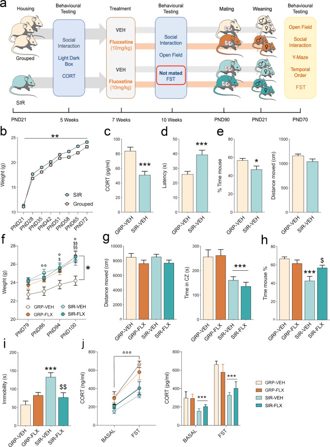
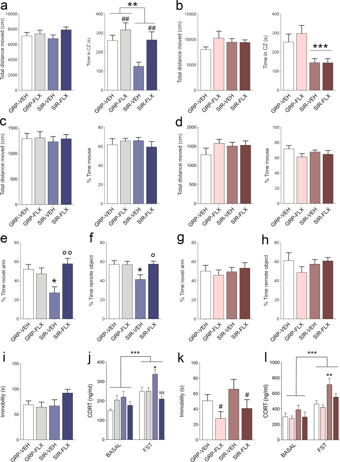
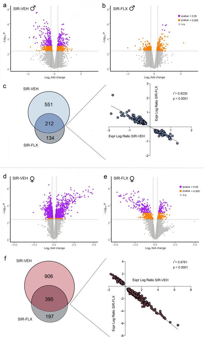
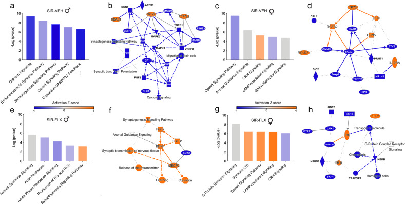
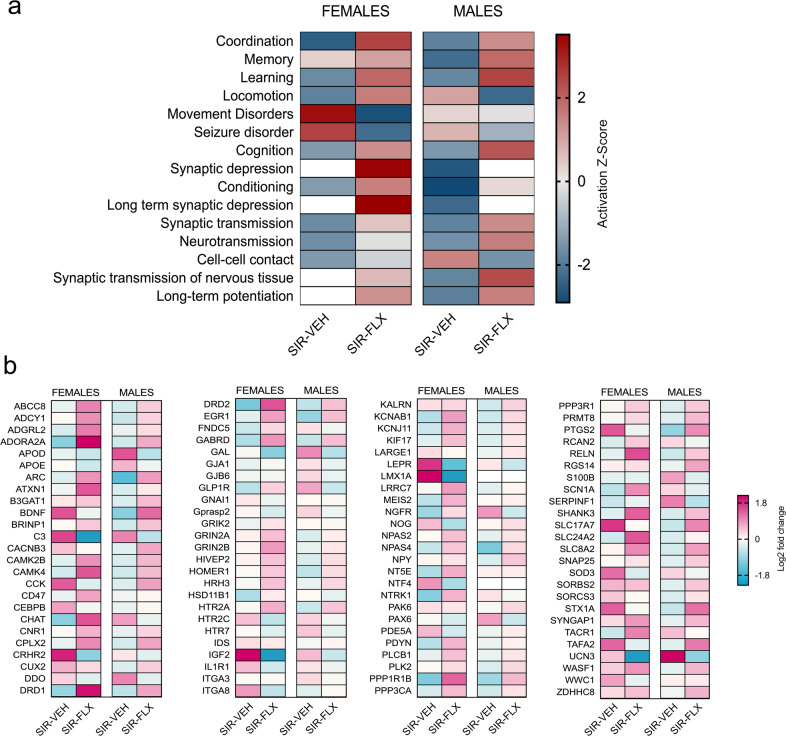
Antenatal psychopathology negatively affects obstetric outcomes and exerts long-term consequences on the offspring's wellbeing and mental health. However, the precise mechanisms underlying these associations remain largely unknown. Here, we present a novel model system in mice that allows for experimental investigations into the effects of antenatal depression-like psychopathology and for evaluating the influence of maternal pharmacological treatments on long-term outcomes in the offspring. This model system in based on rearing nulliparous female mice in social isolation prior to mating, leading to a depressive-like state that is initiated before and continued throughout pregnancy. Using this model, we show that the maternal depressive-like state induced by social isolation can be partially rescued by chronic treatment with the selective serotonin reuptake inhibitor, fluoxetine (FLX). Moreover, we identify numerous and partly sex-dependent behavioral and molecular abnormalities, including increased anxiety-like behavior, cognitive impairments and alterations of the amygdalar transcriptome, in offspring born to socially isolated mothers relative to offspring born to mothers that were maintained in social groups prior to conception. We also found that maternal FLX treatment was effective in preventing some of the behavioral and molecular abnormalities emerging in offspring born to socially isolated mothers. Taken together, our findings suggest that the presence of a depressive-like state during preconception and pregnancy has sex-dependent consequences on brain and behavioral functions in the offspring. At the same time, our study highlights that FLX treatment in dams with a depression-like state can prevent abnormal behavioral development in the offspring.


Conflict of interest statement

The authors declare no competing interests.

Figures

Fig. 1
Fig. 1. Experimental paradigm and behavioral readouts in SIR and GRP-housed female mice.
Physiological and behavioral effects of SIR in female mice prior to conception. a Graphical representation of the experimental design. C57BL6/N female mice were housed in social isolation or in groups from PND21 onwards. After 5 weeks of social isolation or group housing, the animals were subjected to behavioral testing in the light-dark box test and social interaction test as well as to basal CORT measurements. After 7 weeks of social isolation or group housing, each group of SIR and GRP animals were split into two subgroups and treated with either vehicle (drinking water) or FLX (10 mg/kg). After 10 weeks, the animals were subjected to behavioral testing in the open field test and the social interaction test. A subgroup of animals was subjected to the forced swim test and to basal and stress-induced CORT measurements. These animals were excluded from mating. Weight gain was assessed weekly during the whole experimental timeline. The animals were then exposed to a timed mating procedure and their offspring were left undisturbed until adulthood. Maternal FLX treatment was continued throughout mating, pregnancy and the offspring’s pre-weaning period. Adult offspring were subjected to behavioral testing in the open field test, the social interaction test, the Y-maze test of spatial recognition memory, the temporal order test and the forced swim test (FST). Weight gain was monitored throughout development, while basal and stress-induced levels of CORT were measured before and after the FST, respectively. b Weight gain in SIR and GRP females during the first 7 weeks of isolation- or group-housing. **p < 0.01, reflecting the main effect of housing. c Basal plasma levels of CORT in SIR and GRP females at the 5-weeks testing timepoint; ***p < 0.001. d Latency to shuttle to the bright compartment of the light-dark box testing apparatus; ***p < 0.001. e Percent exploration time of an unfamiliar mouse compared to a dummy object and total distance moved during the social interaction test at the 5 weeks testing timepoint; *p < 0.05. f Weight gain in SIR-VEH, SIR-FLX, GRP-VEH and GRP-FLX animals in the three weeks following the start of FLX treatment (SIR-VEH vs GRP-VEH at PND86: °°p < 0.01; SIR-VEH vs GRP-VEH at PND94: °p < 0.05; SIR-FLX vs GRP-VEH at PND94: $p < 0.05; SIR-VEH vs GRP-VEH at PND100: °p < 0.05; SIR-FLX vs GRP-VEH at PND100: $$p < 0.01; GRP-FLX vs GRP-VEH at PND100: §§p < 0.01). g Total distance moved and time spent in the center zone in the open field test at the 10 weeks testing timepoint; ***p < 0.001, reflecting the main effect of SIR. h Percent exploration time of an unfamiliar mouse compared to a dummy object and total distance moved during the social interaction test at the 10 weeks testing timepoint; ***p < 0.001 between SIR-VEH and GRP-VEH; $p < 0.05 between SIR-VEH and SIR-FLX. i Time spent immobile in the FST test at the 10 weeks testing timepoint. $$p < 0.01 between SIR-VEH and SIR-FLX; ***p < 0.001 between SIR-VEH and GRP-VEH. j Basal and stress-induced levels of CORT. °°°p < 0.001, reflecting the main effect of testing condition; ***p < 0.001, reflecting the main effect of housing. N = 40 mice per group in be, N = 20 animals per group in fh, and N = 10 animals per group in ij. All values represent means ± s.e.m.
Fig. 2
Fig. 2. Effects of maternal SIR and FLX treatment on behavior and cognition in the offspring.
a Total distance moved and time spent in the center zone of the open field test in male offspring. **p < 0.01, reflecting the main effect of SIR; ##p < 0.01, reflecting the main effect of FLX treatment. b Total distance moved and time spent in the center zone of the open field test in female offspring. ***p < 0.001, reflecting the main effect of SIR. c Total distance moved and percent exploration time of an unfamiliar mouse compared to a dummy object in the social interaction test in male offspring. d Total distance moved and percent exploration time of an unfamiliar mouse compared to a dummy object in the social interaction test in female offspring. e Relative exploration time (%) of the novel arm in the Y-maze test of spatial recognition memory in male offspring. °°p < 0.01 between SIR-VEH and SIR-FLX; *p < 0.05 between SIR-VEH and GRP-VEH. f Relative time (%) spent exploring the remote object in the temporal order memory test in male offspring; °p < 0.05 between SIR-VEH and SIR-FLX; *p < 0.05 between SIR-VEH and GRP-VEH. g Relative exploration time (%) of the novel arm in the Y-maze test of spatial recognition memory in female offspring. h Relative time (%) spent exploring the remote object in the temporal order memory test in female offspring. i Time spent immobile in the forced swim test (FST) test in male offspring. j Basal and FST-induced levels of CORT in male offspring. *p < 0.05 between SIR-VEH and GRP-VEH; §§§p < 0.001 between SIR-FLX vs SIR-VEH. k Time spent immobile in the forced swim test (FST) test in female offspring. #p < 0.05, reflecting the main effect of FLX treatment. l Basal and FST-induced levels of CORT in female offspring. **p < 0.01 between SIR-VEH and GRP-VEH; N = 10.11 mice per group and sex in ah, N = 12-13 animals per group and sex in il. All values represent means ± s.e.m.
Fig. 3
Fig. 3. Maternal SIR and FLX treatment affect the amygdalar transcriptome in male and female offspring.
Genome-wide transcriptional changes in the amygdala of offspring born to GRP or SIR mothers with or without concomitant FLX treatment. a Volcano plot depicting the differentially expressed genes (DEGs) in SIR-VEH vs GRP-VEH male offspring (FDR q < 0.05: purple; p < 0.005: orange; n.s: gray). b Volcano plot depicting the differentially expressed genes (DEGs) in SIR-FLX vs GRP-VEH male offspring (FDR q < 0.05: purple; p < 0.005: orange; n.s: gray). c The Venn diagram denotes the number of DEGs that are uniquely and commonly affected in SIR-VEH and SIR-FLX male offspring. The linear regression plots the expression log ratio of DEGs commonly affected in SIR-VEH and SIR-FLX offspring. d Volcano plot depicting the differentially expressed genes (DEGs) in SIR-VEH vs GRP-VEH female offspring (FDR q < 0.05: purple; p < 0.005: orange; n.s: gray). e Volcano plot depicting the differentially expressed genes (DEGs) in SIR-FLX vs GRP-VEH female offspring (FDR q < 0.05: purple; p < 0.005: orange; n.s: gray). f The Venn diagram denotes the number of DEGs that are uniquely and commonly affected in SIR-VEH and SIR-FLX male offspring. N = 5 animals per group and sex.
Fig. 4
Fig. 4. Pathway analysis of transcriptomic alterations induced by maternal SIR and FLX treatment.
Ingenuity Pathway Analysis (IPA) was conducted to identify the top canonical signaling pathways affected in male and female offspring born to GRP or SIR mothers with or without concomitant FLX treatment. Orange color = positive activation Z-score (pathways) and increased expression (individual genes), blue color = negative activation Z-score (pathways) and reduced expression (individual genes). For IPA-generated overview of the main biological themes, the most significant entities (canonical pathways, upstream regulators, biological functions and molecules) are shown, with connecting lines representing specific relationships (solid lines represent a direct relationship, slashed lines an indirect relationship and dotted lines and inferred relationship). Arrows represent directionality and activation, while blunt-ended lines represent inhibition. a Top 5 canonical pathways annotated with DEGs in SIR-VEH vs GRP-VEH male offspring. b IPA-generated overview of the main biological themes pertaining to the comparison between SIR-VEH vs GRP-VEH male offspring. c Top 5 canonical pathways annotated with DEGs in SIR-VEH vs GRP-VEH female offspring. d IPA-generated overview of the main biological themes pertaining to the comparison between SIR-VEH vs GRP-VEH female offspring. e Top 5 canonical pathways annotated with DEGs in SIR-FLX vs GRP-VEH male offspring. f IPA-generated overview of the main biological themes pertaining to the comparison between SIR-FLX vs GRP-VEH male offspring. g Top 5 canonical pathways annotated with DEGs in SIR-FLX vs GRP-VEH female offspring. h IPA-generated overview of the main biological themes pertaining to the comparison between SIR-FLX vs GRP-VEH female offspring. The analyses are based on gene expression data obtained from N = 5 animals per group and sex.
Fig. 5
Fig. 5. Comparison analysis of transcriptomic alterations induced by maternal SIR and FLX treatment.
Ingenuity Pathway Analysis (IPA) was used to identify modules of biological functions predicted to be increased or decreased in male and female offspring born to GRP or SIR mothers with or without concomitant FLX treatment. a Heatmap visualization of the top 15 biological functions that are similarly or differentially affected in SIR-VEH and SIR-FLX female and male offspring. The colors of the heatmap reflect the activation Z-score of each biological function in each dataset, and ranges from dark blue (negative activation Z-score) to dark red (positive activation Z-score). b The gene-level heatmap graphically depicts which genes are up- or downregulated in the biological function module ‘memory’ across different datasets. The colors of the heatmap reflect the Log2 fold-change and range from blue (downregulated) to magenta (upregulated). The analyses are based on gene expression data obtained from N = 5 animals per group and sex.

Similar articles

Cited by

References

    1. Graignic-Philippe R, Dayan J, Chokron S, Jacquet AY, Tordjman S. Effects of prenatal stress on fetal and child development: a critical literature review. Neurosci Biobehav Rev. 2014;43:137–162. doi: 10.1016/j.neubiorev.2014.03.022. - DOI - PubMed
    1. Fisher J, Cabral de Mello M, Patel V, Rahman A, Tran T, Holton S, et al. Prevalence and determinants of common perinatal mental disorders in women in low- and lower-middle-income countries: a systematic review. Bull World Health Organ. 2012;90:139G–149G. doi: 10.2471/BLT.11.091850. - DOI - PMC - PubMed
    1. Melville JL, Gavin A, Guo Y, Fan MY, Katon WJ. Depressive disorders during pregnancy: prevalence and risk factors in a large urban sample. Obstet Gynecol. 2010;116:1064–1070. doi: 10.1097/AOG.0b013e3181f60b0a. - DOI - PMC - PubMed
    1. Rogers A, Obst S, Teague SJ, Rossen L, Spry EA, Macdonald JA et al. Association between maternal perinatal depression and anxiety and child and adolescent development: a meta-analysis. JAMA Pediatr. 2020; 10.1001/jamapediatrics.2020.2910. - PMC - PubMed
    1. Loomans EM, van Dijk AE, Vrijkotte TG, van Eijsden M, Stronks K, Gemke RJ, et al. Psychosocial stress during pregnancy is related to adverse birth outcomes: results from a large multi-ethnic community-based birth cohort. Eur J Public Health. 2013;23:485–491. doi: 10.1093/eurpub/cks097. - DOI - PubMed

Publication types