. 2021 Jul;24(7):913-929.
doi: 10.1038/s41593-021-00854-0. Epub 2021 May 17.

Functionally distinct POMC-expressing neuron subpopulations in hypothalamus revealed by intersectional targeting

Affiliations

Functionally distinct POMC-expressing neuron subpopulations in hypothalamus revealed by intersectional targeting

Nasim Biglari et al. Nat Neurosci. 2021 Jul.

Abstract

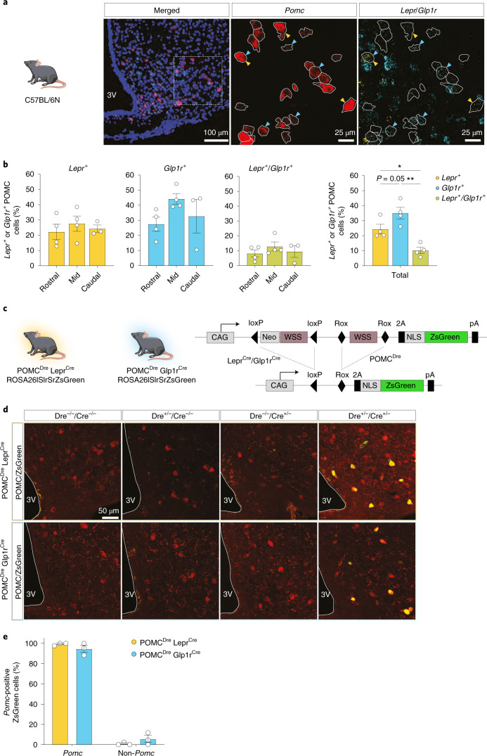
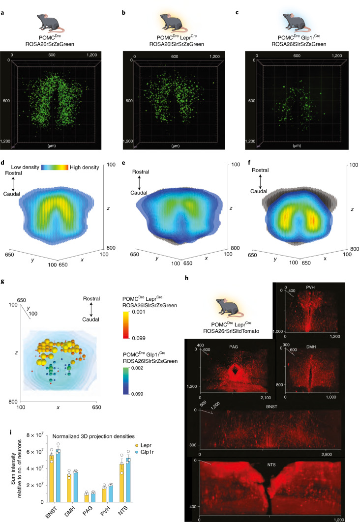
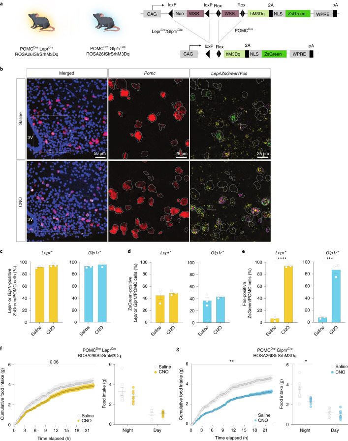
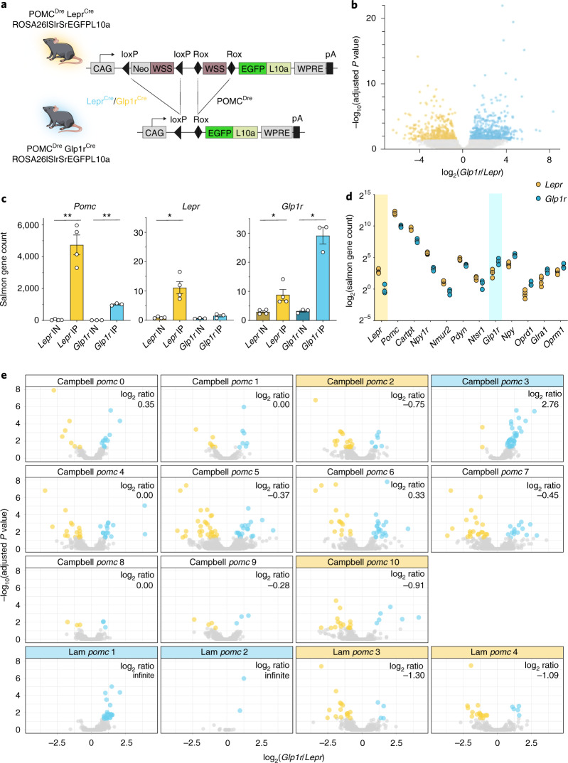
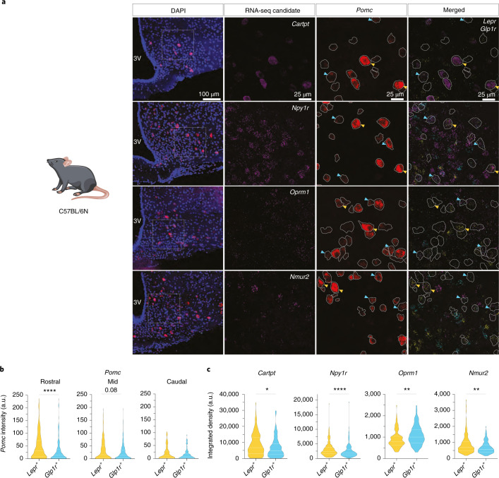
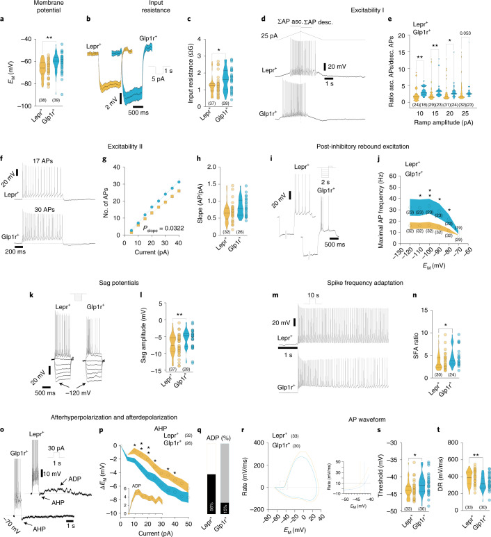
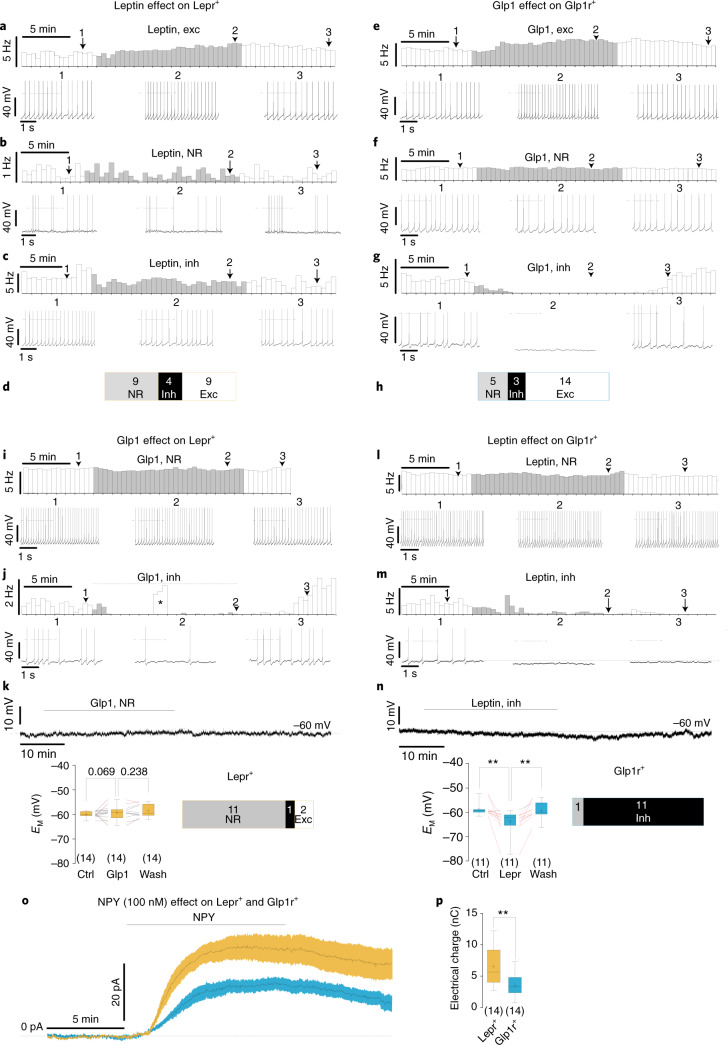
Pro-opiomelanocortin (POMC)-expressing neurons in the arcuate nucleus of the hypothalamus represent key regulators of metabolic homeostasis. Electrophysiological and single-cell sequencing experiments have revealed a remarkable degree of heterogeneity of these neurons. However, the exact molecular basis and functional consequences of this heterogeneity have not yet been addressed. Here, we have developed new mouse models in which intersectional Cre/Dre-dependent recombination allowed for successful labeling, translational profiling and functional characterization of distinct POMC neurons expressing the leptin receptor (Lepr) and glucagon like peptide 1 receptor (Glp1r). Our experiments reveal that POMCLepr+ and POMCGlp1r+ neurons represent largely nonoverlapping subpopulations with distinct basic electrophysiological properties. They exhibit a specific anatomical distribution within the arcuate nucleus and differentially express receptors for energy-state communicating hormones and neurotransmitters. Finally, we identify a differential ability of these subpopulations to suppress feeding. Collectively, we reveal a notably distinct functional microarchitecture of critical metabolism-regulatory neurons.


Conflict of interest statement

The authors declare no competing interests.

Figures

Fig. 1
Fig. 1. The POMCDre driver line successfully targets POMC neurons.
a, Schematic showing POMCDre-dependent recombination in the ROSA26rSrZsGreen reporter line. Excision of rox-flanked stop cassette leads to ZsGreen expression in POMC neurons. b, ZsGreen expression across the rostral, mid and caudal sections of the ARC in POMCDre ROSA26rSrZsGreen mice at 15 weeks of age. c, Dispersed ZsGreen expression in the intermediate and anterior lobes of the pituitary in POMCDre ROSA26rSrZsGreen mice. C1, C2 and C3 depict magnifications of the posterior, intermediate and anterior pituitary, respectively. Scale bar, 150 μm (whole image) and 50 μm (magnified images). d, RNA ISH against Pomc/ZsGreen (top) and Agrp/ZsGreen (bottom) in POMCDre ROSA26rSrZsGreen mice. Magnifications of the boxes are displayed on the right of each image. Scale bars, 50 μm (whole image) and 20 μm (magnified images). 3V, third ventricle. e, Percentage of ZsGreen-positive cells coexpressing or lacking expression of either Pomc or Agrp, quantified from RNA ISH (d). Data are represented as the mean ± s.e.m. (Pomc: 97.57 ± 0.92; Agrp: 1.88 ± 0.36; non-Pomc: 2.43 ± 0.92; non-Agrp: 98.12 ± 0.36; n = 4 mice; a minimum of 13 sections were analyzed for each group).
Fig. 2
Fig. 2. Lepr and Glp1r expression in POMC neurons.
a, Representative microscopic images of RNA ISH against Pomc, Glp1r and Lepr in C57BL/6N mice at 12 weeks of age. First image shows ISH in the ARC with nuclear counterstain (blue, DAPI). Magnifications of the dashed box (right) are shown with the indicated stainings. Pomc-positive neurons are outlined in white. Yellow and cyan arrows indicate Lepr-positive or Glp1r-positive POMC neurons, respectively. Scale bars represent 100 μm in the merged image and 25 μm in the magnifications. b, Percentage of Pomc-positive cells expressing Lepr, Glp1r or both receptors across the rostrocaudal axis. The bar graph on the right depicts the total percentage of POMC neurons coexpressing the receptors as averaged from the individual areas. Left: Lepr+Rostral: 22.18% ± 5.02%, Lepr+Mid: 27.54% ± 4.98%, Lepr+Caudal: 24.21% ± 2.49%; Glp1r+Rostral: 27.44% ± 4.73%, Glp1r+Mid: 44.16% ± 3.48%, Glp1r+Caudal: 32.73% ± 11.15%; Lepr+/Glp1r+Rostral: 8.09% ± 2.44%, Lepr+/Glp1r+Mid: 12.79% ± 3.08%, Lepr+/Glp1r+Caudal: 9.40% ± 3.96%. Right: Lepr+, 24.32% ± 3.31%; Glp1r+, 35.03% ± 3.89%; Glp1r+/Lepr+, 10.17% ± 2.10%. One-way ANOVA, F (1.656, 4.968) = 64.61, P = 0.0003, followed by Tukey’s post hoc test; Glp1r+ versus Lepr+ P = 0.0536, Glp1r+ versus Glp1r+/Lepr+ P = 0.0017, Lepr+ versus Glp1r+/Lepr+ P = 0.0129; n = 4 mice. P values were calculated on the total percentage of subpopulations using one-way repeated-measures ANOVA followed by Tukey’s test. *P ≤ 0.05, **P ≤ 0.01, ***P ≤ 0.001. c, Illustrations of experimental mice and schematic diagram showing Dre- and Cre-dependent recombination of ROSA26lSlrSrZsGreen reporter line. Excision of loxP- or rox-flanked stop cassettes through recombination of both Dre and Cre drivers led to ZsGreen expression in the targeted POMC population. d, Representative microscopic images of immunohistochemical staining against POMC and ZsGreen in the ARC of all resulting genotypes at 15 weeks of age. Scale bar, 50 μm. e, Percentage of ZsGreen-positive cells coexpressing or lacking expression of Pomc, quantified from RNA ISH. POMCDre LeprCre: Pomc, 99.21% ± 0.79%; non-Pomc, 0.79% ± 0.79%; POMCDre Glp1rCre: Pomc, 94.56% ± 3.43%; non-Pomc; 5.44% ± 0.3.43%; n = 3 mice per group; minimum of eight sections analyzed for each. For d and e, data are presented as mean ± s.e.m.
Fig. 3
Fig. 3. POMCLepr+ and POMCGlp1r+ show distinct spatial distribution throughout the ARC.
a–c, Representative 3D reconstruction of the entire POMC population labeled in POMCDre ROSA26rSrZsGreen mice (a), and 3D reconstruction of POMC subpopulations in POMCDre LeprCre ROSA26lSlrSrZsGreen (b) or POMCDre Glp1rCre ROSA26lSlrSrZsGreen (c) mice at 15 weeks of age. Scans were obtained using the LSFM at ×8 total magnification. n = 9 (a), n = 7 (b) and n = 8 (c) mice. df, Isosurface density plots of the entire POMC population (d), and the POMCLepr+ (e) and POMCGlp1r+ (f) subpopulations. Gray shaded areas in e and f depict the entire POMC population. g, Statistical representation of the differences in distribution between the POMCLepr+ and POMCGlp1r+ subpopulations using a two-tailed t-test. P values are plotted as spheres within the space occupied by the POMC neurons (background). The size and color of the spheres indicate the significance values in ranges of yellow to red (POMCLepr+) and green to blue (POMCGlp1r+). h, Representative images of 3D projection densities in POMCDre LeprCre ROSA26rSrlSltdTomato mice in the PVH, PAG, DMH, BNST and NTS. i, Quantification of 3D projection densities shown in h, normalized to the number of neurons. Data are represented as mean ± s.e.m., from n = 3 mice per group. POMCLepr+: BNST: 56,068,002.46 ± 4,157,623.02, DMH: 33,195,450.61 ± 2,618,432.01, PAG: 10,481,156.64 ± 819,984.06, PVH: 18,370,352.69 ± 1149,637.53, NTS: 45,834,835.18 ± 6,188,416.90. POMCGlp1r+; BNST: 63,188,837.7 ± 3,276,156.35, DMH: 37,050,145.65 ± 478,713.82, PAG: 11,722,976.37 ± 369,033.55, PVH: 20,851,215.3 ± 337,219.38, NTS: 5,294,0731.54 ± 3,984,858.12. BNST, Glp1r+ versus Lepr+: P = 0.249747, t = 1.345, df = 4. DMH, Glp1r+ versus Lepr+: P = 0.632027, t = 1.448, df = 4. PAG, Glp1r+ versus Lepr+: P = 0.632027, t = 1.381, df = 4. PVH, Glp1r+ versus Lepr+: P = 0.432578, t = 2.071, df = 4. NTS, Glp1r+ versus Lepr+: P = 0.632027, t = 0.9654, df = 4, unpaired Student’s t-test, Holm–Sidak correction.
Fig. 4
Fig. 4. DREADD-dependent activation of POMCLepr+ or POMCGlp1r+ neurons differentially reduces food intake.
a, Illustrations of experimental mice and schematic diagram showing Dre- and Cre-dependent targeted expression of activatory hM3Dq in either POMCLepr+ or POMCGlp1r+ neurons. Excision of loxP-flanked and rox-flanked stop cassettes through recombination of both Dre and Cre drivers leads to hM3Dq expression in the targeted subpopulation. b, Representative microscopic images of RNA ISH against Pomc, Lepr, ZsGreen (in lieu of hM3Dq) and Fos in POMCDre LeprCre ROSA26lSlrSrhM3Dq males injected with saline or CNO. Images on the left show ISH in the ARC with nuclear counterstain (blue, DAPI). Magnifications of the boxes (right) are shown with the indicated stainings. Pomc-positive neurons are outlined in white. Scale bars, 50 μm (merged image) and 25 μm (magnified images). ce, Percentage of ZsGreen-Pomc-positive cells expressing Lepr or Glp1r (c), percentage of Lepr/Glp1r-Pomc-positive cells expressing ZsGreen (d) and percentage of ZsGreen-Pomc-positive cells expressing Fos (e) in POMCDre LeprCre ROSA26lSlrSrhM3Dq or POMCDre Glp1rCre ROSA26lSlrSrhM3Dq male mice (22–26 weeks old) injected with saline or CNO. CNO, 3 mg kg−1. c: POMCLepr+: saline: 91.84% ± 1.03%, CNO: 94.87% ± 1.62%, saline versus CNO, t = 1.580, P = 0.342863: POMCGlp1r+: saline: 93.52% ± 2.04%, CNO: 96.16% ± 2.31% saline versus CNO, t = 0.8592, P = 0.438695; d: POMCLepr+: saline: 45.46% ± 6.92%, CNO: 49.19% ± 1.92%, saline versus CNO, t = 0.5191, P = 0.631109; POMCGlp1r+: saline: 36.88% ± 5.28%, CNO: 43.24% ± 1.42%, saline versus CNO, t = 1.165, P = 0.522274. e: POMCLepr+: saline: 6.87% ± 2.64%, CNO: 94.03% ± 1.66%, saline versus CNO, t = 28.0, df = 4, PuT = 0.000019; POMCGlp1r+: saline: 8.23% ± 1.13%, CNO: 87.05% ± 6.05%, saline versus CNO, t = 12.82, df = 4, PuT = 0.000214, unpaired Student’s t-test, Holm–Sidak correction; n = 3 mice. f,g, Food intake over a time course of 24 h in POMCDre LeprCre ROSA26lSlrSrhM3Dq (f) and POMCDre Glp1rCre ROSA26lSlrSrhM3Dq male mice (g) starting with the night cycle. Mice were injected with saline at 18:00 and 23:00, followed by a 1-d gap and subsequent CNO injections at 18:00 and 23:00 on the next day. Left: cumulative food intake in mice injected with saline versus CNO; right, total food intake in grams during night and day. f: n = 8; left: saline versus CNO two-way ANOVA, F(1,7) = 4.815, P = 0.0643; right: salineNight: 3.03 ± 0.39, CNONight: 2.79 ± 0.20, saline versus CNO two-way ANOVA followed by Sidak’s test, P = 0.2729, salineDay: 1.05 ± 0.15, CNODay: 1.10 ± 0.07, saline versus CNO two-way ANOVA followed by Sidak’s test, P = 0.9867. g: n = 7, left: saline versus CNO two-way ANOVA, F(1.000, 6.000) = 16.51, P = 0.0066, right: salineNight: 3.42 ± 0.38, CNONight: 2.32 ± 0.12, salineDay: 1.19 ± 0.15, CNODay: 0.98 ± 0.16; saline versus CNO two-way ANOVA followed by Sidak’s test, P = 0.0312; saline versus CNO two-way ANOVA followed by Sidak’s test, P = 0.7992. Data are represented as the mean ± s.e.m. Statistical analyses in ce were performed by unpaired two-tailed Student’s t-test with Holm–Sidak correction for multiple comparisons. For cumulative food intake (f and g left), two-way ANOVA was used; for total food intake (f and g right), two-way ANOVA followed by Sidak’s post hoc test was used. Indices PuT: unpaired t-test. *P ≤ 0.05, **P ≤ 0.01, ***P ≤ 0.001, ***P ≤ 0.0001.
Fig. 5
Fig. 5. POMCLepr+ and POMCGlp1r+ neurons exhibit distinct translational profiles.
a, Illustration of experimental mice and schematic diagram showing Dre- and Cre-dependent targeted expression of EGFPL10a in either POMCLepr+ or POMCGlp1r+ neurons. Excision of loxP-flanked and rox-flanked stop cassettes through recombination of both Dre and Cre drivers led to EGFPL10a expression in the targeted subpopulation. b, Volcano plot of differentially ribotag-enriched transcripts in POMCLepr+ and POMCGlp1r+ neurons. Significantly differentially enriched transcripts (P ≤ 0.05) are indicated in the colored region. Yellow and cyan depict a significantly higher enrichment in POMCLepr+ and POMCGlp1r+ neurons, respectively. P values adjusted for multiple comparisons were calculated by DESeq2 (1.26.0). c, Expression of Pomc, Lepr and Glp1r in input (IN) and IP samples of each subpopulation. Statistics were analyzed using unpaired two-tailed Welch’s t-test. Pomc: Lepr IN: 39.44 ± 19.39, Lepr IP: 4,740.55 ± 618.41, IN vs IP, t = 7.6, P = 0.0047. Glp1r IN: 14.14.55 ± 0.49, Glp1r IP: 1,005.42 ± 40.19, IN vs IP, t = 24.7, P = 0. 0016. Lepr: Lepr IN: 0.92 ± 0.14, Lepr IP: 11.12 ± 2.05, IN vs IP, t = 4.95, P = 0.0155. Glp1r IN: 0.62 ± 0.07, Glp1r IP: 1.50 ± 0.40, IN vs IP, t = 2.18, P = 0.001536. Glp1r: Lepr IN: 2.97 ± 0.38, Lepr IP: 8.80 ± 1.82, IN vs IP, t = 3.15, P = 0.0458. Glp1r IN: 3.23.55 ± 0.35, Glp1r IP: 29.17 ± 2.83, IN vs IP, t = 9.08, P = 0.0108. *P ≤ 0.05, **P ≤ 0.01. d, Significantly differentially enriched genes (P ≤ 0.05) of POMCLepr+ and POMCGlp1r+ neurons, belonging to the GO term ‘neuropeptide-signaling pathways’. Vertical colored area separates higher enrichment in POMCLepr+ neurons (left) from higher enrichment in POMCGlp1r+ neurons (right). e, Overlap analysis of the publicly available single-cell RNA-seq data from mouse hypothalami with our dataset. Here, the volcano plot in b is filtered for the markers reported for each cluster. For POMCDre LeprCre ROSA26lSlrSrEGFPL10a, n = 4 samples of pooled hypothalami from N = 24 mice; for POMCDre Glp1rCre ROSA26lSlrSrEGFPL10a, n = 3 samples of pooled hypothalami from N = 36 mice. Data are represented as the mean ± s.e.m.
Fig. 6
Fig. 6. Differential expression of endogenous mRNAs for identified candidates in POMCLepr+ and POMCGlp1r+ neurons.
a, Representative microscopic images of RNA ISH against Pomc, Glp1r and Lepr together with differentially expressed neuropeptidergic signaling candidate RNAs identified in the ribotag experiments, Cartpt, Npy1r, Oprm1 and Nmur2, in C57BL/6N mice at 12 weeks of age. First image shows ISH in the ARC with nuclear counterstain (blue, DAPI). Magnifications of the boxes (right) are shown with the indicated stainings. Pomc-positive neurons are outlined in white. Yellow and cyan arrows indicate Lepr-positive or Glp1r-positive POMC neurons, respectively. Scale bars represent 100 μm in the merged image and 25 μm in the magnifications. b, Violin plots showing the quantified intensity of Pomc mRNA across the rostrocaudal axis of the ARC in POMCLepr+ or POMCGlp1r+ neurons as assessed from RNA ISH (Fig. 2a). Data are from n = 4 mice, with a minimum of four sections analyzed per animal. PomcRostral, Lepr+: Q1: 7.83, Q2: 31.12, Q3: 71.31; Glp1r+: Q1: 3.92, Q2: 14.86, Q3: 37.05, unpaired Mann–Whitney U-test, Lepr+ versus Glp1r+, U = 21,051, PuT < 0.0001; PomcMid, Lepr+: Q1: 3.02, Q2: 15.71, Q3: 49.22; Glp1r+, Q1: 3.22, Q2: 13.43, Q3: 34.06, unpaired Mann–Whitney U-test, Lepr+ versus Glp1r+, U = 53,910, PuT = 0.0796; PomcCaudal, Lepr+: Q1: 9.46, Q2: 28.3, Q3: 103.5; Glp1r+ Q1: 8.03, Q2: 17.81, Q3: 91.96, unpaired Mann–Whitney U-test, Lepr+ versus Glp1r+, U = 3,467, PuT = 0.06349. a.u., arbitrary units. c, Violin plots showing quantified expression of the RNA-seq candidates measured as integrated density in POMCLepr+ or POMCGlp1r+ neurons as assessed from RNA ISH (a). Cartpt: Lepr+, Q1: 2,977, Q2: 6,729, Q3: 13,244, Glp1r+, Q1: 2,405, Q2: 4,741, Q3: 9,198, Lepr+ versus Glp1r+, UMW = 9,252, PuT = 0.0182; Npy1r: Lepr+, Q1: 1,609, Q2: 2,470, Q3: 3,887; Glp1r+, Q1: 1,154, Q2: 1,802, Q3: 3,069, Lepr+ versus Glp1r+, U = 22,352, PuT < 0.0001; Oprm1: Lepr+, Q1: 631.6, Q2: 800.8, Q3: 1,046; Glp1r+, Q1: 729.2, Q2: 1,002, Q3: 1,360, Lepr+ versus Glp1r+, UMW = 5017, PuT = 0.001; Nmur2: Lepr+, Q1: 475.4, Q2: 689.7, Q3: 1,057; Glp1r+ Q1: 407.3, Q2: 574.9, Q3: 852, Lepr+ versus Glp1r+, UMW = 8,760, PuT = 0.0029. In b and c, solid white lines represent the median (Q2) and dashed white lines represent lower and upper quartiles (Q1 and Q2, respectively). P values were calculated using the unpaired Mann–Whitney (MW) U-test. *P ≤ 0.05, **P ≤ 0.01, ***P ≤ 0.001, ***P ≤ 0.0001.
Fig. 7
Fig. 7. POMCLepr+ and POMCGlp1r+ neurons have distinct intrinsic electrophysiological properties.
a, Membrane potential (EM ) of POMCLepr+ and POMCGlp1r+ neurons (POMCLepr+, n = 38, EM = −66.72 ± 0.20 mV; POMCGlp1r+, n = 39, EM = −61.20 ± 1.29 mV; PMW = 0.001, U = 418). b,c, Input resistance. b, Mean response to 5-pA hyperpolarizing current pulses (POMCLepr+, n = 37; POMCGlp1r+, n = 28) and violin plots (c) showing the input resistance of POMCLepr+ and POMCGlp1r+ neurons (POMCLepr+, n = 37, Ri = −1.24 ± 0.09 GΩ; POMCGlp1r+, n = 28, Ri = −1.61 ± 0.12GΩ; PMW = 0.022, U = 345). d,e, Excitability I. Original recording illustrating a depolarizing ascending (asc.) and descending (desc.) current ramp protocol in POMCLepr+ and POMCGlp1r+ neurons (d) and respective spike-number ratios of the ascending and descending phase of the protocol in POMCLepr+ and POMCGlp1r+ neurons (e; 10 pA: POMCLepr+, n = 24, r = 2.33 ± 0.45; POMCGlp1r+, n = 18, r = 3.35 ± 0.33; PMW = 0.0019, U = 96.5, 15 pA: POMCLepr+, n = 29, r = 2.81 ± 0.54; POMCGlp1r+, n = 23, r = 3.64 ± 0.58; PMW = 0.0033, U = 176.5; 20 pA: POMCLepr+, n = 31, r = 3.06 ± 0.55; POMCGlp1r+, n = 24, r = 3.34 ± 0.56; PMW = 0.024, U = 239.5; 25 pA: POMCLepr+, n = 32, r = 3.28 ± 0.59; POMCGlp1r+, n = 23, r = 3.41 ± 0.68; PMW = 0.0527, U = 245.5). fh, Excitability II. Example responses to 30-pA pulses (f). Number of action potentials (APs) as a function of current pulse amplitude (pA; g; POMCLepr+, n = 32, slope = 0.687 ± 0.073 (AP/pA); POMCGlp1r+, n = 26, slope = 0.817 ± 0.096 (AP/pA); PF = 0.0322, F = 4.62) in POMCLepr+ and POMCGlp1r+ neurons and the respective slopes (h). i,j, Post-inhibitory rebound excitation. Original recordings illustrating the responses to a depolarizing current pulse that followed a prolonged (2 s) hyperpolarizing pre-pulse (i). Mean maximal instantaneous frequency as a function of the pre-pulse potential for POMCLepr+ and POMCGlp1r+ neurons (j; −120mV: POMCLepr+, n = 32, fmax = 14.93 ± 2.97 Hz; POMCGlp1r+, n = 23, fmax = 29.12 ± 7.14 Hz; PMW = 0.0773, U = 264. −110 mV: POMCLepr+, n = 32, fmax = 15.04 ± 2.70 Hz; POMCGlp1r+, n = 23, fmax = 29.83 ± 7.16 Hz; PMW = 0.0374, U = 246. −100 mV: POMCLepr+, n = 32, fmax = 15.29 ± 2.90 Hz; POMCGlp1r+, n = 23, fmax = 31.10 ± 6.38 Hz; PMW = 0.0044, U = 203. −90 mV: POMCLepr+, n = 32, fmax = 14.05 ± 2.53 Hz; POMCGlp1r+, n = 23, fmax = 29.75 ± 6.91 Hz; PMW = 0.0374, U = 246. −80 mV: POMCLepr+, n = 32, fmax = 11.48 ± 1.78 Hz; POMCGlp1r+, n = 22, fmax = 22.14 ± 4.54 Hz; PMW = 0.017, U = 217. −70 mV: POMCLepr+, n = 29, fmax = 7.46 ± 0.54 Hz; POMCGlp1r+, n = 19, fmax = 10.25 ± 1.46 Hz; PMW = 0.2081, U = 215). k,l, Sag potentials during hyperpolarization. Original recordings illustrating the response to five consecutive hyperpolarizing current pulses adjusted to reach −120 mV (k). Violin plots illustrating the sag amplitudes at hyperpolarization to −120mV (l; POMCLepr+, n = 37, EM = −7.46 ± 0.50 mV; POMCGlp1r+, n = 28, EM = −5.69 ± 0.55 mV; PMW = 0.0057, U = 311). m,n, SFA. m, Original traces illustrating the first 5 s of a response to a 10-s depolarizing current pulse in POMCLepr+ and POMCGlp1r+ neurons. Violin plot showing SFA ratios (n) of POMCLepr+ and POMCGlp1r+ neurons (POMCLepr+, n = 30, r = 3.6 ± 0.35; POMCGlp1r+, n = 24, r = 4.66 ± 0.44; PMW = 0.0233, U = 230). oq, Afterhyperpolarization (AHP) and afterdepolarization (ADP). o, Original traces illustrating the slow AHP after 1-s depolarizing stimuli in POMCLepr+ and POMCGlp1r+ neurons, and the ADP in POMCLepr+ neurons. p, AHP amplitude for POMCLepr+ and POMCGlp1r+ neurons as a function of the stimulus amplitude (5 pA, POMCLepr+, n = 32, ΔEM = −1.36 ± 0.19 mV; POMCGlp1r+, n = 26, ΔEM = −1.97 ± 0.30 mV; PuT = 0.084, t = 1.76, df = 56. 10 pA: POMCLepr+, n = 32, ΔEM = −0.77 ± 0.44 mV; POMCGlp1r+, n = 26, ΔEM = −2.20 ± 0.36 mV; PMW = 0.0156, U = 262. 15 pA: POMCLepr+, n = 32, ΔEM = −1.36 ± 0.50 mV; POMCGlp1r+, n = 26, ΔEM = −3.28 ± 0.51 mV; PuT = 0.0098, t = 2.675, df = 56. 20 pA: POMCLepr+, n = 32, ΔEM = −2.01 ± 0.48 mV; POMCGlp1r+, n = 26, ΔEM = −4.38 ± 0.57 mV; PMW= 0.0044, U = 236. 25 pA: POMCLepr+, n = 32, ΔEM = −3.13 ± 0.53 mV; POMCGlp1r+, n = 26, ΔEM = −4.82 ± 0.63 mV; PuT = 0.0438, t = 2.06, df = 56. 30 pA: POMCLepr+, n = 32, ΔEM = −4.06 ± 0.63 mV; POMCGlp1r+, n = 26, ΔEM = −5.82 ± 0.72 mV; PuT = 0.0688, t = 1.86, df = 56. 35 pA: POMCLepr+, n = 32, ΔEM = −4.34 ± 0.59 mV; POMCGlp1r+, n = 26, ΔEM = −6.60 ± 0.66 mV; PuT = 0.0131, t = 2.56, df = 56. 40 pA: POMCLepr+, n = 32, ΔEM = −5.07 ± 0.55 mV; POMCGlp1r+, n = 26, ΔEM = −6.86 ± 0.68 mV; PuT = 0.043, t = 2.07, df = 56. 45 pA: POMCLepr+, n = 32, ΔEM = −5.58 ± 0.67 mV; POMCGlp1r+, n = 26, ΔEM = −7.49 ± 0.73 mV; PMW = 0.0659, U = 298. 50 pA: POMCLepr+, n = 32, ΔEM = −6.29 ± 0.67 mV; POMCGlp1r+, n = 25, ΔEM = −7.66 ± 0.78 mV; PMW = 0.3579, U = 342). Inset shows amplitude of the ADP, which was predominantly observed in POMCLepr+ neurons. q, Percentage of POMCLepr+ and POMCGlp1r+ neurons revealing ADPs after the 1-s excitatory stimuli (POMCLepr+ n = 32; POMCGlp1r+ n = 26). rt, Action potential waveform of POMCLepr+ and POMCGlp1r+ neurons. Mean action potential phase plots; the region of the dashed rectangle is shown in higher resolution on the right (r). Action potential threshold, defined as when the rate in change of EM reaches 10 mV/ms (s; POMCLepr+, n = 33, EM = −43.82 ± 0.43 mV; POMCGlp1r+, n = 30, EM = −42.28 ± 0.55 mV; PMW = 0.0212, U = 328) and depolarization rate (t; POMCLepr+, n = 33, DR = −380.6 ± 14.5 mV/ms; POMCGlp1r+, n = 30, DR = −320.5 ± 15.6 mV/ms; PuT = 0.0064, t = 2.83, df = 61). In all recordings, synaptic input was pharmacologically blocked (Methods). Error bars show ± s.e.m. ***P < 0.001; **P < 0.01; *P < 0.05. Bold lines in violin plots mark the median and light lines represent quartiles. n, number of cells recorded. DR, depolarization rate; r, ratio.
Fig. 8
Fig. 8. Leptin and Glp1 differentially modulate POMCLepr+ and POMCGlp1r+ neurons and exhibit differences in NPY-induced currents.
ah, Effect of leptin on POMCLepr+ neurons (ad) and Glp1 on POMCGlp1r+ neurons (eh). Rate histograms and respective original recordings illustrating the effect of leptin on POMCLepr+ neurons (ac) and the effect Glp1 on POMCGlp1r+ neurons (eg). Each figure shows a single example each of a peptide-excited, a peptide-inhibited and a nonresponsive neuron. d,h, Numbers of peptide-responsive neurons in the respective cell types. in, Effect of Glp1 on POMCLepr+ neurons (ik) and leptin on POMCGlp1r+ neurons (ln). i,j,l,m, Rate histograms and respective original recordings showing single examples of neurons that were nonresponsive or inhibited by the respective peptides. The asterisk in the rate histogram (j) reflects action potentials that were elicited by current protocols. k,n, Current-clamp recordings, in which action-potential-induced synaptic release is suppressed by TTX (1 µM). Top: original recordings. Bottom left: summary and quantification of all recordings. Red lines indicate recordings with significant changes in membrane potentials. The population responses were compared by using one-way ANOVA with Tukey’s post hoc test (k: POMCLepr+, n = 14, control (ctrl) versus Glp1 P = 0.0692, Glp1 versus wash P = 0.238, ctrl versus wash P = 0.0218, F = 6.40; n: POMCGlp1r+, n = 12, ctrl versus leptin P = 0.0067, leptin versus wash P = 0.008, ctrl versus wash P = 0.722; F = 8.72; **P < 0.01). Box plots were generated according to Tukey’s test, where ‘+’ illustrates the mean. Bottom right: numbers of peptide-responsive neurons in the respective cell types. o,p, Effect of NPY on POMCLepr+ neurons and POMCGlp1r+ neurons. o, Voltage-clamp recordings. NPY induced inward currents in POMCLepr+ neurons (n = 14; yellow) and POMCGlp1r+ neurons (n = 14; blue), shown as the mean ± s.e.m. p, Electrical charge that flowed during 10 min of NPY application (POMCLepr+, n = 14, minima = 2.73, Q1 = 4.03, Q2 = 5.68, Q3 = 9.19, maxima = 12.24, mean ± s.e.m. = 6.54 ± 0.80 nC; POMCGlp1r+, n = 14, minima = 0.78, Q1 = 2.34, Q2 = 3.274, Q3 = 4.82, maxima = 7.32, mean ± s.e.m. = 3.53 ± 0.48 nC; PuT = 0.0034, t = 3.23, df = 26, two-tailed unpaired Student’s t-test. **P < 0.01). In the box plots, a ‘+’ sign show the mean and the horizontal line is the median. The whiskers were calculated according to the Tukey method. In all recordings, synaptic input was pharmacologically blocked (Methods). Peptides were bath applied at the indicated concentrations: leptin (100 nM), Glp1 (300 nM) and NPY (100 nM). Responsiveness of individual neurons was defined by the 3-σ criterion (Methods). n, number of cells recorded. exc, excited; inh, inhibited; NR, not responsive. Q1, Q2 and Q3 represent the lower quartile, median and upper quartile, respectively.
Extended Data Fig. 1
Extended Data Fig. 1. The POMCDre transgene shows stable integration into the genome and causes no metabolic phenotype as compared to wildtype controls.
a, Schematic diagram of the targeting strategy for the POMCDre BAC construct. b, RNA in situ hybridization against ZsGreen in POMCDre ROSA26rSrZsGreen mice. Scale bars represent 100 μm. ap = area postrema, NTS = nucleus tractus solitarius, DMX = dorsal motor nucleus. c, Quantification of copy number PCR of Pomc exon 1, performed on genomic DNA from the first four generations (f1-f4) of the POMCDre mouse line in comparison to wildtype (wt) animals. For wt, f1, f2, f3, f4; n = 4, 4, 3, 2, 3, respectively. For wt vs. f1 p = 0.0004, wt vs. f2 p = 0.0003, wt vs. f3 p = 0.0016, wt vs. f4 p = 0.0031. d, Quantification of ZsGreen-positive POMC neurons in the ARC of POMCDre ROSA26rSrZsGreen mice at indicated time points. For age groups 5, 10, 15, 20, 25 and 31 weeks; n = 3, 3, 6, 4, 3, 3, respectively. p = 0.0002. e-f, Body weight curve of POMCDre males (e, Control NCD n = 19, Control HFD n = 23, POMCDre NCD n = 15, POMCDre HFD n = 4) and females (f, Control NCD n = 12, Control HFD n = 8, POMCDre NCD n = 19, POMCDre HFD n = 7) versus control littermates on NCD and HFD. g-n, Glucose tolerance test (g, h), body composition (i, j), food intake (k, l), and energy expenditure (m, n) of POMCDre males and females on NCD. g, Control n = 20, POMCDre n = 16, h, Control n = 12, POMCDre n = 19, i, Control n = 17, POMCDre n = 14, j, Control n = 12, POMCDre n = 19, k, Control n = 15, POMCDre n = 16, l, Control n = 11, POMCDre n = 19, m, Control n = 17, POMCDre n = 14, n, Control n = 12, POMCDre n = 19. n indicates the number of mice. Data are represented as mean ± SEM. p-values were calculated by one-way-ANOVA (d) followed by Dunnett’s (c), two-way-ANOVA (g, h, m, n) or unpaired two-tailed Student’s t-test followed by Holm-Sidak correction for multiple comparisons (i, j, k, l). *p ≤ 0.05, **p ≤ 0.01, ***p ≤ 0.001. Further statistical details are given in Source data extended Fig. 1. Source data extended Fig. 1. Source data
Extended Data Fig. 2
Extended Data Fig. 2. Schematic diagram of the B9-36 targeting, breeding scheme for experimental mice, whole brain images displaying ZsGreen expression and the comparison of coronal sections in 2D vs 3D.
a, Schematic diagram of the B9-36 targeting and Southern blot strategy for transgene expression dependent on intersectional Cre- and Dre-recombinase activity. Filled triangles: loxP sites; Filled diamonds: rox sites; SAH: short arm of homology; LAH: long arm of homology; WSS: Westphal Stop Sequence; NEO: neomycin resistance gene; CAG: chicken-β-actin promotor; WPRE: woodchuck hepatitis post-transcriptional regulatory element; 2 A: self-cleaving T2A peptide sequence. b, Breeding scheme for obtaining experimental mice, depicting the triple transgenic mice and their littermate controls. c, Left to right: Whole brain imaging of PomcDre+ LeprCre− ROSA26rSrlSlZSGreen+/−, PomcDre− GLP1RCre+ ROSA26rSrlSlZSGreen+/−, PomcDre+ LeprCre+ ROSA26rSrlSlZSGreen+/− 15-week-old mice, respectively. ZsGreen signal specific to the POMClepr+ in the ARC is displayed in the boxed area within the last image on the right. No ectopic expression was observed in only-Cre+ or only-Dre+ mice as depicted in the images on the left. Whole-brain scans were acquired using the LSFM with total magnification of 1.6X. d, Bilateral, coronal view of the distribution pattern of POMCLepr+ and POMCGlp1r+ neurons in POMCDre LeprCre ROSA26lSlrSrZsGreen and POMCDre Glp1rCre ROSA26lSlrSrZsGreen mice. Coronal cross-sections were generated by extracting the 3D coordinates of transgenically labelled POMCLepr+ and POMCGlp1r+ neurons in the given section. Data was merged from n = 3 mice/group. e, Unilateral, coronal view of the distribution pattern of POMCLepr+ and POMCGlp1r+ neurons labelled via RNA in situ hybridization. Data was merged from n = 3 mice/group.
Extended Data Fig. 3
Extended Data Fig. 3. Cre/Dre-dependent tdTomato transgenic lines used for projection analysis of POMCLepr+ and POMCGlp1r+ neurons in 2D sections.
a, Illustration of experimental mice and schematic diagram showing Dre- and Cre-dependent targeted expression of tdTomato in either POMCLepr+ or POMCGlp1r+ neurons. Excision of loxP and rox-flanked stop cassettes through recombination of both Cre and Dre drivers leads to tdTomato expression in the targeted subpopulation. b, Representative microscopic images of immunofluorescent staining against tdTomato and POMC in 12-week-old POMCDre LeprCre ROSA26rSrlSltdTomato and POMCDre Glp1rCre ROSA26rSrlSltdTomato in the anterior bed nucleus of the stria terminalis (BNST), the periaqueductal gray (PAG), the dorsomedial hypothalamic area (DMH) and the paraventricular nucleus of the hypothalamus (PVH). Scale bar represents 150 μm. 3 V = third ventricle, ac = anterior commissure, AQ = cerebral aqueduct. c-d, Quantification of POMC (c) and tdTomato fiber density (d) assessed as raw integrated density of immunofluorescent staining depicted in (b). c, n = 5 mice and a minimum of 3, 3, 2, 1section(s) per mouse were analyzed for the BNST, PVH, DMH and PAG. d, n = 5 mice and a minimum of 3,3,2,1 sections per mouse were analyzed for the BNST, PVH, DMH and PAG. In d and e, data are represented as mean ± SEM. *p ≤ 0.05. Further statistical details are given in Source data extended Fig. 2 Source data extended Fig. 2. Source data
Extended Data Fig. 4
Extended Data Fig. 4. Visualized 3D projection densities of POMCGlp1r+ neurons.
Representative images of 3D-rendered tdTomato fiber density in POMCDre Glp1rCre ROSA26lSlrSrtdTomato mice in the paraventricular nucleus of the hypothalamus (PVH), the periaqueductal gray (PAG), the dorsomedial hypothalamic area (DMH), the anterior bed nucleus of the stria terminalis (BNST) and the nucleus tractus solitarius (NTS).
Extended Data Fig. 5
Extended Data Fig. 5. Cre/Dre-dependent hM3Dq transgenic lines allow for specific neuronal activation solely in triple transgenic mice.
a-b, Southern blots of R26lSlrSrhM3Dq mouse line with the ROSA26 (a) and NEO probe (b). The asterisk indicates the clone chosen for blastocyst injection. (c) Representative microscopic images of RNA in situ hybridization against Pomc, Glp1r, ZsGreen (in lieu of hM3Dq) and Fos in POMCDre Glp1rCre ROSA26lSlrSrhM3Dq mice injected with saline (top) or CNO (bottom). Images on the left show ISH in the ARC with nuclear counterstain (blue, DAPI). Magnifications of the dashed boxes are displayed on the right showing the indicated stainings. Pomc-positive neurons have been traced with a white outline. Scale bars represent 50 μm in the merged image and 25 μm in the magnifications. 3 V = third ventricle. d-e, Percentage of Pomc-positive cells expressing Fos in the different CNO-injected genotype controls (d) and triple transgenic animals injected with saline or CNO (e) of POMCDre LeprCre ROSA26lSlrSrhM3Dq and POMCDre Glp1rCre ROSA26lSlrSrhM3Dq mice. d, n = 4 mice, e, n = 5 and 6 mice for groups with and without CNO, respectively. f-g, Percentage of Lepr+ or Glp1r+ cells in (f) POMCDre LeprCre ROSA26lSlrSrhM3Dq and (g) POMCDre Glp1rCre ROSA26lSlrSrhM3Dq male mice as quantified from RNA in situ hybridization. n = 3 mice. h-j, Percentage of ZsGreen-Pomc-positive cells expressing Lepr or Glp1r (h), percentage of Lepr/Glp1r-Pomc-positive cells expressing ZsGreen (i) and percentage of ZsGreen-Pomc-positive cells expressing Fos (j) in POMCDre LeprCre ROSA26lSlrSrhM3Dq or POMCDre Glp1rCre ROSA26lSlrSrhM3Dq females (22–26 weeks old) injected with saline or CNO. j; POMCLepr+, saline n = 3, CNO n = 4, saline vs CNO p < 0.000001, POMCGlp1r+, saline n = 3, CNO n = 3, saline vs CNO p = 0.000002. n indicates number of mice. Data are represented as mean ± SEM. CNO = 3 mg/kg. Statistical analyses on (b) were assessed by two-way-ANOVA. All other statistical analyses were performed by unpaired two-tailed Student’s t-test with (h-j) or without (e-g) Holm-Sidak correction for multiple comparisons. ***p ≤ 0.001, ****p ≤ 0.0001. Further statistical details are given in Source data extended Fig. 3 Source data extended Fig. 3. Source data
Extended Data Fig. 6
Extended Data Fig. 6. Metabolic phenotyping of Cre/Dre-dependent ROSA26lSlrSrhM3Dq in males and food intake measurements in female mice.
a-b, Cumulative food intake over a time course of 24 hours in all saline-injected genotype controls (a) and pooled saline- or CNO-injected control animals vs. saline-injected POMCDre LeprCre ROSA26lSlrSrhM3Dq and POMCDre Glp1rCre ROSA26lSlrSrhM3Dq male mice (b) starting with the night cycle. Mice were injected with saline at 18:00 and 23:00, followed by one-day gap and subsequent CNO injections at 18:00 and 23:00 on the next day. Data of all genotype controls in (a) is shown as pooled saline-injected Control in (b), data of saline-injected triple transgenic animals (a) are shown in Fig. 4f and g as control group. (a) Cre-/Dre-, Cre-/Dre+, LeprCre+/Dre-, Glp1rCre+/Dre-, n = 8, 9, 4 and 5 mice respectively. (b) Control Saline n = 25 and Control CNO n = 26 mice. c-h, Energy expenditure (c,d), RER (e,f) and locomotion (g,h) in POMCDre LeprCre ROSA26lSlrSrhM3Dq (c,e,g) and POMCDre Glp1rCre ROSA26lSlrSrhM3Dq male mice (d,f,h) vs. controls at 12–14 weeks of age. Mice were injected with saline at 17:00, 22:00 and 07:00 followed by CNO at 17:00, 22:00 and 07:00 on the next day. Scatter plots on the right show the average values during night and day as averaged from left graph. Values of control animals were pooled from all corresponding genotype controls. Sal=Saline. c and e, Control n = 29, POMCDre LeprCre n = 13. d and f, Control n = 38, POMCDre Glp1rCre n = 12. g, Control n = 31, POMCDre LeprCre n = 14. h Control n = 41, POMCDre Glp1rCre n = 12. Eright; Night: ControlSaline vs POMCLepr+CNO, p = 0.016, Day: ControlSaline vs POMCLepr+CNO, p = 0.0014, POMCLepr+Saline vs POMCLepr+CNO, p = 0.0065, Day: ControlSaline vs POMCLepr+CNO, p = 0.001. Fright; Night: ControlSaline vs POMCGlp1r+CNO, p = 0.001, ControlCNO vs POMCGlp1r+CNO, p = 0.0049, Day: ControlSaline vs POMCGlp1r+CNO, p = 0.035, POMCGlp1r+Saline vs POMCGlp1r+CNO, p = 0.028. i-j, Food intake over a time course of 24 hours in POMCDre LeprCre ROSA26lSlrSrhM3Dq (i) and POMCDre Glp1rCre ROSA26lSlrSrhM3Dq female mice (j) starting with the night cycle. Mice were injected with saline at 18:00 and 23:00, followed by one day gap and subsequent CNO injections at 18:00 and 23:00 on the next day. Left: Cumulative food intake in mice injected with saline vs. CNO. Right: Total food intake during night and day. n = 9 mice/group (i), n = 10 mice/group (j). CNO = 3 mg/kg. Data are represented as mean ± SEM. All p-values were calculated by two-way-ANOVA. For cumulative food intake (i-j, left) two-way-ANOVA was used, for total food intake (i-j, right) two-way-ANOVA followed by Sidak’s post-hoc test. In c-h significance was calculated exclusively on scatter plots and ANOVA was followed by Tukey’s post-hoc test, where relevant. *p ≤ 0.05, **p ≤ 0.01, ***p ≤ 0.001. Further statistical details are given in Source data extended Fig. 4 Source data extended Fig. 4. Source data
Extended Data Fig. 7
Extended Data Fig. 7. Vgat and Vglut2 expression in POMCLepr+ and POMCGlp1r+ neurons.
a, Representative microscopic images of RNA in situ hybridization against ZsGreen and Vgat or Vglut2 in POMCDre LeprCre ROSA26lSlrSrhM3Dq (left) and POMCDre Glp1rCre ROSA26lSlrSrhM3Dq (right) male mice. Scale bars represent 100 μm in the merged image and 25 μm in the magnifications. b, Quantification of the percentage overlap of Vgat or Vglut2 with ZsGreen-expressing cells. For POMCLepr+ and POMCGlp1r+ n = 4 and 5 mice, respectively. Data are represented as mean ± SEM. Statistical analyses on B were performed by two-way ANOVA with Sidak’s post-hoc test. **p ≤ 0.01. Further statistical details are given in Source data extended Fig. 5. Source data extended Fig. 5 Source data.
Extended Data Fig. 8
Extended Data Fig. 8. ZsGreen expression in pituitary of POMCDre LeprCre ROSA26lSlrSrZsGreen and POMCDre Glp1rCre ROSA26lSlrSrZsGreen mice.
a,b, Representative microscopic images of RNA in situ hybridization against ZsGreen in pituitary of POMCDre LeprCre ROSA26lSlrSrZsGreen (a) and POMCDre Glp1rCre ROSA26lSlrSrZsGreen (b) mice, respectively. Scale bars represent 150 μm in the merged image and 50 μm in the magnifications. c,d, Corticosterone levels in sera of CNO treated POMCDre LeprCre ROSA26lSlrSrhM3Dq (c) and POMCDre Glp1rCre ROSA26lSlrSrhM3Dq (d) male and female mice. Mice were fasted for 2 hours and injected with CNO 1 hour into the fast prior to serum collection. CNO = 3 mg/kg. C males; Controls n = 7, Cre+Dre+ n = 11, C females; Controls n = 8, Cre+Dre+ n = 8. D males; Controls n = 8, Cre+Dre+ n = 7, D females; Controls n = 10, Cre+Dre+ n = 11. Data are represented as mean ± SEM. Statistical analyses on (c-d) were performed by unpaired two-tailed Student’s t test with Holm-Sidak correction for multiple comparisons. Further statistical details are given in Source data extended Fig. 6 Source data extended Fig. 6.
Extended Data Fig. 9
Extended Data Fig. 9. Validation of the Dre-Cre dependent ROSA26lSlrSrEGFPL10a transgenic line.
a-b, Southern blots of ROSA26lSlrSrEGFPL10a mouse line with the ROSA26 (a) and Neo probe (b). The asterisk indicates the clone chosen for blastocyst injection. c, Representative microscopic images of immunofluorescent staining against POMC and EGFP in the ARC of all resulting genotypes at 12 weeks of age. Scale bar represents 50 μm. 3 V = third ventricle. d, Principal component analysis of all RNA input and IP samples. e-g, GO term representations of the differentially enriched genes between the POMCLepr+ and POMCGlp1r+ clusters mapping the percentage of regulated genes to the total GO term gene count against significant gene count and adjusted p-value per term. h, UMAP plot of 773 POMC neurons identified in normal chow diet fed mice. Cells that are closer in position in the plot have a similar genetic profile. In (e-g) p-values were calculated using the DESeq2 1.26.0 pipeline with adjustments for multiple comparisons.
Extended Data Fig. 10
Extended Data Fig. 10. Spontaneous firing frequency, cell capacitance and significantly differentially enriched genes encoding ion channels and sub-units potentially contributing to different electrophysiological profiles of POMCLepr+ and POMCGlp1r+ neurons.
Violin plots illustrating spontaneous firing frequencies (a), whole-cell capacitance (b), threshold currents for the generation of action potentials (c), and the repolarisation rate of action potentials (d) of POMCLepr+ and POMCGlp1r+ neurons. e, Genes encoding sodium-potassium-ATPases. f, Genes encoding voltage-gated Na+ channels and subunits modulating voltage-gated Na+ channels. g, Genes encoding voltage-gated K+ channels and subunits modulating voltage-gated K+ channels. h, Gene Hcn1 hyperpolarization-activated cyclic-nucleotide-gated cation channels 1. i, Gene Cacnb4 encoding a subunit modulating L-Type voltage-gated Ca2+ channels. j, Gene Kcnq5 and Kcnmb4 encoding a M-current K+ channel and the beta-subunit 4 modulating large conductance Ca2+ activated K+ channels, respectively. Data are represented as mean ± SEM. (a-d) p-values were calculated using the unpaired Mann-Whitney-U test. e-j, Selected from differentially ribotag-enriched transcripts analyzed in Fig. 5b (p ≤ 0.05). For POMCDre LeprCre ROSA26lSlrSrEGFPL10a n = 4 hypothalami pooled from N = 24 mice, for POMCDre Glp1rCre ROSA26lSlrSrEGFPL10a n = 3 hypothalami pooled from N = 36 mice. p-values were calculated by DESeq2 1.26.0. Statistical details are given in Source data extended Fig. 7 Source data extended Fig. 7.

References

    1. Gautron L, Elmquist JK, Williams KW. Neural control of energy balance: translating circuits to therapies. Cell. 2015;161:133–145. - PMC - PubMed
    1. Cowley MA, et al. Leptin activates anorexigenic POMC neurons through a neural network in the arcuate nucleus. Nature. 2001;411:480–484. - PubMed
    1. Boston BA, Blaydon KM, Varnerin J, Cone RD. Independent and additive effects of central POMC and leptin pathways on murine obesity. Science. 1997;278:1641–1644. - PubMed
    1. Balthasar N, et al. Leptin receptor signaling in POMC neurons is required for normal body weight homeostasis. Neuron. 2004;42:983–991. - PubMed
    1. Barsh G, et al. Neuroendocrine regulation by the Agouti/Agrp-melanocortin system. Endocr. Res. 2000;26:571. - PubMed

Publication types

Substances