. 2021 May;27(3):323-328.
doi: 10.5152/dir.2021.20190.

Value of accurate diagnosis for metastatic supraclavicular lymph nodes in breast cancer: assessment with neck US, CT, and 18F-FDG PET/CT

Affiliations

Value of accurate diagnosis for metastatic supraclavicular lymph nodes in breast cancer: assessment with neck US, CT, and 18F-FDG PET/CT

Jeeyeon Lee et al. Diagn Interv Radiol. 2021 May.

Abstract

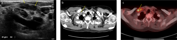
Purpose: Neck ultrasonography (US), computed tomography (CT), and 18F-fluorodeoxyglucose (FDG) positron emission tomography/computed tomography (PET/CT) are all known to be useful imaging modalities for detecting supraclavicular lymph node (SCN) metastasis in breast cancer. The authors compared the diagnostic values of neck US, CT, and PET/CT in the detection of SCN metastasis in breast cancer.

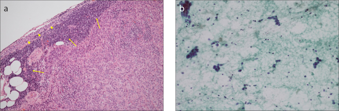
Methods: SCN metastases identified in neck US, CT, or PET/CT during follow-up visits of patients with breast cancer were pathologically confirmed with the use of US-guided fine-needle aspiration cytology. The clinicopathological factors of the patients were analyzed, and the statistical parameters including sensitivity, specificity, positive and negative predictive values, false-positive and false-negative rates, and accuracy of neck US, CT, and PET/CT were compared.

Results: Among 32 cases of suspicious SCNs, 24 were pathologically confirmed as metastasis of breast cancer. The sensitivity of US + CT was 91.7%, which was the same as that of PET/CT, while the sensitivity rates of US alone and CT alone were 87.5% and 83.3%, respectively. Accuracy was 99.8% in PET/CT alone and 98.1% in US + CT. The false-negative rate was 0.1% in US + PET/CT, while it was 0.2% in PET/CT and US + CT, 0.3% in US alone and 0.4% in CT alone.

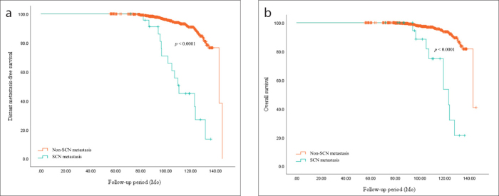
Conclusion: PET/CT can be the first choice for detecting SCN metastases in breast cancer. However, if PET/CT is unavailable for any reason, US + CT could be a good second option to avoid false-negative results.


Conflict of interest statement

Conflict of interest disclosure

The authors declared no conflicts of interest.

Figures

Figure 1. a–c
Figure 1. a–c
During the follow-up of a 55-year-old female patient with breast cancer, two suspicious supraclavicular lymph nodes (SCNs) on the right side of the neck were identified in US, chest CT, and 18F-FDG PET/CT. These lymph nodes were diagnosed as metastatic lymph nodes through fine-needle aspiration cytology. During the neck US shown in (a), two abutting suspicious SCNs were found in the lateral portion of the right carotid artery. The enlarged lymph nodes are hypoechoic with cortical thickening and loss of fatty hilum (arrows). Chest CT image (b) shows the presence of suspicious SCNs. Two abutted, enlarged SCNs on the right side of the neck show heterogeneous enhancement (arrow). In 18F-FDG PET/CT shown in (c), metastatic SCNs are seen on the right side of the neck (arrow) with a SUVmax 10.9.
Figure 2. a, b
Figure 2. a, b
Histopathologic findings of a metastatic SCN in breast cancer. Panel (a) shows histologic section of a lymph node demonstrating tumor cell deposits replacing the normal cells (arrows). In addition, normal lymphoid tissues are observed on the periphery (arrowheads) (×40, hematoxylin and eosin staining). In the smear of fine-needle aspiration cytology method (b), the lymph node shows clusters of pleomorphic malignant cells (×200, Papanicolaou stain).
Figure 3. a, b
Figure 3. a, b
Comparison of oncologic outcomes between metastatic supraclavicular lymph node (SCN) and non-metastatic SCN in breast cancer. In panel (a), distant metastasis-free survival was significantly superior in patients with breast cancer who were not diagnosed with SCN metastasis (p < 0.0001). In panel (b), overall survival was also significantly superior in patients with breast cancer who did not have SCN metastasis (p < 0.0001).

References

    1. Rue M, Vilaprinyo E, Lee S, et al. Effectiveness of early detection on breast cancer mortality reduction in catalonia (spain) BMC Cancer. 2009;9:326. doi: 10.1186/1471-2407-9-326. - DOI - PMC - PubMed
    1. Taylor R, Gregory M, Sexton K, et al. Breast cancer mortality and screening mammography in new zealand: Incidence-based and aggregate analyses. J Med Screen. 2019;26:35–43. doi: 10.1177/0969141318776039. - DOI - PubMed
    1. McPhail S, Johnson S, Greenberg D, Peake M, Rous B. Stage at diagnosis and early mortality from cancer in england. Brit J Cancer. 2015;112(Suppl 1):S108–115. doi: 10.1038/bjc.2015.49. - DOI - PMC - PubMed
    1. Narod SAIJ, Miller AB. Why have breast cancer mortality rates declined? J Cancer Policy. 2015;5:8–17. doi: 10.1016/j.jcpo.2015.03.002. - DOI
    1. Pedersen AN, Moller S, Steffensen KD, et al. Supraclavicular recurrence after early breast cancer: A curable condition? Breast Cancer Res Treat. 2011;125:815–822. doi: 10.1007/s10549-010-0918-8. - DOI - PubMed

Substances