. 2021 Aug;49(3):295-307.
doi: 10.1007/s10827-021-00790-9. Epub 2021 May 18.

Modeling the interaction among three cerebellar disorders of eye movements: periodic alternating, gaze-evoked and rebound nystagmus

Affiliations

Modeling the interaction among three cerebellar disorders of eye movements: periodic alternating, gaze-evoked and rebound nystagmus

Ari A Shemesh et al. J Comput Neurosci. 2021 Aug.

Abstract

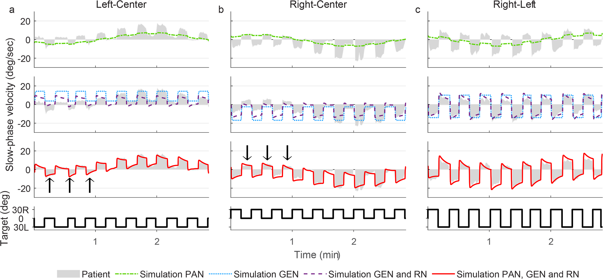
A woman, age 44, with a positive anti-YO paraneoplastic cerebellar syndrome and normal imaging developed an ocular motor disorder including periodic alternating nystagmus (PAN), gaze-evoked nystagmus (GEN) and rebound nystagmus (RN). During fixation there was typical PAN but changes in gaze position evoked complex, time-varying oscillations of GEN and RN. To unravel the pathophysiology of this unusual pattern of nystagmus, we developed a mathematical model of normal function of the circuits mediating the vestibular-ocular reflex and gaze-holding including their adaptive mechanisms. Simulations showed that all the findings of our patient could be explained by two, small, isolated changes in cerebellar circuits: reducing the time constant of the gaze-holding integrator, producing GEN and RN, and increasing the gain of the vestibular velocity-storage positive feedback loop, producing PAN. We conclude that the gaze- and time-varying pattern of nystagmus in our patient can be accounted for by superposition of one model that produces typical PAN and another model that produces typical GEN and RN, without requiring a new oscillator in the gaze-holding system or a more complex, nonlinear interaction between the two models. This analysis suggest a strategy for uncovering gaze-evoked and rebound nystagmus in the setting of a time-varying nystagmus such as PAN. Our results are also consistent with current ideas of compartmentalization of cerebellar functions for the control of the vestibular velocity-storage mechanism (nodulus and ventral uvula) and for holding horizontal gaze steady (the flocculus and tonsil).

Keywords: Adaptation; Cerebellum; Gaze-evoked nystagmus; Paraneoplastic; Periodic alternating nystagmus; Rebound nystagmus; Superposition.


Conflict of interest statement

Conflicts of interest/Competing interests: The authors declare that they have no conflict of interest

Figures

Fig. 1
Fig. 1
A conceptual scheme of the patterns of abnormal nystagmus that arise from isolated abnormalities in the vestibular or gaze-holding systems Each column shows simulated eye position (top) and slow-phase velocity (bottom) plotted against time for each pattern of nystagmus. Left column: Periodic alternating nystagmus (PAN), generated by an oscillatory vestibular system. Middle column: Gaze-evoked nystagmus (GEN) generated by an impaired “leaky” gaze-holding integrator. Right column: Rebound nystagmus (RN), which decays over time after return to straight ahead following GEN. An adaptive mechanism tends to cancel the GEN over time. Note the much longer time scale for PAN.
Fig. 2
Fig. 2
A schematic block diagram of the model. The two elements highlighted in red correspond with the two locations of the hypothetical lesions. Block transfer functions are characterized using the Laplace transform with s, the Laplace variable. The shaded (green) segment at the bottom represents the model of the vestibulo-ocular reflex (VOR) that incorporates a positive feedback loop for velocity-storage (low-pass filter with time constant close to that of the cupula, τVS) and negative feedback loop for central adaptation (perfect integrator with time constant τA). The overall time constant of the VOR is governed by the feedback loop gain (KVS) which under certain circumstances can drive the vestibular system into oscillatory behavior (PAN). The vestibular system responds to inputs arising from the semicircular canals, modeled as a high-pass filter with time constant τC. The shaded (blue) segment at the top represents the model of the gaze-holding system consisting of a low-pass filter with time constant τNI (neural integrator) with a positive feedback loop (position bias) used for adaptation and produces RN. The positive feedback includes a low-pass filter with a time constant of τPB that integrates an efference copy of eye position and shifts the null eye position slowly towards the previously held eccentric eye position. In the upper left, the saccades generator is triggered by the error signal derived by subtracting eye position from target position. An eye velocity signal arising from the saccade generator (“pulse“) projects via the direct pathway to the ocular motor neurons and ocular plant to overcome orbital viscosity (its gain equals the dominant time constant of the plant, τE1). The pulse also projects to the ocular motor integrator which in turn generates a position command (“step”) that also projects to the ocular motor neurons and ocular plant to counteract elastic restoring forces. The parameters values used in the model are listed in Table 1.
Fig. 3
Fig. 3
PAN during fixation of a straight-ahead target Horizontal slow-phase velocity plotted against time. Patient data is represented by the envelope of the gray shaded area and the model simulation by dashdot line (green). Target position, which is straight ahead, is on the bottom trace. Parameter values used in the simulation are listed in Table 1. The R-squared value was 0.98.
Fig. 4
Fig. 4
Effect of changing gaze on the pattern of nystagmus. Slow-phase velocity is on the Y-axis, and time on the X-axis. Target position is on the bottom trace. The patient changes gaze approximately every 10 sec. Column A. Patient looks between 30 deg to the left and straight ahead. Column B. Patient looks between 30 deg to the right and straight ahead. Column C. Patient looks between 30 deg to the left and 30 deg to the right. Slow-phase velocity of the patient is represented by the envelope of the gray shaded areas. Top row. Dashdot line (green) is a simulation using changes in the vestibular velocity-storage mechanism alone to produce PAN. Second row. Dotted line (blue) is a simulation of GEN and dashed line (purple) is a simulation of GEN and RN together. Third row. The solid line (red) is a simulation of the full model to produce PAN, GEN and RN. Arrow heads show where the effect of RN can be appreciated. Parameters for simulations are shown in Table 1. By presenting the vestibular and gaze-holding components of the model in separate plots before combining them in the complete model, one can interpret how slow-phase velocity changes with time due to superposition of the three types of nystagmus. Note that some deviations between simulations and data may be due to deviations on eye position during the recordings. While we simulated perfect gaze holding the subject would sometimes change gaze or blink during the recording. The R-squared value for column A, B and C was 0.92, 0.91 and 0.91, respectively.
Fig. 5
Fig. 5
Effect of sustained eccentric fixation Slow-phase velocity is on the Y-axis, and time on the X-axis. Target position is on the bottom trace. Periods of eccentric fixation at 30 degrees right or left are approximately one minute. Slow-phase velocity of the patient is shown by the envelope of the gray shaded areas. Dashdot line (green) is a simulation using changes in the vestibular velocity-storage mechanism alone to produce PAN. Dotted line (blue) is a simulation of GEN and dashed line (purple) is a simulation of GEN and RN together. The solid line (red) is a simulation of the full model to produce PAN, GEN and RN. Thin arrow heads indicate RN and thick arrow heads indicate GEN. Parameters for the simulations are shown in Table 1. The R-squared value was 0.89 compared with 0.24 for figure 3 PAN set of parameters.
Fig. 6
Fig. 6
Superposition effects on different PAN amplitudes and different oculomotor integrator time constants Slow-phase velocity is on the left Y-axis, target position is on the right Y-axis, and time on the X-axis. Slow-phase velocity is depicted as a blue dotted line for left-beating nystagmus (corresponding to rightward slow-phase velocity) and a red dashdot line for right-beating nystagmus (corresponding to leftward slow-phase velocity). Black line corresponds with the slow-phase velocity caused by PAN if gaze did not shift. Panel A shows model simulation of PAN with a small peak velocity (APAN was tuned to 6 deg/sec) and a severely impaired oculomotor integrator (τNI was set to 2.5 sec). Boxes 1 and 2 highlight the effect of gaze shifts around the time of the PAN transition or change in direction (null). Boxes 3 and 4 show the PAN transition without the interference of the gaze shifts. Panel B shows a model simulation of large peak velocity PAN (APAN was tuned to 12 deg/sec) and mildly impaired oculomotor integrator (τNI was set to 5 sec). Boxes 5 and 6 highlight how PAN at its peak can mask GEN, since it is right beating nystagmus during right gaze and left beating during left gaze. Boxes 7 and 8 show how GEN is revealed unambiguously around the time of the PAN null, with right beating nystagmus during right gaze and left beating during left gaze. Panel C shows another simulation with the same parameters of panel B but with different timing of the target jumps relative to the PAN cycle. Boxes 9 and 10 highlight how PAN at its peak can mask RN, with right-beating nystagmus after right gaze and left-beating after left gaze. Boxes 11 and 12 show how RN is revealed unambiguously around the time of the PAN null, with left-beating nystagmus after right gaze and right-beating after left gaze.

Similar articles

Cited by

References

    1. Baloh RW, Honrubia V, Konrad HR (1976) Periodic alternating nystagmus. Brain J Neurol 99:11–26. 10.1093/brain/99.1.11 - DOI - PubMed
    1. Bögli SY, Straumann D, Schuknecht B, et al. (2020) Cerebellar Rebound Nystagmus Explained as Gaze-Evoked Nystagmus Relative to an Eccentric Set Point: Implications for the Clinical Examination. Cerebellum Lond Engl 10.1007/s12311-020-01118-6 - DOI - PubMed
    1. Cannon SC, Robinson DA (1987) Loss of the neural integrator of the oculomotor system from brain stem lesions in monkey. J Neurophysiol 57:1383–1409 - PubMed
    1. Cohen B, Helwig D, Raphan T (1987) Baclofen and velocity storage: a model of the effects of the drug on the vestibulo-ocular reflex in the rhesus monkey. J Physiol 393:703–725 - PMC - PubMed
    1. Furman JM, Wall C, Pang DL (1990) Vestibular function in periodic alternating nystagmus. Brain J Neurol 113 ( Pt 5):1425–1439. 10.1093/brain/113.5.1425 - DOI - PubMed

Publication types