

Ultra-High Field Magnetic Resonance Imaging of the Retrobulbar Optic Nerve, Subarachnoid Space, and Optic Nerve Sheath in Emmetropic and Myopic Eyes
- PMID: 34003892
- PMCID: PMC7873495
- DOI: 10.1167/tvst.10.2.8
Ultra-High Field Magnetic Resonance Imaging of the Retrobulbar Optic Nerve, Subarachnoid Space, and Optic Nerve Sheath in Emmetropic and Myopic Eyes
Abstract
Purpose: We aimed to image the optic nerve, subarachnoid space and optic nerve sheath in emmetropes and myopes ultra-high field (7-Tesla) magnetic resonance imaging (MRI). We targeted the retrobulbar distance of approximately 3 mm behind the eyeball, an area of clinical interest because of optic nerve sheath distensibility and pressure-related enlargement.
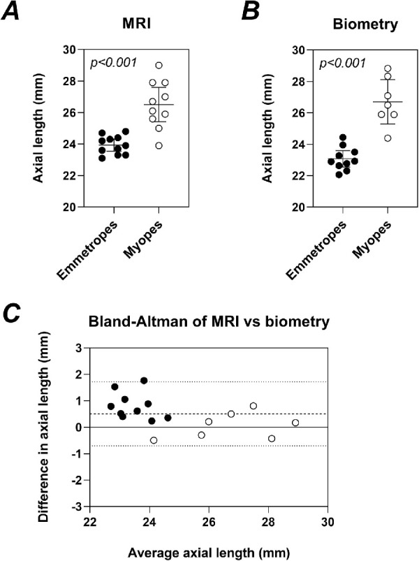
Methods: Eleven emmetropes (+0.75 to -0.50D, aged 20-41 years) and 10 myopes (-4.5 to -12D, aged 21-37 years) participated. Cross-sectional area of the optic nerve, subarachnoid space and optic nerve sheath at approximately 3 mm behind the eye were measured from two-dimensional T2-weighted coronal oblique MRI images obtained through the left optic nerve. Axial length of the left eye was measured from T2-weighted axial MRI images. In nine emmetropes and seven myopes, the optic nerve head was imaged with optical coherence tomography to compare retrobulbar and intraocular measures.
Results: Retrobulbar optic nerve, subarachnoid space and optic nerve sheath dimensions differed between myopes and emmetropes. Myopes tended to have smaller optic nerve and subarachnoid space. Longer MRI-derived axial length was associated with smaller optic nerve area (P = 0.03). Bruch's membrane opening area did not predict retrobulbar optic nerve area (P = 0.48).
Conclusions: This study demonstrates the feasibility of using 7-Tesla MRI to measure optic nerve, subarachnoid space, and optic nerve sheath dimensions behind the eye. In healthy adults, the retrobulbar optic nerve and subarachnoid space size are influenced by the degree of myopia.
Translational relevance: ultra-high field MRI is a practical tool for assessing the morphometry of the optic nerve and surrounding anatomy behind the eye.
Conflict of interest statement
Disclosure:
Figures


Similar articles
-
Imaging retrobulbar subarachnoid space around optic nerve by swept-source optical coherence tomography in eyes with pathologic myopia.Invest Ophthalmol Vis Sci. 2011 Dec 28;52(13):9644-50. doi: 10.1167/iovs.11-8597. Invest Ophthalmol Vis Sci. 2011. PMID: 22076987
-
Posture-Dependent Collapse of the Optic Nerve Subarachnoid Space: A Combined MRI and Modeling Study.Invest Ophthalmol Vis Sci. 2021 Apr 1;62(4):26. doi: 10.1167/iovs.62.4.26. Invest Ophthalmol Vis Sci. 2021. PMID: 33877263 Free PMC article.
-
Geometry of the Retinal Nerve Fibers From Emmetropia Through to High Myopia at Both the Temporal Raphe and Optic Nerve.Invest Ophthalmol Vis Sci. 2019 Nov 1;60(14):4896-4903. doi: 10.1167/iovs.19-27539. Invest Ophthalmol Vis Sci. 2019. PMID: 31752019
-
[Reproducibility and comparison study of two methods evaluating the optic nerve subarachnoid space sectional area using MRI].Zhonghua Yan Ke Za Zhi. 2017 Jun 11;53(6):440-444. doi: 10.3760/cma.j.issn.0412-4081.2017.06.009. Zhonghua Yan Ke Za Zhi. 2017. PMID: 28606266 Chinese.
-
Collapse or distention of the perioptic space in children - What does it mean to pediatric radiologists? Comprehensive review of perioptic space evaluation.Clin Imaging. 2024 Jul;111:110150. doi: 10.1016/j.clinimag.2024.110150. Epub 2024 Apr 10. Clin Imaging. 2024. PMID: 38723403 Review.
Cited by
-
Measuring axial length of the eye from magnetic resonance brain imaging.BMC Ophthalmol. 2022 Feb 5;22(1):54. doi: 10.1186/s12886-022-02289-y. BMC Ophthalmol. 2022. PMID: 35123441 Free PMC article.
-
3D Reconstruction of a Unitary Posterior Eye by Converging Optically Corrected Optical Coherence and Magnetic Resonance Tomography Images via 3D CAD.Transl Vis Sci Technol. 2022 Jul 8;11(7):24. doi: 10.1167/tvst.11.7.24. Transl Vis Sci Technol. 2022. PMID: 35895054 Free PMC article.
-
The impact of horizontal eye movements versus intraocular pressure on optic nerve head biomechanics: A tridimensional finite element analysis study.Heliyon. 2023 Feb 11;9(2):e13634. doi: 10.1016/j.heliyon.2023.e13634. eCollection 2023 Feb. Heliyon. 2023. PMID: 36865452 Free PMC article.
-
Effect of Eye Globe and Optic Nerve Morphologies on Gaze-Induced Optic Nerve Head Deformations.Invest Ophthalmol Vis Sci. 2024 Jul 1;65(8):48. doi: 10.1167/iovs.65.8.48. Invest Ophthalmol Vis Sci. 2024. PMID: 39083312 Free PMC article.
-
Effects of head-down tilt on optic nerve sheath diameter in healthy subjects.Ophthalmic Physiol Opt. 2023 Nov;43(6):1531-1539. doi: 10.1111/opo.13200. Epub 2023 Jul 3. Ophthalmic Physiol Opt. 2023. PMID: 37401194 Free PMC article.
References
-
- Hansen HC, Helmke K.. The subarachnoid space surrounding the optic nerves. An ultrasound study of the optic nerve sheath. Surg Radiol Anat. 1996; 18: 323–328. - PubMed
-
- Liu D, Kahn M.. Measurement and relationship of subarachnoid pressure of the optic nerve to intracranial pressures in fresh cadavers. Am J Ophthalmol. 1993; 116: 548–556. - PubMed
-
- Anderson DR. Fine structure and function of ocular tissues. The optic nerve. Int Ophthalmol Clin. 1973; 13: 229–242. - PubMed
-
- Anderson DR, Hoyt WF.. Ultrastructure of intraorbital portion of human and monkey optic nerve. Arch Ophthalmol. 1969; 82: 506–530. - PubMed
-
- Hansen HC, Helmke K.. Validation of the optic nerve sheath response to changing cerebrospinal fluid pressure: ultrasound findings during intrathecal infusion tests. J Neurosurg. 1997; 87: 34–40. - PubMed
Publication types
MeSH terms
LinkOut - more resources
Full Text Sources
Other Literature Sources
