Ocular Surface Microbiome Alterations Are Found in Both Eyes of Individuals With Unilateral Infectious Keratitis
- PMID: 34003904
- PMCID: PMC7884290
- DOI: 10.1167/tvst.10.2.19
Ocular Surface Microbiome Alterations Are Found in Both Eyes of Individuals With Unilateral Infectious Keratitis
Abstract
Purpose: To analyze the ocular surface microbiome (OSM) profile in both eyes of individuals with unilateral keratitis.
Methods: In this prospective, cross-sectional study, the conjunctival OSM of adults (>18 years old) presenting to an ophthalmic emergency department with acute unilateral keratitis and controls without an acute infectious process was sampled. Samples underwent DNA amplification and 16S sequencing using Illumina MiSeq 250 and were analyzed using Qiime. Statistical analysis was performed using a two-sided Student t-test, diversity indices, and principal coordinate analysis. The main outcome measures included relative abundance and α and β diversity.
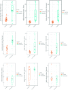
Results: Bacterial DNA was recovered from all 34 eyes of 17 individuals with keratitis (mean age, 49.3 ± 17.5 years) and 16 eyes of controls (mean age, 56.6 ± 17.0 years). In the two culture-positive eyes, 16S aligned with culture results. Significant differences in α diversities were noted when comparing both eyes of individuals with keratitis to control eyes (all P < 0.05), but no significant differences between the eyes of an individual with keratitis. Principal coordinate analysis plots confirmed this finding, demonstrating separation between either eye of patients with keratitis and controls (both P < 0.01), however not between eyes in patients with unilateral keratitis. Both eyes of individuals with keratitis had greater abundance of Pseudomonas compared with controls both on compositional analysis and linear discriminant analysis.
Conclusions: Alterations in the OSM profile are detected in both eyes of individuals with unilateral keratitis compared with controls. Beyond the causative organism, a greater abundance of potential pathogens and lesser abundance of commensal organisms were found.
Translational relevance: The OSM profile is altered in both eyes of individuals with unilateral keratitis, which may lend insight into the role of the microbiome in the pathophysiology of disease.
Conflict of interest statement
Disclosure:
Figures


Similar articles
-
Alterations in the conjunctival surface bacterial microbiome in bacterial keratitis patients.Exp Eye Res. 2021 Feb;203:108418. doi: 10.1016/j.exer.2020.108418. Epub 2020 Dec 23. Exp Eye Res. 2021. PMID: 33359511
-
Alterations of the bacterial ocular surface microbiome are found in both eyes of horses with unilateral ulcerative keratitis.PLoS One. 2023 Sep 8;18(9):e0291028. doi: 10.1371/journal.pone.0291028. eCollection 2023. PLoS One. 2023. PMID: 37682941 Free PMC article.
-
Ocular microbial dysbiosis and its linkage with infectious keratitis patients in Northwest China: A cross-sectional study.Microb Pathog. 2023 Nov;184:106371. doi: 10.1016/j.micpath.2023.106371. Epub 2023 Sep 22. Microb Pathog. 2023. PMID: 37741304
-
Infectious interface keratitis (IIK) following lamellar keratoplasty: A literature review.Ocul Surf. 2019 Oct;17(4):635-643. doi: 10.1016/j.jtos.2019.08.001. Epub 2019 Aug 12. Ocul Surf. 2019. PMID: 31415815
-
Critical insights into the ocular surface microbiome: the need to standardize.New Microbiol. 2024 Nov;47(3):201-216. New Microbiol. 2024. PMID: 39560032 Review.
Cited by
-
Changes in the ocular surface microbiome of patients with coronavirus disease 2019 (COVID-19).Front Microbiol. 2024 Jul 8;15:1389139. doi: 10.3389/fmicb.2024.1389139. eCollection 2024. Front Microbiol. 2024. PMID: 39040901 Free PMC article.
-
Distinct Ocular Surface Microbiome in Keratoconus Patients Correlate With Local Immune Dysregulation.Invest Ophthalmol Vis Sci. 2025 Jan 2;66(1):60. doi: 10.1167/iovs.66.1.60. Invest Ophthalmol Vis Sci. 2025. PMID: 39869087 Free PMC article.
-
Toward the Characterization of the Human Core Ocular Surface Microbiome.Invest Ophthalmol Vis Sci. 2025 Jul 1;66(9):40. doi: 10.1167/iovs.66.9.40. Invest Ophthalmol Vis Sci. 2025. PMID: 40657968 Free PMC article.
-
A Comprehensive Review of Microbial Biofilms on Contact Lenses: Challenges and Solutions.Infect Drug Resist. 2024 Jun 26;17:2659-2671. doi: 10.2147/IDR.S463779. eCollection 2024. Infect Drug Resist. 2024. PMID: 38947374 Free PMC article. Review.
References
-
- AAO Cornea/External Disease Preferred Practice Patterns Panel. Bacterial keratitis preferred practice patterns 2013. American Academy of Ophthalmology. Available at: www.aao.org/preferred-practice-pattern/bacterial-keratitis-ppp–2013. Accessed October 4, 2018.
-
- Eng BH, Gritz DC, Kumar AB, et al. .. Epidemiology of ulcerative keratitis in northern California. Arch Ophthalmol. 2010; 128: 1022–1028. - PubMed
-
- Stapleton F, Edwards K, Keay L, et al. .. Risk factors for moderate and severe microbial keratitis in daily wear contact lens users. Ophthalmology. 2012; 119(8): 1516–1521. - PubMed
Publication types
MeSH terms
Grants and funding
LinkOut - more resources
Full Text Sources
Other Literature Sources

