

Ocular Blood Flow in Preterm Neonates: A Preliminary Report
- PMID: 34003907
- PMCID: PMC7900851
- DOI: 10.1167/tvst.10.2.22
Ocular Blood Flow in Preterm Neonates: A Preliminary Report
Abstract
Purpose: Retinopathy of prematurity (ROP) is a vision-threatening complication occurring in pre-term neonates. The standard of care entails regular monitoring by dilated ophthalmoscopy examinations, which entail stress and potential morbidity. In this pilot study, we used plane-wave ultrasound (PWUS) to image, measure, and assess the association of blood-flow velocities in the retrobulbar vessels with ROP stages ranging from stage 0 (immature vessels without ROP) to stage 3.
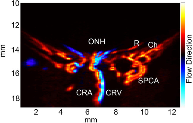
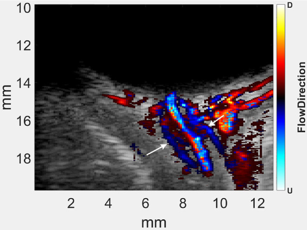
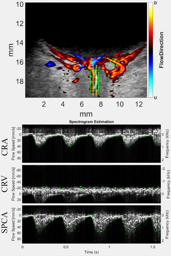
Methods: Both eyes of 14 preterm neonates at risk for ROP were examined by 18 MHz PWUS. All but two subjects had a follow-up examination. PWUS was acquired for 1.5 seconds at 3000 compound B-scans/sec. Data were postprocessed to form color-flow images and spectrograms depicting flow velocity in the central retinal artery (CRA), central retinal vein (CRV), and the short posterior ciliary arteries (SPCA). Flow parameters derived from spectrograms were compared by ROP stage.
Results: ROP stage was found to correlate with flow velocities. Velocities were significantly elevated with respect to non-ROP eyes in all vessels at stage 3 and in the SPCAs at stage 2.
Conclusions: PWUS measurement of blood flow may provide a quantitative, clinically important, and easily tolerated means for detecting and assessing the risk of ROP in preterm neonates. We speculate that the observed increase in flow velocity results from elevated vascular endothelial growth factor (VEGF) in ROP eyes.
Translational relevance: PWUS offers a gentle, nonmydriatic method for monitoring neonates at risk for ROP that would complement ophthalmoscopy.
Conflict of interest statement
Disclosure:
Figures
References
-
- Terry TL. Extreme prematurity and fibroblastic overgrowth of persistent vascular sheath behind each crystalline lens: I. Preliminary report. Am J Ophthalmol. 1942; 25: 203–204. - PubMed
-
- Smith LE. Igf-1 and retinopathy of prematurity in the preterm infant. Biol Neonate. 2005; 88: 237–244. - PubMed
-
- Provis JM, Leech J, Diaz CM, et al. .. Development of the human retinal vasculature: cellular relations and VEGF expression. Exp Eye Res. 1997; 65: 555–568. - PubMed
Publication types
MeSH terms
Substances
Grants and funding
LinkOut - more resources
Full Text Sources
Other Literature Sources
