

Intraoperative Retinal Changes May Predict Surgical Outcomes After Epiretinal Membrane Peeling
- PMID: 34003921
- PMCID: PMC7910632
- DOI: 10.1167/tvst.10.2.36
Intraoperative Retinal Changes May Predict Surgical Outcomes After Epiretinal Membrane Peeling
Abstract
Purpose: To investigate whether intraoperative retinal changes during epiretinal membrane (ERM) peeling affect anatomic or functional outcomes after surgery.
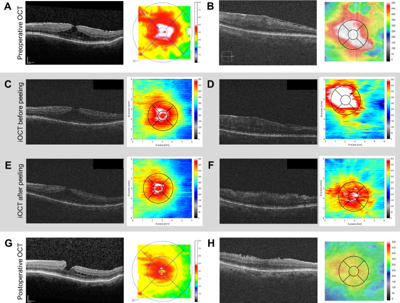
Methods: We measured retinal thickness using an intraoperative optical coherence tomography (iOCT) device in patients undergoing pars plana vitrectomy with membrane peeling for idiopathic ERM. Changes in intraoperative central macular thickness (iCMT) were compared with postoperative improvements in CMT and best-corrected visual acuity (VA).
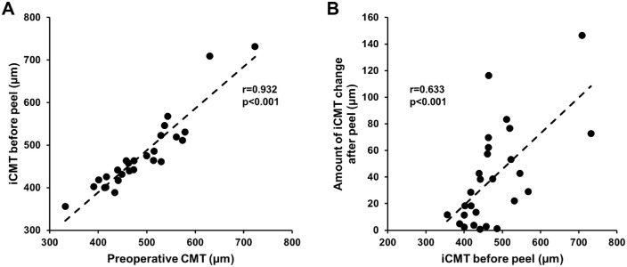
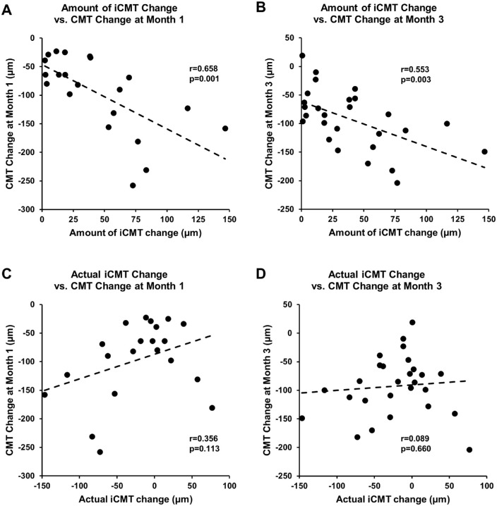
Results: Twenty-seven eyes from 27 patients (mean age 68 years) underwent iOCT-assisted ERM peeling surgery. Before surgery, mean VA was logMAR 0.50 ± 0.36 (Snellen 20/63), and mean baseline CMT was 489 ± 82 µm. Mean iCMT before peeling was 477 ± 87 µm, which correlated well with preoperative CMT (P < 0.001). Mean change in iCMT was -39.6 ± 37 µm (range -116 to +77 µm). After surgery, VA improved to logMAR 0.40 ± 0.38 (Snellen 20/50) at month 1 and logMAR 0.27 ± 0.23 (Snellen 20/37) at month 3, whereas CMT decreased to 397 ± 44 µm and 396 ± 51 µm at months 1 and 3. Eyes that underwent greater amount of iCMT change (absolute value of iCMT change) were associated with greater CMT reduction at month 1 (P < 0.001) and month 3 (P = 0.010), whereas those with greater intraoperative thinning (actual iCMT change) showed a trend toward better VA outcomes at months 1 (P = 0.054) and 3 (P = 0.036).
Conclusions: Intraoperative changes in retinal thickness may predict anatomic and visual outcomes after idiopathic ERM peeling surgery.
Translational relevance: Our study suggests that intraoperative retinal tissue response to ERM peeling surgery measured by iOCT may be a prognostic indicator for restoration of retinal architecture and for visual acuity outcomes.
Conflict of interest statement
Disclosure:
Figures

Similar articles
-
Anatomical, Functional, and Prognostic Results of Vitrectomy in Epiretinal Membranes Secondary to Retinal Vein Occlusions.Ophthalmologica. 2025;248(1):29-39. doi: 10.1159/000542770. Epub 2024 Nov 25. Ophthalmologica. 2025. PMID: 39586293 Free PMC article.
-
Epiretinal Membrane Surgery Using Intraoperative OCT-Guided Membrane Removal in the DISCOVER Study versus Conventional Membrane Removal.Ophthalmol Retina. 2021 Dec;5(12):1254-1262. doi: 10.1016/j.oret.2021.02.013. Epub 2021 Feb 27. Ophthalmol Retina. 2021. PMID: 33647472 Free PMC article.
-
Vitrectomy With Epiretinal Membrane Peeling Alone Verses Combined With Internal Limiting Membrane Peeling For Idiopathic Epiretinal Membrane.J Ayub Med Coll Abbottabad. 2020 Oct-Dec;32(4):450-453. J Ayub Med Coll Abbottabad. 2020. PMID: 33225642 Clinical Trial.
-
Visual Outcomes of Pars Plana Vitrectomy with Epiretinal Membrane Peeling in Patients with Asteroid Hyalosis: A Matched Cohort Study.Ophthalmic Res. 2017;58(1):35-39. doi: 10.1159/000468990. Epub 2017 May 3. Ophthalmic Res. 2017. PMID: 28463846
-
THE EFFECT OF INTERNAL LIMITING MEMBRANE PEELING ON IDIOPATHIC EPIRETINAL MEMBRANE SURGERY, WITH A REVIEW OF THE LITERATURE.Retina. 2017 May;37(5):873-880. doi: 10.1097/IAE.0000000000001263. Retina. 2017. PMID: 27617536 Review.
Cited by
-
Optical coherence tomography retinal imaging: narrative review of technological advancements and clinical applications.Ann Transl Med. 2025 Apr 30;13(2):17. doi: 10.21037/atm-24-211. Epub 2025 Apr 29. Ann Transl Med. 2025. PMID: 40438521 Free PMC article. Review.
-
Classification and management of myopic traction maculopathy: a vitrectomy-based consensus from taiwanese experts.Int Ophthalmol. 2025 May 5;45(1):174. doi: 10.1007/s10792-025-03526-1. Int Ophthalmol. 2025. PMID: 40323482
-
Clinical significance of signal shadowing during intraoperative optical coherence tomography-assisted vitreoretinal surgery.Sci Rep. 2024 Mar 5;14(1):5393. doi: 10.1038/s41598-024-56125-y. Sci Rep. 2024. PMID: 38443491 Free PMC article.
-
Idiopathic Epiretinal Membrane Surgery in Patients Aged Over 80 Years: Efficacy and Safety.Clin Ophthalmol. 2023 Nov 3;17:3365-3372. doi: 10.2147/OPTH.S437815. eCollection 2023. Clin Ophthalmol. 2023. PMID: 37941775 Free PMC article.
References
-
- Moisseiev E, Davidovitch Z, Kinori M, Loewenstein A, Moisseiev J, Barak A.. Vitrectomy for idiopathic epiretinal membrane in elderly patients: surgical outcomes and visual prognosis. Curr Eye Res. 2012; 37(1): 50–54. - PubMed
-
- Pichi F, Alkabes M, Nucci P, Ciardella AP.. Intraoperative SD-OCT in macular surgery. Ophthalmic Surg Lasers Imaging. 2012; 43(6 Suppl): S54–S60. - PubMed
-
- Kim JH, Kim YM, Chung EJ, Lee SY, Koh HJ.. Structural and functional predictors of visual outcome of epiretinal membrane surgery. Am J Ophthalmol. 2012; 153(1): 103–110.e1. - PubMed
Publication types
MeSH terms
Grants and funding
LinkOut - more resources
Full Text Sources
Other Literature Sources
Miscellaneous
