. 2021 Mar 1;10(3):11.
doi: 10.1167/tvst.10.3.11.

Clinical 3D Imaging of the Anterior Segment With Ultrasound Biomicroscopy

Affiliations

Clinical 3D Imaging of the Anterior Segment With Ultrasound Biomicroscopy

Richard W Helms et al. Transl Vis Sci Technol. .

Abstract

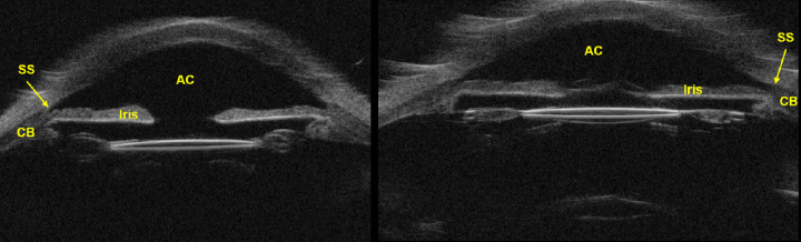
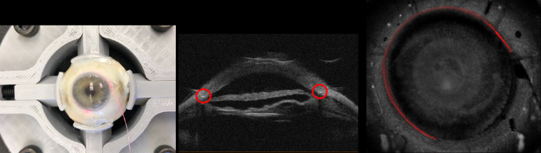
Purpose: Ultrasound biomicroscopy (UBM) is an important ophthalmic imaging modality due to its ability to see behind pigmented iris and to visualize anterior chamber when the eye's transparency is compromised. We created a three-dimensional UBM (3D-UBM) system and acquired example images to illustrate its potential.

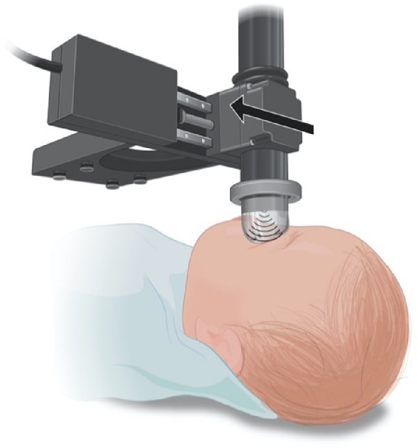
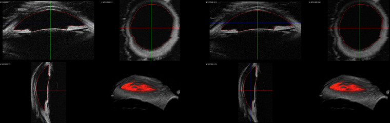
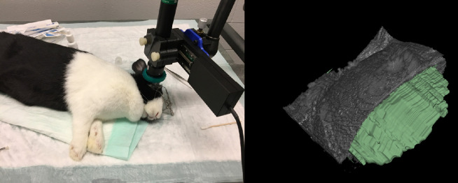
Methods: A commercial 50-MHz two-dimensional UBM (2D-UBM) system was attached to a precision translation stage and translated across the eye to acquire an image volume. The stage was mounted on a surgical microscope, which enabled safe, stable positioning. Image processing steps included image alignment, noise reduction, and calibration. 3D visualization included alignment of the optic axis, multiplanar reformatting at arbitrary orientations, and volume rendering with optimized transfer functions. Scans were performed on cadaver and rabbit eyes.

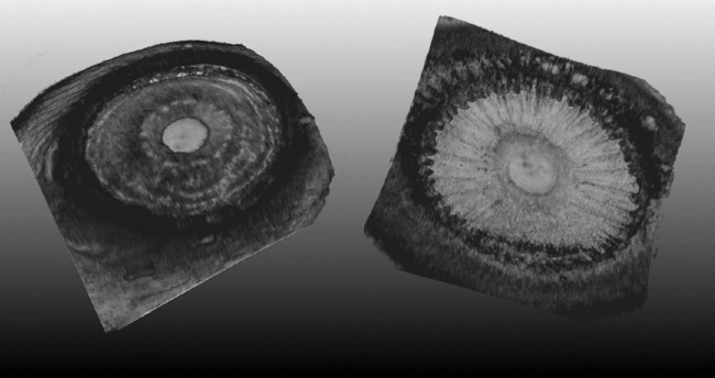
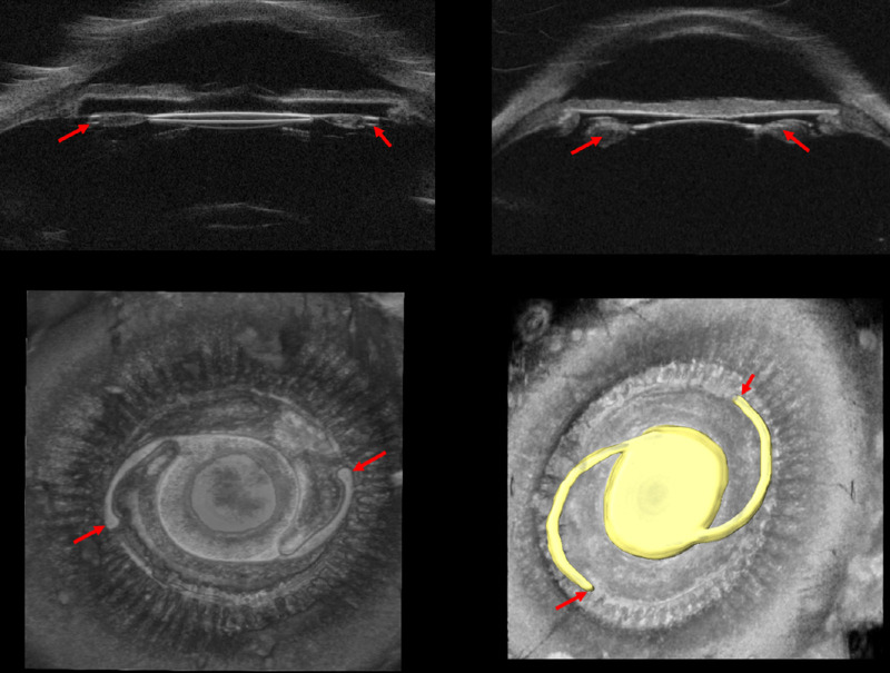
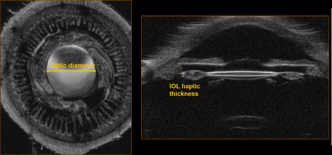
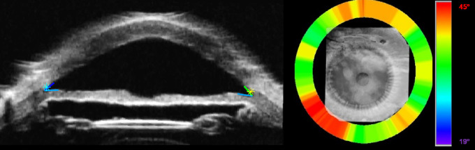
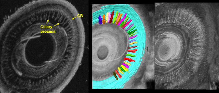
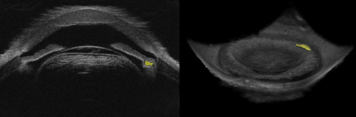
Results: 3D-UBM allowed visualization of the anterior segment tissues within a 3D anatomical context, unlike 2D-UBM. En face views and interactive slicer operations suggested an ability to plan and assess treatments, including lens placement and microcatheter cannulation of Schlemm's canal. Interactive software allowed us to make accurate measurements of tissue structures (e.g., iridocorneal angles, cyst volumes). In addition, unique measurements of ciliary tissues included single ciliary process volumes of 0.234 ± 0.093 mm3 with surface areas of 3.02 ± 1.07 mm2 and ciliary muscle volume of 67.87 mm3.

Conclusions: 3D-UBM imaging of the anterior segment can be used to enable unique visualization and quantification of anterior segment structures.

Translational relevance: 3D-UBM provides informative 3D imaging of tissues in the eye that are invisible to light to potentially provide physicians with improved diagnosis, treatment planning, and treatment assessment as compared to conventional 2D-UBM.


Conflict of interest statement

Disclosure: R.W. Helms, None; A.T. Minhaz, None; D.L. Wilson, None; F.H. Örge, None

Figures

Figure 1.
Figure 1.
Schematic view of the ultrasound probe attached to the translation stage. The probe and stage are suspended below a surgical microscope (not shown). The probe is coupled to the eye using the ClearScan probe cover and translated across the eye in a direction perpendicular to the probe sweep.
Figure 2.
Figure 2.
Conventional 2D-UBM images of ex vivo human eye. 2D-UBM allows visualization of important anatomical structures, such as the anterior chamber (AC), scleral spur (SS), iris, and structures posterior to the iris, such as the ciliary body (CB).
Figure 3.
Figure 3.
Anterior (left) and posterior (right) iris in an ex vivo human eye. The anterior cornea is cropped away to provide unobstructed view of the iris from both the anterior and posterior perspective. The anterior view allows complete 360° visualization of the iridocorneal angle. The posterior view allows visualization of the ciliary body, including individual ciliary processes. See Supplementary Movie S1 for an interactive 3D viewing of this eye.
Figure 4.
Figure 4.
Structures important for treatment planning and assessment are easily visualized. Positioning of IOL using conventional 2D-UBM does not provide any meaningful visualization of the IOL haptics (red arrow); shown are an AcrySof single-piece IOL (top left) and a three-piece IOL (top right). 3D-UBM shows the exact location of IOLs (bottom right, yellow), location of the haptics (bottom, red arrow), and condition of surrounding tissues. Supplementary Movie S2 provides an interactive view of IOL haptics enabled by 3D-UBM.
Figure 5.
Figure 5.
Cadaver eye with iTrack microcatheter placed in Schlemm's canal. (A) Color photograph shows the catheter inserted externally at 3 o'clock and threaded counterclockwise. The tip of the catheter is indicated by a red light emitted from the tip through the sclera. (B) Conventional UBM shows the catheter as a bright spot within the Schlemm's canal. (C) 3D-UBM volume of the same eye with the cornea cropped away and the catheter segmented to show its location within the canal. Between 9 and 1 o'clock, the catheter appears less bright, as it is slightly deeper in the tissue.
Figure 6.
Figure 6.
Cadaver eye with an AcrySof single-piece IOL (optic diameter 6 mm, thickness 0.43 mm) confirming dimensions measured in 3D-UBM. The cadaver eye was imaged in 3D, producing the two cross-sections shown above. The size of the lens was measured at eight different locations from the 3D image and compared with that of the explanted lens. Optic diameter was measured at 6.14 ± 0.05 mm (error, 2.33%) and corrected IOL haptic thickness was 0.40 mm (error, 6.97%).
Figure 7.
Figure 7.
Manual (left) and automated (right) segmentation of the anterior chamber of a cadaver eye with the boundary in red. The anterior chamber volume from the manual segmentation was measured at 264 mm3; from the fully automated segmentation, it was 267 mm3.
Figure 8.
Figure 8.
Automated trabecular iris angle (TIA) measurements. (A) Rotational view image with scleral spur and angles identified. (B) Angle heat map showing variation around the eye. Note the tendency for increased angle between 7 and 8 o'clock. The color map depicts angle variations from 19° (blue) to 45° (red).
Figure 9.
Figure 9.
En face view and segmentation of ciliary muscle (blue) and ciliary processes (multicolor). The en face view allows unique visualization of the ciliary body and processes not possible with conventional 2D-UBM. The anterior–posterior rendering clearly shows the ciliary body. Segmentations reveal the unique 3D biometrics of tissues that are important in glaucoma.
Figure 10.
Figure 10.
Cadaver eye with cyst-like formation following cyclophotocoagulation (CPC). After application of CPC, the formation (yellow) appeared near the iris and ciliary body. The formation was manually segmented, giving a volume of 0.38 mm3. With 3D-UBM, we can visualize its entire volume and location relative to other anterior segment tissues.
Figure 11.
Figure 11.
In vivo imaging of rabbit anterior chamber. Rabbits were sedated, and the probe was attached to a flexible arm and positioned over the rabbit's eye. The AC was scanned using 100 frames. The AC was manually segmented (3D Slicer software) to determine AC volume and depth. This figure shows the segmented AC (green) with part of the cornea cropped away.

References

    1. He M, Wang D, Jiang Y. Overview of ultrasound biomicroscopy. J Curr Glaucoma Pract. 2012; 6(1): 25–53. - PMC - PubMed
    1. Mannino G, Abdolrahimzadeh B, Calafiore S, Anselmi G, Mannino C, Lambiase A. A review of the role of ultrasound biomicroscopy in glaucoma associated with rare diseases of the anterior segment. Clin Ophthalmol. 2016; 10: 1453–1459. - PMC - PubMed
    1. Silverman RH. High-resolution ultrasound imaging of the eye – a review. Clin Exp Ophthalmol. 2009; 37(1): 54–67. - PMC - PubMed
    1. Li M, Chen Y, Chen X, et al. .. Differences between fellow eyes of acute and chronic primary angle closure (glaucoma): an ultrasound biomicroscopy quantitative study. PLoS One. 2018; 13(2): e0193006. - PMC - PubMed
    1. Ramasubramanian V, Glasser A. Objective measurement of accommodative biometric changes using ultrasound biomicroscopy. J Cataract Refract Surg. 2015; 41(3): 511–526. - PMC - PubMed

Publication types