

Correlation of Morphology and Function of Flecks Using Short-Wave Fundus Autofluorescence and Microperimetry in Patients With Stargardt Disease
- PMID: 34003952
- PMCID: PMC7991959
- DOI: 10.1167/tvst.10.3.18
Correlation of Morphology and Function of Flecks Using Short-Wave Fundus Autofluorescence and Microperimetry in Patients With Stargardt Disease
Abstract
Purpose: The purpose of this study was to evaluate the functional relevance of longitudinal changes in hyperautofluorescent areas and flecks in Stargardt disease (STGD1) using short-wavelength autofluorescence (SW-AF) imaging.
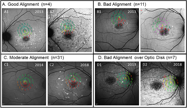
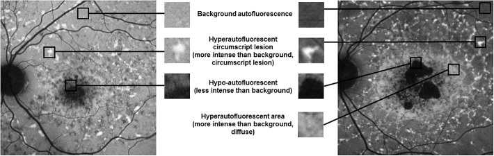
Methods: In this prospective, longitudinal study, 31 patients with STGD1 (56 eyes) underwent microperimetry (MP) and SW-AF imaging twice in 3 to 5 years. A total of 760 MP test points were included in the statistical analysis based on stable fixation and accurate alignment of SW-AF and MP. Autofluorescence intensity was qualitatively assessed in all MP test points. Small circumscriptive hyperautofluorescent lesions were defined as flecks. Longitudinal imaging characteristics observed on SW-AF were classified into the following categories: appearing, disappearing, and stable flecks, stable hyperautofluorescent, and stable background autofluorescence. The relationship between SW-AF intensity changes and MP changes was analyzed using a linear mixed model corrected for baseline sensitivity.
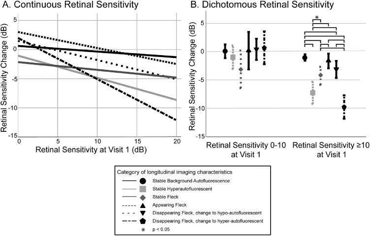
Results: Retinal sensitivity declined most in locations without change in SW-AF intensity. Functional decline per year was significantly larger in flecks that disappeared (-0.72 ± 1.30 dB) compared to flecks that appeared (-0.34 ± 0.65 dB), if baseline sensitivity was high (≥10 dB; P < 0.01). The correlation between the change observed on SW-AF and the sensitivity change significantly depended on the sensitivity at baseline (P = 0.000).
Conclusions: Qualitative longitudinal assessment of SW-AF poorly reflected the retinal sensitivity loss observed over the course of 3 to 5 years.
Translational relevance: When aiming to assess treatment effect on lesion level, a multimodal end point including MP focused on hyperautofluorescent lesions appears essential but needs further studies on optimizing MP grids, eye-tracking systems, and alignment software.
Conflict of interest statement
Disclosure:
Figures
References
-
- Allikmets R, Singh N, Sun H, et al. .. A photoreceptor cell-specific ATP-binding transporter gene (ABCR) is mutated in recessive Stargardt macular dystrophy. Nat Genet. 1997; 15: 236–246. - PubMed
-
- Weng J, Mata NL, Azarian SM, Tzekov RT, Birch DG, Travis GH.. Insights into the function of Rim protein in photoreceptors and etiology of Stargardt's disease from the phenotype in abcr knockout mice. Cell. 1999; 98: 13–23. - PubMed
Publication types
MeSH terms
Substances
LinkOut - more resources
Full Text Sources
Other Literature Sources
Miscellaneous
