

Smart Eye Camera: A Validation Study for Evaluating the Tear Film Breakup Time in Human Subjects
- PMID: 34004005
- PMCID: PMC8083120
- DOI: 10.1167/tvst.10.4.28
Smart Eye Camera: A Validation Study for Evaluating the Tear Film Breakup Time in Human Subjects
Abstract
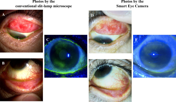
Purpose: This study aimed to demonstrate the efficacy of a "Smart Eye Camera (SEC)" in comparison with the efficacy of the conventional slit-lamp microscope by evaluating their diagnostic functionality for dry eye disease (DED) in clinical cases.
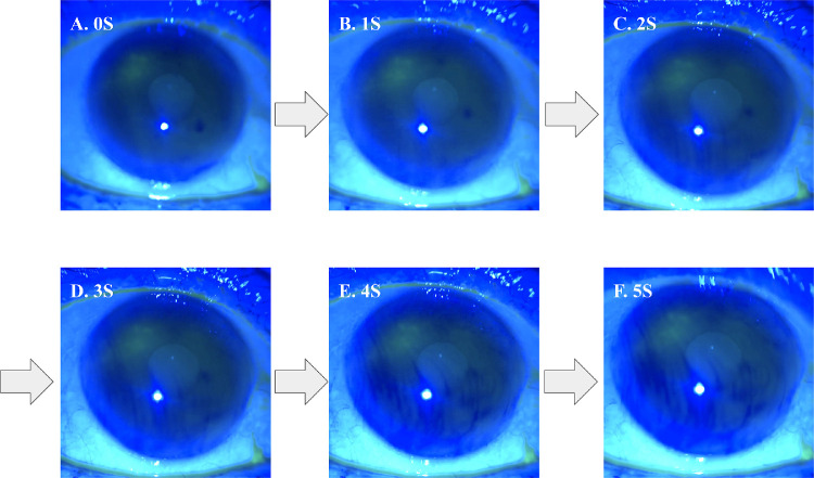
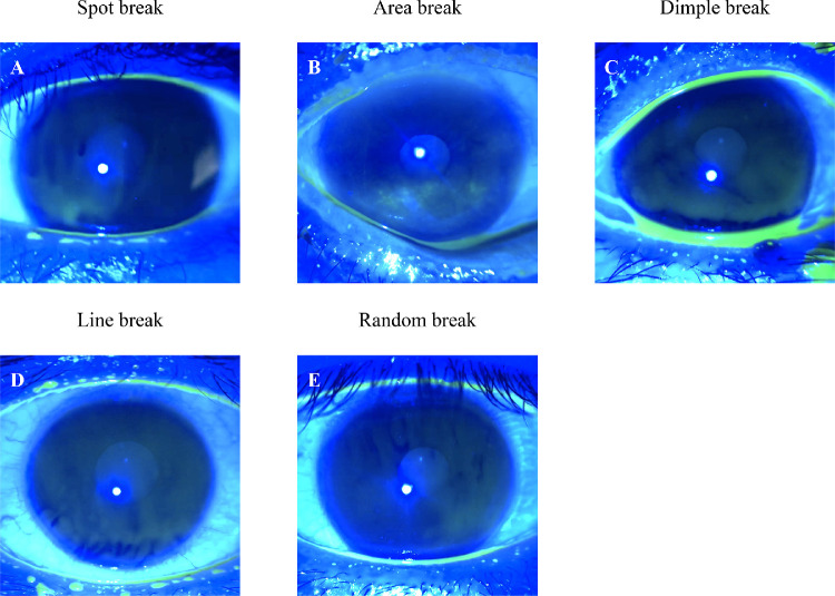
Methods: This retrospective study included 106 eyes from 53 adult Japanese patients who visited the Ophthalmology outpatient clinics in Keio University Hospital from June 2019 to March 2020. Tear film breakup time (TFBUT) and corneal fluorescence score (CFS) measurements for the diagnosis of DED were compared between the conventional slit-lamp microscope and SEC.
Results: The objective findings of DED showed that there was a strong correlation between the conventional slit-lamp microscope and SEC with respect to TFBUT and CFS results (Spearman's r = 0.887, 95% confidence interval [CI] = 0.838-0.922, and r = 0.920, 95% CI = 0.884-0.945, respectively). The interobserver reliability between the conventional slit-lamp microscope and SEC showed a moderate agreement (weighted Kappa κ = 0.527, 95% CI = 0.517-0.537 and κ = 0.550, 95% CI = 0.539-0.561 for TFBUT and CFS, respectively). The diagnostic performance of the SEC for Asia Dry Eye Society diagnostic criteria showed a sensitivity of 0.957 (95% CI = 0.841-0.992), specificity of 0.900 (95% CI = 0.811-0.927), positive predictive value of 0.880 (95% CI = 0.774-0.912), and negative predictive value of 0.964 (95% CI = 0.869-0.993). Moreover, the area under the receiver operating characteristic curve was 0.928 (95% CI = 0.849-1.000).
Conclusions: Compared with the conventional slit-lamp microscope, SEC has sufficient validity and reliability for diagnosing DED in the clinical setting.
Translational relevance: The SEC can portably evaluate TFBUT in both basic research and clinical care.
Conflict of interest statement
Disclosure:
Figures

Similar articles
-
Artificial intelligence to estimate the tear film breakup time and diagnose dry eye disease.Sci Rep. 2023 Apr 10;13(1):5822. doi: 10.1038/s41598-023-33021-5. Sci Rep. 2023. PMID: 37037877 Free PMC article.
-
Clinical observation of tear film breakup time with a novel smartphone-attachable technology.BMC Ophthalmol. 2023 May 10;23(1):204. doi: 10.1186/s12886-023-02932-2. BMC Ophthalmol. 2023. PMID: 37165312 Free PMC article.
-
Interobserver Reliability of Tear Break-Up Time Examination Using "Smart Eye Camera" in Indonesian Remote Area.Clin Ophthalmol. 2023 Jul 24;17:2097-2107. doi: 10.2147/OPTH.S412233. eCollection 2023. Clin Ophthalmol. 2023. PMID: 37521149 Free PMC article.
-
A novel and innovative paper-based analytical device for assessing tear lactoferrin of dry eye patients.Ocul Surf. 2019 Jan;17(1):160-166. doi: 10.1016/j.jtos.2018.11.001. Epub 2018 Nov 3. Ocul Surf. 2019. PMID: 30399438 Review.
-
New Perspectives on Dry Eye Definition and Diagnosis: A Consensus Report by the Asia Dry Eye Society.Ocul Surf. 2017 Jan;15(1):65-76. doi: 10.1016/j.jtos.2016.09.003. Epub 2016 Oct 8. Ocul Surf. 2017. PMID: 27725302 Review.
Cited by
-
Artificial intelligence to estimate the tear film breakup time and diagnose dry eye disease.Sci Rep. 2023 Apr 10;13(1):5822. doi: 10.1038/s41598-023-33021-5. Sci Rep. 2023. PMID: 37037877 Free PMC article.
-
Validation and optimization of smart eye camera as teleophthalmology device for the reduction of preventable and treatable blindness in Nigeria.Eye (Lond). 2025 Apr;39(5):925-930. doi: 10.1038/s41433-024-03489-0. Epub 2024 Dec 2. Eye (Lond). 2025. PMID: 39623111
-
A Case of Traumatic Hyphema Diagnoses by Telemedicine Between a Remote Island and the Mainland of Tokyo.Cureus. 2024 Jul 22;16(7):e65153. doi: 10.7759/cureus.65153. eCollection 2024 Jul. Cureus. 2024. PMID: 39176324 Free PMC article.
-
Successful Transition From Topical Ophthalmic Drops to Cream Formulation in the Management of Mild Allergic Conjunctivitis: A Case Report.Cureus. 2025 Feb 28;17(2):e79818. doi: 10.7759/cureus.79818. eCollection 2025 Feb. Cureus. 2025. PMID: 40166528 Free PMC article.
-
Feasibility of Tear Meniscus Height Measurements Obtained with a Smartphone-Attachable Portable Device and Agreement of the Results with Standard Slit Lamp Examination.Diagnostics (Basel). 2024 Feb 1;14(3):316. doi: 10.3390/diagnostics14030316. Diagnostics (Basel). 2024. PMID: 38337832 Free PMC article.
References
-
- Tsubota K, Yokoi N, Shimazaki J, et al. .. Members of the Asia Dry Eye Society. New perspectives on dry eye definition and diagnosis: a consensus report by the Asia Dry Eye Society. Ocul Surf. 2017; 15(1): 65–76. - PubMed
-
- Shimazaki J. Definition and diagnostic criteria of dry eye disease: historical overview and future directions. Invest Ophthalmol Vis Sci. 2018; 59(14): DES7–DES12. - PubMed
-
- Kojima T, Dogru M, Kawashima M, Nakamura S, Tsubota K.. Advances in the diagnosis and treatment of dry eye [published online ahead of print Jan 29, 2020]. Prog Retin Eye Res, https://doi.or/10.1016/j.preteyeres.2020.100842. - DOI - PubMed
Publication types
MeSH terms
LinkOut - more resources
Full Text Sources
