. 2021 Jun 10;184(12):3109-3124.e22.
doi: 10.1016/j.cell.2021.04.023. Epub 2021 May 17.

Small RNAs are modified with N-glycans and displayed on the surface of living cells

Affiliations

Small RNAs are modified with N-glycans and displayed on the surface of living cells

Ryan A Flynn et al. Cell. .

Erratum in

Abstract

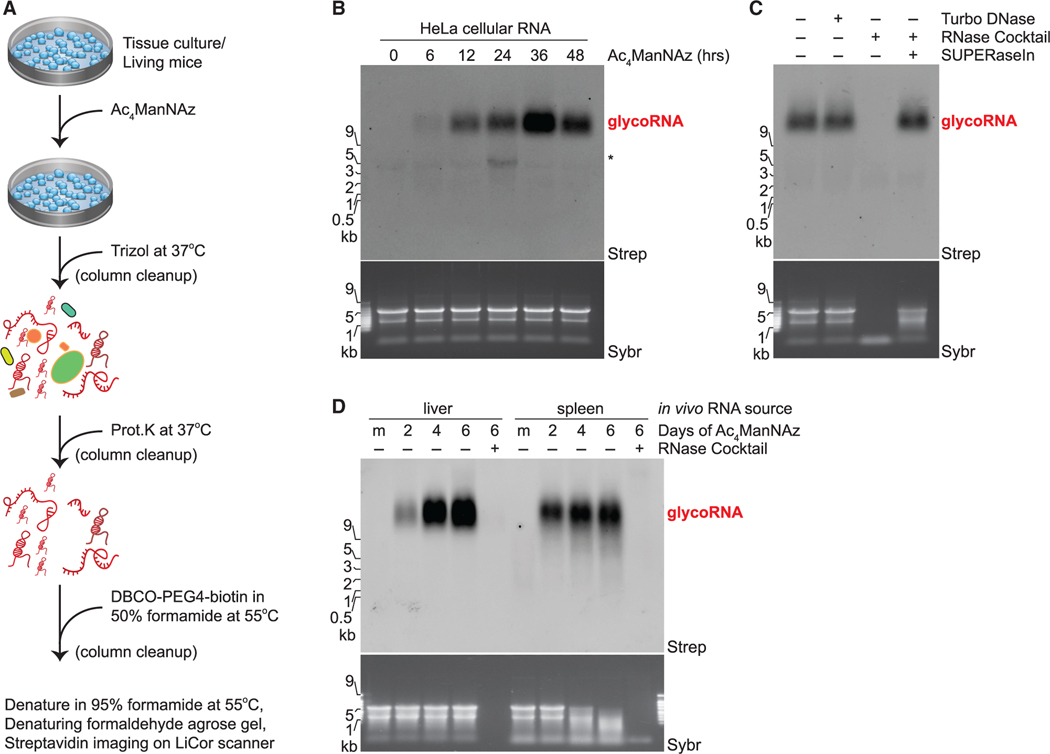
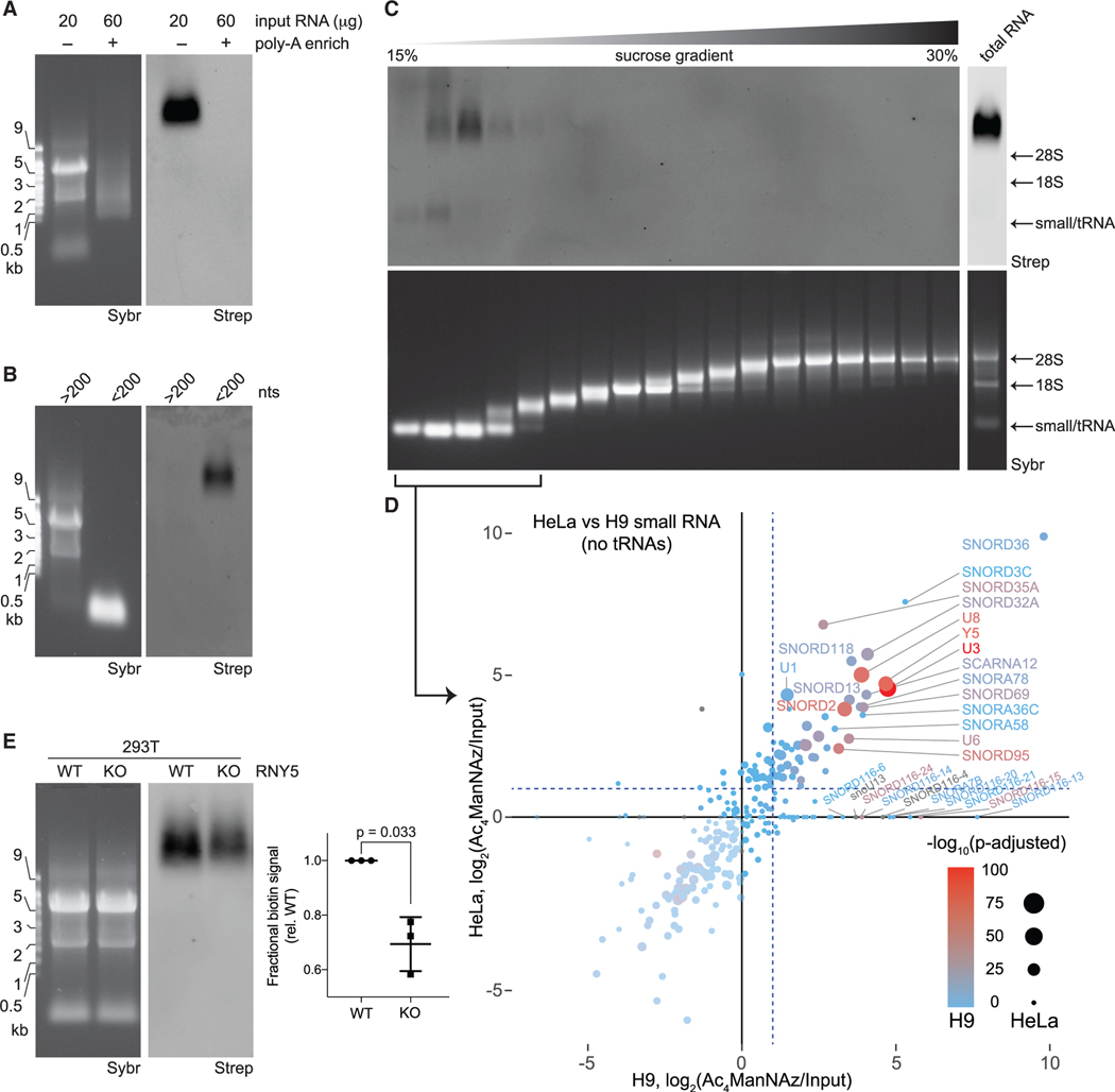
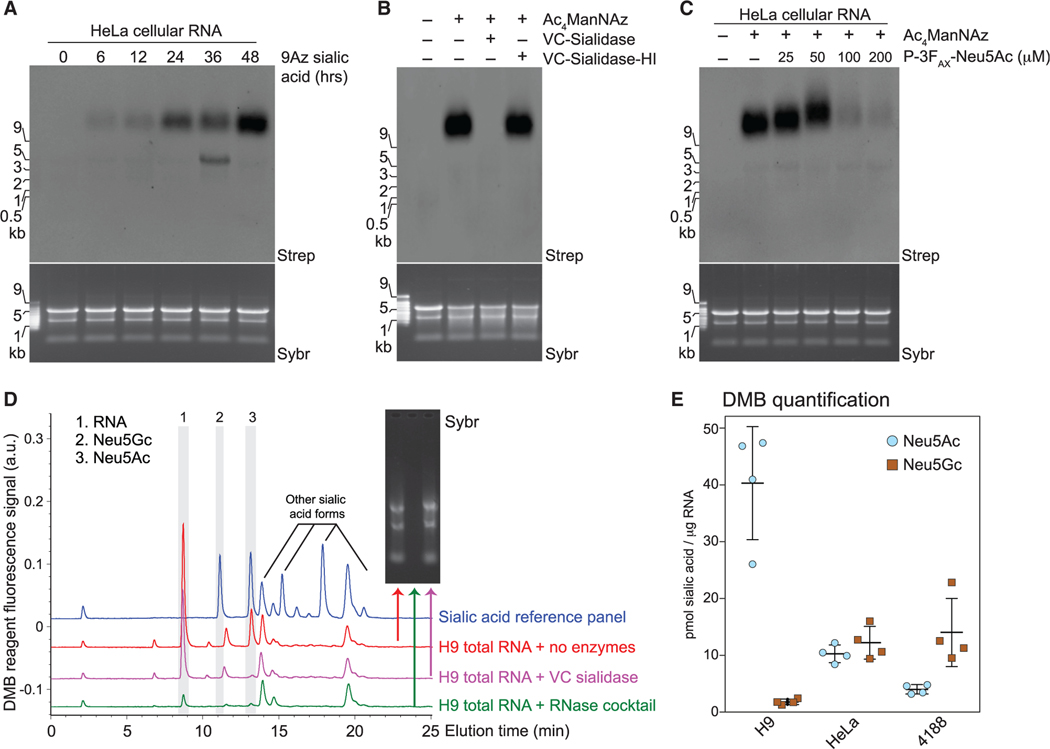
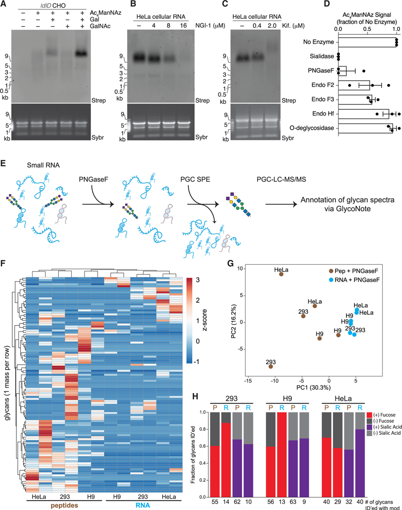
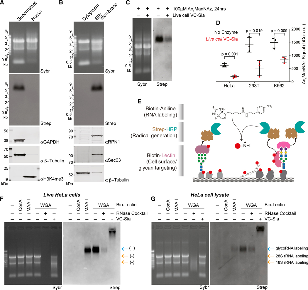
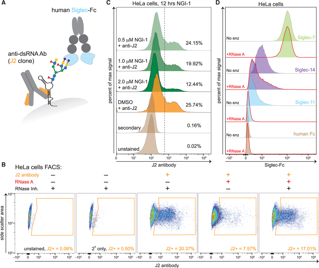
Glycans modify lipids and proteins to mediate inter- and intramolecular interactions across all domains of life. RNA is not thought to be a major target of glycosylation. Here, we challenge this view with evidence that mammals use RNA as a third scaffold for glycosylation. Using a battery of chemical and biochemical approaches, we found that conserved small noncoding RNAs bear sialylated glycans. These "glycoRNAs" were present in multiple cell types and mammalian species, in cultured cells, and in vivo. GlycoRNA assembly depends on canonical N-glycan biosynthetic machinery and results in structures enriched in sialic acid and fucose. Analysis of living cells revealed that the majority of glycoRNAs were present on the cell surface and can interact with anti-dsRNA antibodies and members of the Siglec receptor family. Collectively, these findings suggest the existence of a direct interface between RNA biology and glycobiology, and an expanded role for RNA in extracellular biology.

Keywords: RNA biology; glycoRNA; glycobiology; siglec; small RNAs.


Conflict of interest statement

Declaration of interests C.R.B. is co-founder and SAB member of Redwood Bioscience (a subsidiary of Catalent), Enable Biosciences, Palleon Pharmaceuticals, InterVenn Bio, OliLux Bio, and Lycia Therapeutics and a member of the Board of Directors of Eli Lilly.

Figures

Figure 1.
Figure 1.. Ac4ManNAz, a glycan reporter, incorporates into mammalian cellular RNA
(A) Schematic of RNA extraction protocol. Ac4ManNAz, peracetylated N-azidoacetylmannosamine; Prot.K, proteinase K; DBCO, dibenzocyclooctyne. (B) RNA blotting of RNA from HeLa cells treated with 100 μM Ac4ManNAz for the indicated amount of time. After RNA purification, Ac4ManNAz was conjugated to DBCO-biotin, visualized with streptavidin-IR800 (Strep), and imaged on an infrared scanner. Before RNA transfer to the membrane, total RNA was stained and imaged with SYBR Gold (Sybr) to interrogate quality and loading. All subsequent blots were prepared in this manner, and Ac4ManNAz is always used at 100 μM. The regions where glycoRNAs are present (red text) and non-specific labeling (*) is noted. (C) RNA Blot of Ac4ManNAz-labeled HeLa RNA treated in vitro with Turbo DNase or RNase cocktail (A/T1) +/− SUPERaseIn (RNase inhibitor). (D) RNA Blot of murine RNA after in vivo Ac4ManNAz delivery via intraperitoneal injection on indicated days at 300 mg Ac4ManNAz/kg/day. RNA from the liver and spleen were analyzed. Mock (m) mice were injected with DMSO only. RNase treatment was performed on extracted RNA. See also Figure S1.
Figure 2.
Figure 2.. Small, non-polyadenylated, and conserved transcripts comprise the pool of cellular glycoRNA
(A) Blotting of total or poly-adenylated (poly-A) enriched RNA from HeLa cells treated with Ac4ManNAz. (B) Blotting of total RNA from HeLa cells treated with Ac4ManNAz after differential precipitation fractionation using silica-based columns. (C) Blotting of total RNA from H9 human embryonic stem cells (H9) treated with Ac4ManNAz after sucrose density gradient (15%–30% sucrose) fractionation. An input profile is displayed to the right of the gradient. (D) Scatterplot analysis Ac4ManNAz-enriched RNAs purified from the small RNA fractions of (C) from HeLa and H9 cells. Reads mapping to snRNA, snoRNAs, and Y RNAs are shown. Significance scores (-log10(adjusted p value) are overlaid for HeLa cells as the size of each data point and for H9 cells as the color of each data point. (E) Representative blot of total RNA from wild-type (WT) or Y5 knockout (KO) 293T cells treated with Ac4ManNAz. Inset: quantification of the blot in (E) from biological triplicates. p value calculated by a paired, two-tailed t test. See also Figures S2 and S3 and Tables S1 and S2.
Figure 3.
Figure 3.. Glycans modifying RNA contain sialic acid
(A) Blotting of RNA from HeLa cells treated with 1.75 mM 9-azido sialic acid for indicated times. (B) Blotting of Ac4ManNAz-labeled HeLa cell RNA treated with Vibrio cholerae (VC) sialidase or heat-inactivated sialidase (VC-sialidase-HI). (C) Blotting of RNA from HeLa cells treated with Ac4ManNAz and the indicated concentrations of P-3FAX-Neu5Ac. (D) Unlabeled total RNA from H9 cells was isolated, reacted with the indicated enzyme (no enzymes, RNase cocktail, or Sialidase treatment), cleaned up to remove cleaved metabolites, and processed with the fluorogenic 1,2-diamino-4,5-methylenedioxybenzene (DMB) probe. HPLC analysis quantified the presence and abundance of specific sialic acids. Inset, Sybr gel image of the total RNA for each condition. The main sialic acid peaks are 2 and 3. The identity of peak 1 is unknown, but it is RNase-sensitive. (E) Quantification of DMB results (D) from 4,188, H9, and HeLa cells from four biological replicates. See also Figure S3.
Figure 4.
Figure 4.. A distinct set of N-glycans are enriched with glycoRNAs
(A) Blotting of RNA from ldlD CHO cells labeled with Ac4ManNAz, Galactose (Gal, 10 μM), N-acetylgalactosamine (GalNAc, 100 μM), or all for 24 h. (B) Blotting of RNA from HeLa cells treated with Ac4ManNAz and indicated concentrations of NGI-1, an inhibitor of OST, for 24 h. (C) Blotting as in (B) but with the indicated concentrations of kifunensine. (D) Quantification of Ac4ManNAz signal after treatment of Ac4ManNAz-labeled HeLa cell RNA with the indicated enzymes in vitro each for 1 h at 37°C in biological triplicate. (E) Schematic of the method used to release glycans from RNA samples and subsequently purify free glycans for mass spectrometry analysis. (F) Unsupervised clustering analysis of glycans (rows) released from peptide and RNA fractions (columns) of 293, H9, or HeLa cells via PNGaseF cleavage. Glycans had to be found biological replicates of at least one of the six samples to be included. (G) Principal component analysis of peptide- and RNA PNGaseF-release glycans. (H) Bar plots of the fraction of glycans containing fucose (red) or sialic acid (purple) modifications that were released from peptides or RNA samples. Numbers below are the absolute numbers of glycans found with each of the modifications from a given dataset. See also Figure S4 and Table S3.
Figure 5.
Figure 5.. glycoRNAs are on the external surface of living cells
(A) Blotting of RNA and proteins after subcellular fractionation designed to robustly purify nuclei. Non-nuclear proteins GAPDH and β-tubulin and nuclear histone 3 lysine 4 trimethylation (H3K4me3) are visualized by western blot. (B) Blotting of RNA and proteins after subcellular fractionation designed to separate soluble cytosol from membranous organelles. Membrane proteins RPN1, Sec63, and soluble β-tubulin are visualized by western blot. (C) Blotting of RNA from HeLa cells labeled with 100 μM Ac4ManNAz for 24 h and then exposed to fresh media containing 100 μM Ac4ManNAz with or without 150 nM VC-Sia for 60 min at 37°C (D) Quantification of the experiment in (C) across biological triplicates and from 293T or K562 cells treated in the same manner. p value calculated by a paired, two-tailed t test. (E) Schematic of the lectin-based proximity labeling of RNA on cell surfaces. Living cells are stained with a biotinylated lectin that recruits streptavidin-HRP that is in turn able to generate nitrene radicals from biotin-aniline after the addition of hydrogen peroxide. RNA from these cells is then extracted and analyzed for biotin labeling that reveals if that RNA was in proximity to the lectin. (F) Blotting of total RNA samples generated as described in (E). Lanes 5 and 6 were processed in vitro (after purifying RNA) with RNase cocktail or VC-Sia to demonstrate any sensitivity of the biotin-aniline signal to these enzymes. (G) Blotting of total RNA samples similar to (F) however cells were first lysed in a hypotonic buffer, destroying cellular membranes that are normally impermeable to nitrene radicals. Labeling of rRNA is evident here but not in (F). See also Figure S5.
Figure 6.
Figure 6.. Cell surface glycoRNAs contribute to the binding of select Siglec proteins
(A) Cartoon model of a glycoRNA on the cells surface depicted with two glycans identified in the PNGaseF release experiment. Prediction locations of binding for the anti-dsRNA antibody (J2) and Siglec-Fc proteins are highlighted. (B) Fluorescence-activated cell sorting (FACS) analysis of single HeLa cells pre-treated with the indicated enzymes or inhibitors and then stained with the J2 antibody. Gated region (orange) indicates the population shifted toward high J2 binding. (C) FACS analysis of single HeLa cells pre-treated with the OST inhibitor NGI-1 for 12 h at the indicated concentrations. Dashed vertical line denotes a J2-high population, and for each sample, the fraction of cells within this region is shown as a percentage. (D) FACS analysis of single HeLa cells pre-treated with RNase then stained with the indicated Siglec-Fc reagents. See also Figure S6.

Comment in

References

    1. Abu-Remaileh M, Wyant GA, Kim C, Laqtom NN, Abbasi M, Chan SH, Freinkman E, and Sabatini DM (2017). Lysosomal metabolomics reveals V-ATPase- and mTOR-dependent regulation of amino acid efflux from lysosomes. Science 358, 807–813. - PMC - PubMed
    1. Agard NJ, Prescher JA, and Bertozzi CR (2004). A strain-promoted [3 + 2] azide-alkyne cycloaddition for covalent modification of biomolecules in living systems. J. Am. Chem. Soc 126, 15046–15047. - PubMed
    1. Ahlin E, Mathsson L, Eloranta M-L, Jonsdottir T, Gunnarsson I, Rönnblom L, and Rönnelid J. (2012). Autoantibodies associated with RNA are more enriched than anti-dsDNA antibodies in circulating immune complexes in SLE. Lupus 21, 586–595. - PubMed
    1. Aizawa S, Fujiwara Y, Contu VR, Hase K, Takahashi M, Kikuchi H, Kabuta C, Wada K, and Kabuta T. (2016). Lysosomal putative RNA transporter SIDT2 mediates direct uptake of RNA by lysosomes. Autophagy 12, 565–578. - PMC - PubMed
    1. Angata T. (2014). Associations of genetic polymorphisms of Siglecs with human diseases. Glycobiology 24, 785–793. - PubMed

Publication types