. 2021 May 25;35(8):109179.
doi: 10.1016/j.celrep.2021.109179. Epub 2021 May 13.

Mapping the SARS-CoV-2 spike glycoprotein-derived peptidome presented by HLA class II on dendritic cells

Affiliations

Mapping the SARS-CoV-2 spike glycoprotein-derived peptidome presented by HLA class II on dendritic cells

Robert Parker et al. Cell Rep. .

Abstract

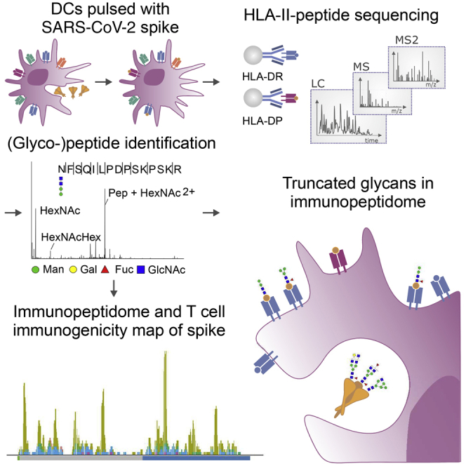
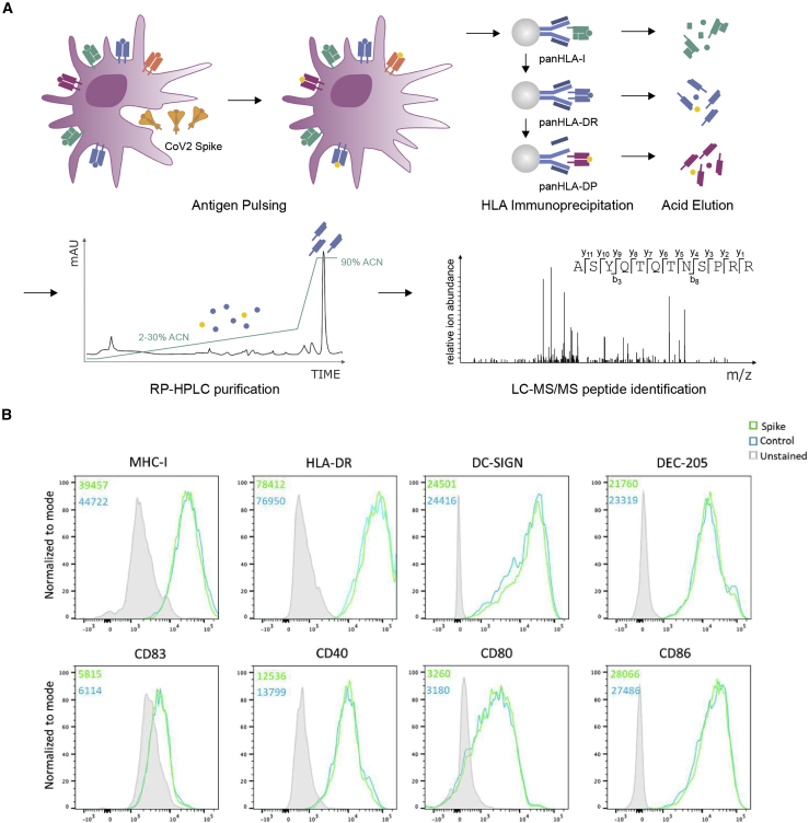
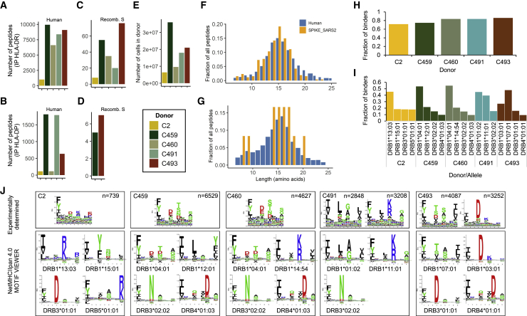
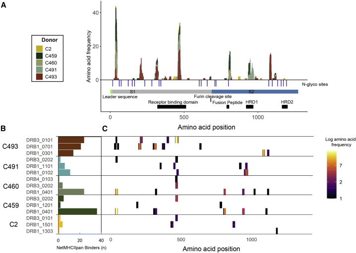
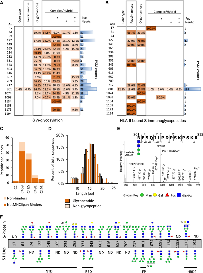
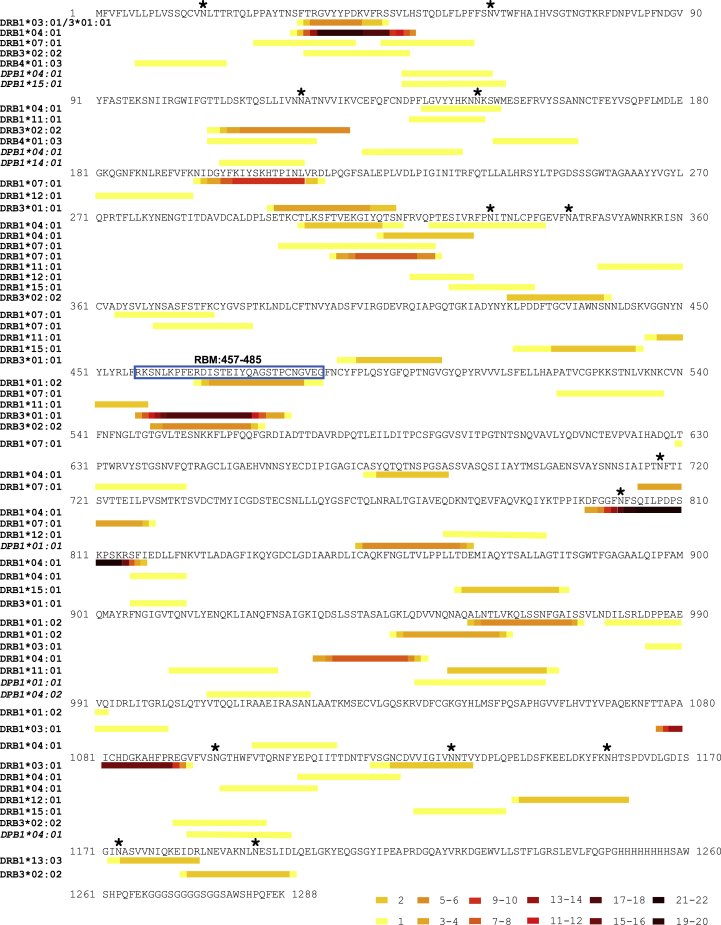
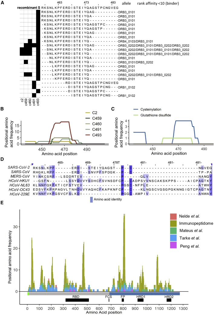
Understanding and eliciting protective immune responses to severe acute respiratory syndrome coronavirus 2 (SARS-CoV-2) is an urgent priority. To facilitate these objectives, we profile the repertoire of human leukocyte antigen class II (HLA-II)-bound peptides presented by HLA-DR diverse monocyte-derived dendritic cells pulsed with SARS-CoV-2 spike (S) protein. We identify 209 unique HLA-II-bound peptide sequences, many forming nested sets, which map to sites throughout S including glycosylated regions. Comparison of the glycosylation profile of the S protein to that of the HLA-II-bound S peptides reveals substantial trimming of glycan residues on the latter, likely induced during antigen processing. Our data also highlight the receptor-binding motif in S1 as a HLA-DR-binding peptide-rich region and identify S2-derived peptides with potential for targeting by cross-protective vaccine-elicited responses. Results from this study will aid analysis of CD4+ T cell responses in infected individuals and vaccine recipients and have application in next-generation vaccine design.

Keywords: HLA class II; HLA-II; LC-MS; SARS-CoV-2; antigen presentation; dentritic cells; glycopeptides; glycoslyation; human leukocyte antigen; immunopeptidomics.


Conflict of interest statement

Declaration of interests N.T. is directing immunopeptidomics research at Enara Bio part-time and serves on the Scientific Advisory Boards of Enara Bio and T-Cypher Bio. N.T. is consultant to Hoffman-La Roche and Grey Wolf Therapeutics. All other authors declare no conflict of interest.

Figures

None
Graphical abstract
Figure 1
Figure 1
Generation of MDDCs for HLA-II immunopeptidomic profiling (A) Schematic depicting the workflow used to generate the recombinant S protein pulsed MDDC immunopeptidome. MDDCs were pulsed with S or control protein for 18 h, and sequential HLA IPs were carried out as indicated. Peptides were eluted from HLA molecules, purified by preparative HPLC, and subjected to a LC-MS/MS identification workflow. (B) MDDCs were harvested following overnight protein pulse and stained with antibodies to identify DCs and assess their expression of HLA-I, HLA-DR, DC-SIGN, DEC-205, CD83, CD40, CD80, and CD86 and then analyzed by flow cytometry. Histogram plots are gated on live CD11c+CD4+ DCs and show expression of each indicated marker on cells treated with S or control protein. Unstained cells are shown in filled gray histograms. Inset numbers indicate mean fluorescence intensity for each sample. See also Figure S2.
Figure 2
Figure 2
Immunopeptidomic profiling of HLA-DR and HLA-DP-bound peptides presented by S-pulsed MDDCs (A and B) Number of unique peptide sequences identified in each HLA-DR (A) or HLA-DP (B) immunoprecipitation sample. (C and D) Number of unique peptide sequences that map to S identified in the HLA-DR (C) or HLA-DP (D) samples. (E) Total number of MDDCs harvested from each donor. (F and G) Length distribution of peptide sequences identified as mapping to human proteins (blue) or S protein (orange). (H) Proportion of peptide sequences predicted to bind to each donor’s HLA-DR and -DP alleles by NetMHCIIpan 4.0. (I) Proportion of total predicted HLA-DR binders stratified by allele for each donor. (J) All 12–20 mers in each sample were clustered using the online (unsupervised) GibbsCluster algorithm. Each cluster is represented by a sequence logo, which corresponds to at least one of the HLA-DRB alleles expressed by the donor MDDCs. Amino acids are represented by their single letter code; the more frequently an amino acid occurs a position within peptides, the larger the letter is displayed. The number (n) of peptides within each cluster is indicated along with the number of outlier peptides removed, and clusters are presented with the specific sequence motifs for donor DR alleles as reported by NetMHCIIpan 4.0. See also Figures S4 and S5 and Table S1.
Figure 3
Figure 3
Distribution of HLA-II-presented regions within the S glycoprotein (A) Stacked histogram of positional amino acid frequency in S for all peptide sequences identified in the immunopeptidome stratified and colored by donor. Horizontal bars depict the S1/2 regions, and blue vertical lines indicate N-linked glycosylation sites identified in the purified protein detected by digestion with PNGase F in the presence of heavy water, which leads to a detectable specific mass shift of 2 Da when an N-linked glycan has been removed. The furin cleavage site, receptor binding domain, fusion peptide, and heptad repeat domains (HRD) 1 and 2 are depicted as black boxes. (B) Number of S peptide sequences predicted to bind to each donor HLA-DR allele by NetMHCIIpan 4.0. C. Position of the identified DR-bound peptides across the S sequence, depicted as a heatmap of amino acid coverage and stratified by the donor and predicted DR allele of origin on the y axis. See also Figures S4 and S5 and Tables S1 and S2.
Figure 4
Figure 4
Glycosylation site and glycopeptide analysis of recombinant S protein and HLA-II-bound S peptides (A and B) Table showing distribution of glycans identified at each glycosylation site grouped according to the main N-glycan types based on intact glycopeptide analysis. (A) glycopeptides generated by in vitro digestion of the recombinant S protein and (B) HLA-II-bound S glycopeptides detected in the immunopeptidome of S-pulsed MDDCs. The heatmap colors indicate the relative frequency of each glycan composition present. The total number of peptide spectral matches (PSM) is reported (blue bars). (C) Donor-specific overview of the number of glycosylated peptide sequences and proportion predicted to bind to the donor HLA-DR allele profile. (D) Histogram illustrating the amino acid length distribution of glycopeptides and non-modified peptide sequences of S. (E) Example of an annotated fragment spectrum of an HLA-II-bound peptide carrying a paucimannosidic-type N-glycan in position N801. (F) Depiction of the most abundant glycan identified at each N-glycosylation sites of the S polypeptide chain prior to MDDC pulsing (top) and of the HLA-II-bound S peptides (bottom). The main domains of S are indicated. Man, mannose; Gal, galactose; Fuc, fucose; NeuAc, N-acetylneuraminic acid; GlcNac, N-Acetylglucosamine G. See also Tables S2, S3, and S4.
Figure 5
Figure 5
HLA-II peptide mapping across the S protein and RBM A map illustrating the location in the SARS-CoV-2 protein sequence of all the S-derived peptides of 9–25 amino acids in length identified in the HLA-II-bound immunopeptidome of S-pulsed MDDCs. HLA-DR-bound peptides are grouped according to the HLA-DR allele to which they had the highest predicted binding affinity (as determined using NetMHCIIpan 4.0), and HLA-DP-bound peptides are grouped by donor HLA-DPB1 type. Within each group, nested peptide sets are indicated in heatmap form, where the color represents the frequency of each amino acid position within the peptide group. Where a single peptide amino acid sequence was identified multiple times with different sequence modifications the unique amino acid sequence was included only once in positional frequency calculations. Glycosylation sites in S identified in the Byonic analysis of the immunopeptidome are indicated with an asterisk, and the nested set covering the RBM is boxed.
Figure 6
Figure 6
A nested set of unique peptides in the immunopeptidome map to positions 457–485 in the RBD of the full-length S protein (A) The black squares on the left indicate the donor(s) in which each peptide was identified. On the right of each peptide sequence, the HLA-DR allele to which it showed the highest predicted affinity of binding (NetMHCIIpan4.0), and the regarding rank score is indicated. (B) Line plots showing the frequency with which each amino acid position was represented by a peptide identification across the RBD of the S protein in each donor. (C) Line plots showing the frequency with which each amino acid position was represented by a cysteine-modified peptide identification (cysteinylated peptides and glutathione-modified peptides) across the RBD of the S protein. (D) Alignment of the S protein sequences of the SARS-CoV-2, SARS-CoV, MERS-CoV, and 4 endemic human coronaviruses (obtained from Uniprot). Positions relative to the RBD amino acids 457–485 in S are shown. The color indicates the degree of amino acid identify. (E) Stacked histogram plot showing the peptides identified as being recognized by CD4+ T cells in each of the four T cell epitope mapping studies and those in the immunopeptidome across the S protein sequence. The height of each bar represents the frequency at which each amino acid was detected in the relevant dataset. The domain structure for S is indicated below the plot as horizontal bars (S1/2 regions: gray/blue); and the furin cleavage site (FCS), receptor binding domain (RBD), fusion peptide (FP), and heptad repeat domains (HRD) 1 and 2 are depicted as black boxes.

Update of

References

    1. Altmann D.M., Boyton R.J. SARS-CoV-2 T cell immunity: Specificity, function, durability, and role in protection. Sci. Immunol. 2020;5 eabd6160. - PubMed
    1. Alves M.J., Kawahara R., Viner R., Colli W., Mattos E.C., Thaysen-Andersen M., Larsen M.R., Palmisano G. Comprehensive glycoprofiling of the epimastigote and trypomastigote stages of Trypanosoma cruzi. J. Proteomics. 2017;151:182–192. - PubMed
    1. Baum L.G., Cobb B.A. The direct and indirect effects of glycans on immune function. Glycobiology. 2017;27:619–624. - PubMed
    1. Braun J., Loyal L., Frentsch M., Wendisch D., Georg P., Kurth F., Hippenstiel S., Dingeldey M., Kruse B., Fauchere F. SARS-CoV-2-reactive T cells in healthy donors and patients with COVID-19. Nature. 2020;587:270–274. - PubMed
    1. Brouwer P.J.M., Caniels T.G., van der Straten K., Snitselaar J.L., Aldon Y., Bangaru S., Torres J.L., Okba N.M.A., Claireaux M., Kerster G. Potent neutralizing antibodies from COVID-19 patients define multiple targets of vulnerability. Science. 2020;369:643–650. - PMC - PubMed

Substances