

WFS1 protein expression correlates with clinical progression of optic atrophy in patients with Wolfram syndrome
- PMID: 34006618
- PMCID: PMC8685651
- DOI: 10.1136/jmedgenet-2020-107257
WFS1 protein expression correlates with clinical progression of optic atrophy in patients with Wolfram syndrome
Abstract
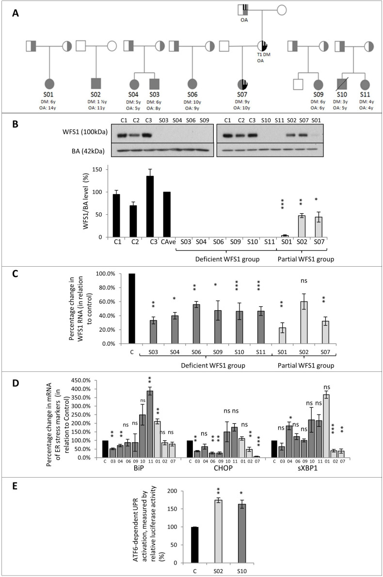
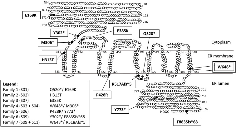
Background: Wolfram syndrome (WFS) is a rare disorder characterised by childhood-onset diabetes mellitus and progressive optic atrophy. Most patients have variants in the WFS1 gene. We undertook functional studies of WFS1 variants and correlated these with WFS1 protein expression and phenotype.
Methods: 9 patients with a clinical diagnosis of WFS were studied with quantitative PCR for markers of endoplasmic reticulum (ER) stress and immunoblotting of fibroblast protein extracts for WFS1 protein expression. Luciferase reporter assay was used to assess ATF-6 dependent unfolded protein response (UPR) activation.
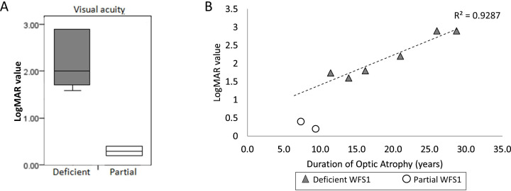
Results: 6 patients with compound heterozygous nonsense mutations in WFS1 had no detectable WFS1 protein expression; 3 patients with missense variants had 4%, 45% and 48% WFS1 protein expression. One of these also had an OPA1 mutation and was reclassified as autosomal dominant optic atrophy-plus syndrome. There were no correlations between ER stress marker mRNA and WFS1 protein expression. ERSE-luciferase reporter indicated activation of the ATF6 branch of UPR in two patients tested. Patients with partial WFS1 expression showed milder visual acuity impairment (asymptomatic or colour blind only), compared with those with absent expression (registered severe vision impaired) (p=0.04). These differences remained after adjusting for duration of optic atrophy.
Conclusions: Patients with WFS who have partial WFS1 protein expression present with milder visual impairment. This suggests a protective effect of partial WFS1 protein expression on the severity and perhaps progression of vision impairment and that therapies to increase residual WFS1 protein expression may be beneficial.
Keywords: diabetes mellitus; genetics, medical; neurodegenerative diseases.
© Author(s) (or their employer(s)) 2022. Re-use permitted under CC BY. Published by BMJ.
Conflict of interest statement
Competing interests: None declared.
Figures
Similar articles
-
Molecular characterization of WFS1 in patients with Wolfram syndrome.J Mol Diagn. 2003 May;5(2):88-95. doi: 10.1016/s1525-1578(10)60457-6. J Mol Diagn. 2003. PMID: 12707373 Free PMC article.
-
Identification of p.A684V missense mutation in the WFS1 gene as a frequent cause of autosomal dominant optic atrophy and hearing impairment.Am J Med Genet A. 2011 Jun;155A(6):1298-313. doi: 10.1002/ajmg.a.33970. Epub 2011 Apr 28. Am J Med Genet A. 2011. PMID: 21538838 Free PMC article.
-
Wolfram syndrome in the Japanese population; molecular analysis of WFS1 gene and characterization of clinical features.PLoS One. 2014 Sep 11;9(9):e106906. doi: 10.1371/journal.pone.0106906. eCollection 2014. PLoS One. 2014. PMID: 25211237 Free PMC article.
-
Wolfram syndrome 1 and Wolfram syndrome 2.Curr Opin Pediatr. 2012 Aug;24(4):512-7. doi: 10.1097/MOP.0b013e328354ccdf. Curr Opin Pediatr. 2012. PMID: 22790102 Review.
-
Wolfram Syndrome Type I Case Report and Review-Focus on Early Diagnosis and Genetic Variants.Medicina (Kaunas). 2024 Jun 28;60(7):1064. doi: 10.3390/medicina60071064. Medicina (Kaunas). 2024. PMID: 39064493 Free PMC article. Review.
Cited by
-
Genotype and clinical characteristics of patients with Wolfram syndrome and WFS1-related disorders.Front Genet. 2023 Jun 21;14:1198171. doi: 10.3389/fgene.2023.1198171. eCollection 2023. Front Genet. 2023. PMID: 37415600 Free PMC article.
-
Multimorbidity due to novel pathogenic variants in the WFS1/RP1/NOD2 genes: autosomal dominant congenital lamellar cataract, retinitis pigmentosa and Crohn's disease in a British family.BMJ Open Ophthalmol. 2023 Jul;8(1):e001252. doi: 10.1136/bmjophth-2023-001252. BMJ Open Ophthalmol. 2023. PMID: 37493686 Free PMC article.
-
WFS1 autosomal dominant variants linked with hearing loss: update on structural analysis and cochlear implant outcome.BMC Med Genomics. 2023 Apr 11;16(1):79. doi: 10.1186/s12920-023-01506-x. BMC Med Genomics. 2023. PMID: 37041640 Free PMC article.
-
NAD depletion mediates cytotoxicity in human neurons with autophagy deficiency.Cell Rep. 2023 May 30;42(5):112372. doi: 10.1016/j.celrep.2023.112372. Epub 2023 Apr 21. Cell Rep. 2023. PMID: 37086404 Free PMC article.
-
Neuro-Ophthalmologic Variability in Presentation of Genetically Confirmed Wolfram Syndrome: A Case Series and Review.Brain Sci. 2023 Jul 5;13(7):1030. doi: 10.3390/brainsci13071030. Brain Sci. 2023. PMID: 37508961 Free PMC article.
References
-
- Inoue H, Tanizawa Y, Wasson J, Behn P, Kalidas K, Bernal-Mizrachi E, Mueckler M, Marshall H, Donis-Keller H, Crock P, Rogers D, Mikuni M, Kumashiro H, Higashi K, Sobue G, Oka Y, Permutt MA. A gene encoding a transmembrane protein is mutated in patients with diabetes mellitus and optic atrophy (Wolfram syndrome). Nat Genet 1998;20:143–8. 10.1038/2441 - DOI - PubMed
-
- Tranebjaerg L, Barrett T, Rendtorff ND. WFS1-Related Disorders. GeneReviews((R)). Seattle (WA): University of Washington, Seattle University of Washington, Seattle. GeneReviews is a registered trademark of the University of Washington, Seattle. All rights reserved 1993.
-
- Astuti D, Sabir A, Fulton P, Zatyka M, Williams D, Hardy C, Milan G, Favaretto F, Yu-Wai-Man P, Rohayem J, López de Heredia M, Hershey T, Tranebjaerg L, Chen J-H, Chaussenot A, Nunes V, Marshall B, McAfferty S, Tillmann V, Maffei P, Paquis-Flucklinger V, Geberhiwot T, Mlynarski W, Parkinson K, Picard V, Bueno GE, Dias R, Arnold A, Richens C, Paisey R, Urano F, Semple R, Sinnott R, Barrett TG. Monogenic diabetes syndromes: locus-specific databases for Alström, Wolfram, and thiamine-responsive megaloblastic anemia. Hum Mutat 2017;38:764–77. 10.1002/humu.23233 - DOI - PMC - PubMed
Publication types
MeSH terms
Substances
Grants and funding
LinkOut - more resources
Full Text Sources
Other Literature Sources