. 2021 May 18;12(6):502.
doi: 10.1038/s41419-021-03735-3.

Notch3 inhibits cell proliferation and tumorigenesis and predicts better prognosis in breast cancer through transactivating PTEN

Affiliations

Notch3 inhibits cell proliferation and tumorigenesis and predicts better prognosis in breast cancer through transactivating PTEN

Yong-Qu Zhang et al. Cell Death Dis. .

Abstract

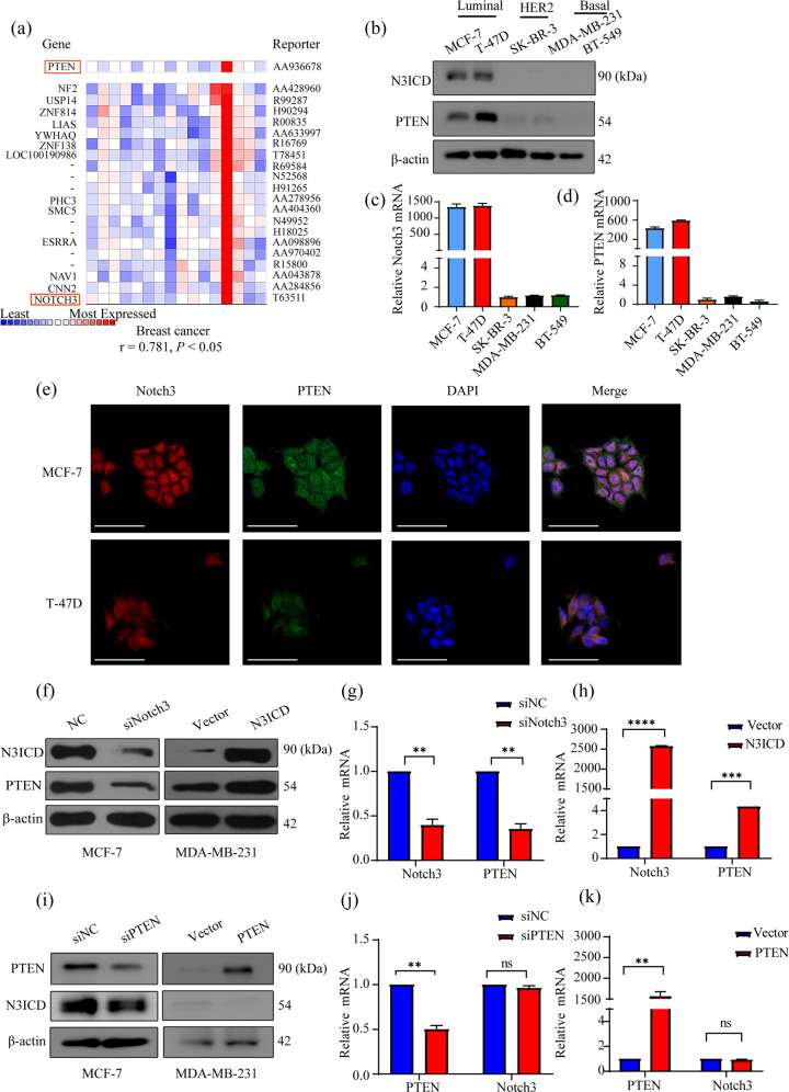
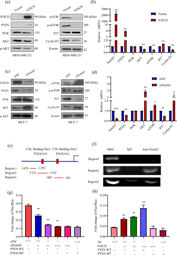
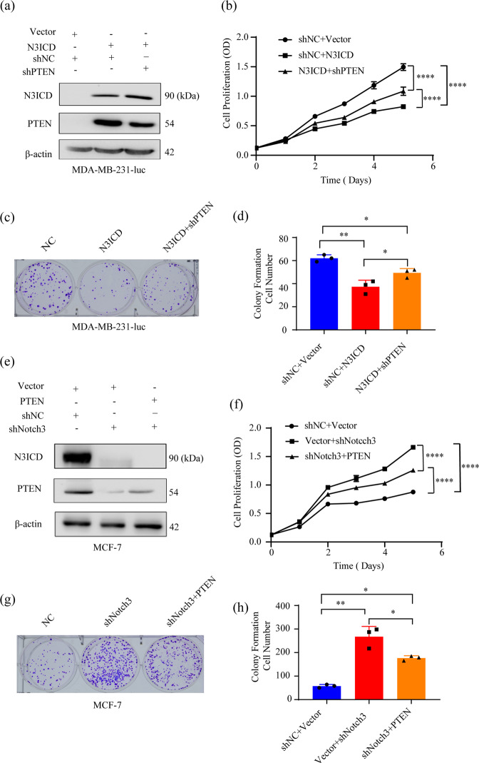
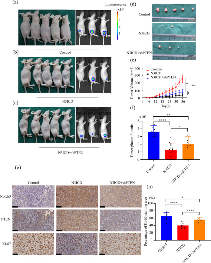
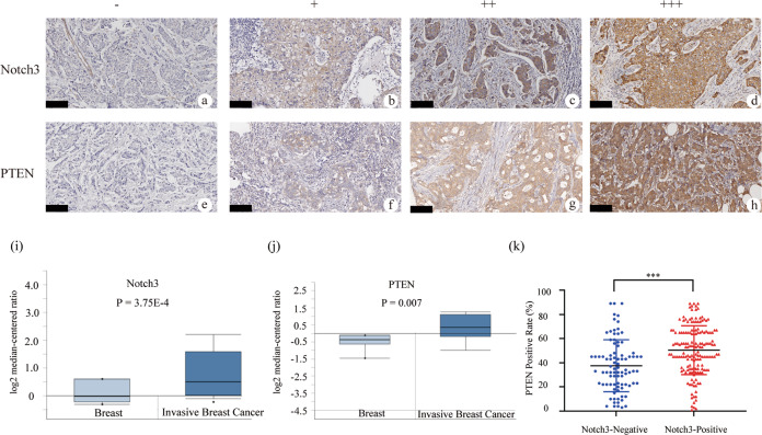
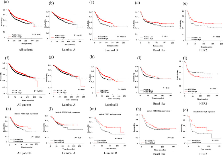
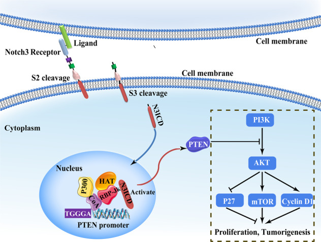
Notch receptors (Notch1-4) play critical roles in tumorigenesis and metastasis of malignant tumors, including breast cancer. Although abnormal Notch activation is related to various tumors, the importance of single receptors and their mechanism of activation in distinct breast cancer subtypes are still unclear. Previous studies by our group demonstrated that Notch3 may inhibit the emergence and progression of breast cancer. PTEN is a potent tumor suppressor, and its loss of function is sufficient to promote the occurrence and progression of tumors. Intriguingly, numerous studies have revealed that Notch1 is involved in the regulation of PTEN through its binding to CBF-1, a Notch transcription factor, and the PTEN promoter. In this study, we found that Notch3 and PTEN levels correlated with the luminal phenotype in breast cancer cell lines. Furthermore, we demonstrated that Notch3 transactivated PTEN by binding CSL-binding elements in the PTEN promoter and, at least in part, inhibiting the PTEN downstream AKT-mTOR pathway. Notably, Notch3 knockdown downregulated PTEN and promoted cell proliferation and tumorigenesis. In contrast, overexpression of the Notch3 intracellular domain upregulated PTEN and inhibited cell proliferation and tumorigenesis in vitro and in vivo. Moreover, inhibition or overexpression of PTEN partially reversed the promotion or inhibition of cell proliferation induced by Notch3 alterations. In general, Notch3 expression positively correlated with elevated expression of PTEN, ER, lower Ki-67 index, and incidence of involved node status and predicted better recurrence-free survival in breast cancer patients. Therefore, our findings demonstrate that Notch3 inhibits breast cancer proliferation and suppresses tumorigenesis by transactivating PTEN expression.


Conflict of interest statement

The authors declare no competing interests.

Figures

Fig. 1
Fig. 1. Notch3 is expressed in the luminal subtype and modulates PTEN expression in breast cancer cell lines.
a Heat-map representing the correlation of the mRNA of PTEN, Notch3, and some other genes obtained from Weigelt Breast dataset based on the analyses of about 10,335 genes in ONCOMINE. There was a positive correlation between the mRNA level of Notch3 and PETN, the correlation coefficient r = 0.781, P < 0.05 (b) Notch3 and PTEN expression in distinct subtype of breast cancer cell lines detected by western blotting. c, d Relative Notch3 and PTEN mRNA levels in breast cancer cell lines quantified by real-time PCR. e The immunofluorescence staining of PTEN and Notch3 in MCF-7 and T-47D cells. Nuclei were counterstained with DAPI. f, g The levels of Notch3 and PTEN protein and mRNA in MCF-7 cells were detected by western blotting or real-time PCR, respectively, following Notch3 knockdown by siRNA. f, h Notch3 and PTEN protein and mRNA expression levels in MDA-MB-231 cells were measured by western blotting or real-time PCR, respectively, following expression of pCLE-N3ICD. i, j The levels of Notch3 and PTEN protein and mRNA in MCF-7 cells were detected by western blotting or real-time PCR, respectively, following PTEN knockdown by siRNA. i, k The levels of Notch3 and PTEN protein and mRNA in MDA-MB-231 cells were detected by western blotting or real-time PCR, respectively, following ectopic expression of PTEN in MDA-MB-231 cells. The experiments were repeated three times. The scale bar represents 50 μm. *P < 0.05, **P < 0.01, ***P < 0.001, ****P < 0.0001.
Fig. 2
Fig. 2. Notch3 inhibits the AKT-mTOR pathway by binding to the CSL element of the PTEN promoter.
Notch3ICD overexpression increased PTEN and p27 expression in MDA-MB-231 cells but inhibited the protein (a) and mRNA (b) expression levels of AKT-mTOR-Cyclin D1 pathway components. Notch3 knockdown inhibited PTEN and p27 expression in MCF-7 cells but induced the protein (c) and mRNA (d) expression of AKT-mTOR-Cyclin D1pathway components. e Schematic diagram of the PTEN promoter regions amplified by different primer sets in the ChIP assays. Regions 2 and 3 each contain a CSL-binding element. Region 1, which does not contain the CSL-binding element, was used as a negative control. f For the ChIP assays, Notch3-binding PCR products were detected in regions 2 and 3 using an anti-Notch3 antibody. g Notch3 was knocked down in MCF-7 cells by siRNA, which were co-transfected with a wild-type or mutant PTEN promoter-containing firefly luciferase construct and pRL-SV40 as the control plasmid containing Renilla luciferase. The ratio of firefly luciferase to Renilla luciferase values was used to determine promoter activity. Each sample was performed in triplicate. h N3ICD overexpression in MDA-MB-231 cells co-transfected with a wild-type or mutant PTEN promoter-containing Firefly luciferase construct and pRL-SV40 as the control plasmid containing Renilla luciferase. The ratio of Firefly luciferase to Renilla luciferase was used to determine the promoter activity. Each sample was performed in triplicate. *P < 0.05, **P < 0.01.
Fig. 3
Fig. 3. Ectopic Notch3 expression inhibits proliferation and tumorigenesis in vitro, which is attenuated by PTEN silencing.
a Notch3 and PTEN expression in MDA-MB-231-luc cells measured by western blotting following stable N3ICD overexpression, with or without PTEN knockdown. β-actin served as an internal control. b Stable N3ICD expression inhibited MDA-MB-231-luc cell growth in vitro. This effect was attenuated by PTEN silencing with shRNA. c N3ICD overexpression inhibited colony formation, which was reversed by stable PTEN knockdown with shRNA. Representative pictures and quantitative data from the colony formation assays are presented. d Representative micrographs and data for Matrigel-coated assays. The stably transfected N3ICD MDA-MB-231-luc were co-transfected with shPTEN or shNC as a negative control. e Notch3 and PTEN expression in MCF-7 cells measured by western blotting following stable Notch3 knockdown, with or without stable PTEN overexpression. f Stable Notch3 knockdown induced MCF-7 cell growth in vitro, which was attenuated by PTEN overexpression. g Stable Notch3 knockdown promoted the colony formation, which was reversed by stable PTEN overexpression. Representative pictures and quantitative data from the colony formation assays are presented. h Representative micrographs for Matrigel-coated. Stably transfected shNotch3 MCF-7 cells were co-transfected with PTEN plasmid or negative control vector. Three independent experiments were performed, and all the data were analyzed using the two-sided t-test. *P < 0.05, **P < 0.01, ***P < 0.001, ****P < 0.0001.
Fig. 4
Fig. 4. Notch3 inhibits cell proliferation by regulating PTEN in vivo.
ac Control vector, Notch3-expressing, and N3ICD + shPTEN MDA-MB-231-Luc cells (2 × 106 cells) were injected subcutaneously into the right thigh of immunodeficient NU/NU mice (n = 5). Tumor size was evaluated every three days. d Primary tumors from mice inoculated with vector, N3ICD, or N3ICD + shPTEN transfected MDA-MB-231-luc cells were collected 36 days post-inoculation. e Time course of tumor growth is shown. f Quantitation of bioluminescence using the IVIS Kinetic Imaging System. g Immunohistochemical staining for Notch3, PTEN, and Ki-67 was performed on dissected tumors. h Quantitative analysis of Ki-67 staining was performed. Original magnification, 200x. The scale bar indicates 50 μm. *P < 0.05, **P < 0.01, ***P < 0.001, ****P < 0.0001.
Fig. 5
Fig. 5. Notch3 expression is associated with PTEN in clinical breast cancer specimens.
a Representative image of Notch3-negative (−) (b) Notch3-weak positive (+) (c) Notch3-medium positive (++) (d) Notch3-strong positive (+++) (e) Representative image of PTEN-negative (−) (f) PTEN-weak positive (+) (g) PTEN-medium positive (++) (h) PTEN-strong positive (+++) specimens, original magnification, 400x. Comparison of the mRNA level of (i) Notch3 and (j) PTEN in Finak Breast dataset by ONCOMINE analysis. k The positive correlation between Notch3 and PTEN expression was determined using the two-sided t-test. The scale bar for 100 μm. *** P < 0.001.
Fig. 6
Fig. 6. High Notch3 and PTEN mRNA expression predicts a better RFS in breast cancer patients.
High expression of Notch3 (ae) or PTEN (fh) resulted in a better RFS among different breast cancer subtypes. PTEN levels had no effect on RFS among the basal-like (i) and HER2-positive (j) subtypes. A better RFS was observed for breast cancer patients expressing both high Notch3 and high PTEN levels with all patients, luminal B or HER2-positive subtypes (k, m, o) but not the luminal A or basal-like subtype (l, n).
Fig. 7
Fig. 7. A hypothetical model for the regulation of cell proliferation and tumorigenesis by the Notch3ICD/PTEN axis.
Notch3 is subjected to S2 and S3 cleavage, which generates Notch3ICD. Notch3ICD binds directly to the CSL-binding elements in the PTEN promoter, resulting in transactivation of PTEN and its downstream signaling pathway and inhibition of breast cancer cell proliferation and tumorigenesis.

Similar articles

Cited by

References

    1. Ferlay J, et al. Estimating the global cancer incidence and mortality in 2018: GLOBOCAN sources and methods. Int. J. Cancer. 2019;144:1941–1953. doi: 10.1002/ijc.31937. - DOI - PubMed
    1. Chen W, et al. Cancer statistics in China, 2015. CA Cancer J. Clin. 2016;66:115–132. doi: 10.3322/caac.21338. - DOI - PubMed
    1. Curigliano G, et al. De-escalating and escalating treatments for early-stage breast cancer: the St. Gallen international expert consensus conference on the primary therapy of early breast cancer 2017. Ann. Oncol. 2017;28:1700–1712. doi: 10.1093/annonc/mdx308. - DOI - PMC - PubMed
    1. Fan L, et al. Breast cancer in China. Lancet Oncol. 2014;15:e279–289. doi: 10.1016/S1470-2045(13)70567-9. - DOI - PubMed
    1. Baker A, et al. Notch-1-PTEN-ERK1/2 signaling axis promotes HER2+ breast cancer cell proliferation and stem cell survival. Oncogene. 2018;37:4489–4504. doi: 10.1038/s41388-018-0251-y. - DOI - PMC - PubMed

Publication types