. 2021 May 18;12(1):2922.
doi: 10.1038/s41467-021-23158-0.

Rheology of rounded mammalian cells over continuous high-frequencies

Affiliations

Rheology of rounded mammalian cells over continuous high-frequencies

Gotthold Fläschner et al. Nat Commun. .

Abstract

Understanding the viscoelastic properties of living cells and their relation to cell state and morphology remains challenging. Low-frequency mechanical perturbations have contributed considerably to the understanding, yet higher frequencies promise to elucidate the link between cellular and molecular properties, such as polymer relaxation and monomer reaction kinetics. Here, we introduce an assay, that uses an actuated microcantilever to confine a single, rounded cell on a second microcantilever, which measures the cell mechanical response across a continuous frequency range ≈ 1-40 kHz. Cell mass measurements and optical microscopy are co-implemented. The fast, high-frequency measurements are applied to rheologically monitor cellular stiffening. We find that the rheology of rounded HeLa cells obeys a cytoskeleton-dependent power-law, similar to spread cells. Cell size and viscoelasticity are uncorrelated, which contrasts an assumption based on the Laplace law. Together with the presented theory of mechanical de-embedding, our assay is generally applicable to other rheological experiments.


Conflict of interest statement

The authors D.M.-M, D.J.M., and G.F. declare the following competing interests: D.M.-M. and D.J.M. have filed two patents related to the technology of the microoscillator-based device for mass and mechanical measurements (PCT/EP2015/000350) and its applications (EP3108283A1). D.M.-M., G.F., and D.J.M. have filed a patent related to the environmental chamber (PCT/EP2016/001243). C.I.R. and N.S. declare no competing interests.

Figures

Fig. 1
Fig. 1. Working principle and conceptual proof of high-frequency rheology over a continuous frequency range.
a A rounded cell (green) confined between two parallel microcantilevers. A blue laser actuates the master microcantilever while a red laser reads the motion of the slave microcantilever and measures the cell mechanical properties. b For morphological characterization the setup is combined with an optical microscope. A chamber controlling humidity, temperature, and gas atmosphere maintains cell culture conditions during the experiments. To arrange both microcantilevers in a parallel-plate assay, the slave microcantilever is mounted on a wedge. c Lumped-mass model of configuration shown in a. The motions of both microcantilevers, which have effective masses mm and ms, are determined by transfer functions gm(f) and gs(f), which describe their respective coupling to their cantilever chips. Both cantilevers are coupled to each other via the transfer function of the cell gc(f). d Amplitude (left) and phase (right) measurements of a rounded HeLa cell exposed to glutaraldehyde and hardening over 25 min. At 0 min, glutaraldehyde was added to the medium to a final concentration of 1% (vol/vol). e Simulation of amplitude (left) and phase (right) measurements in a lumped-mass system (inset). Microcantilevers are modeled as Kelvin–Voigt elements, the cell as a simple spring (Methods). The curves are generated for a gradual increase of the cellular spring constant.
Fig. 2
Fig. 2. Extracting viscoelastic properties by mechanical de-embedding framework.
a To extract the mechanical properties of the cell, the setup is characterized using three sets of laser positions MM, MS, and SS. Top left, differential interference contrast (DIC) image of a rounded HeLa cell confined between master (left) and slave (right) microcantilever. Scale bar, 10 µm. DIC images (false colored) show the actuating blue laser having a spot size of 6 µm and red readout laser having a spot size of 21 µm. b Diagram showing the position of blue actuating and red readout laser spots on the cantilever. c Numerically acquired amplitude responses of a triangular cantilever actuated and readout at the free end (zblue=zred=0.9L, dashed line) and actuated at the base end and readout at the free end (zblue=0.1L,zred=0.9L, black line). Together with the corresponding phase curves (Supplementary Fig. 5), a correction for the laser position was extracted (Methods, Supplementary Fig. 6). d Procedure to extract the mechanical properties of the cell. Top, experimental acquisition of amplitude (black) and phase (red) response curves of the microcantilever. Middle, mathematically shifting the position of the blue laser actuating the cantilever to coincide with the point of cantilever readout gives a corrected amplitude (black, dashed) and phase (red, dashed). Bottom, applying the transfer function gc(f) to extract the storage (red, Ecort) and loss (black, Ecort) moduli. e Model used for FEM simulations. Microcantilever dimension, stiffness, damping, and cell size are taken from the experiment. Colors encode deflections along the microcantilever actuated at 19 kHz (Methods). The model generates amplitude and phase responses to test the mechanical de-embedding framework (Supplementary Fig. 7a, b). f Extracting gc (black data points imaginary part, red data points real part) by applying the method summarized in d to amplitude and phase response curves (Supplementary Fig. 7a, b) generated in e. The results are compared to gc(f) calculated from simulating of a deformed cell excluding both cantilevers (thin green and black lines). Only deviations <1% are found for this particular rheological (Maxwell) model (other rheological models shown in Supplementary Fig. 7c). g Simulation of a single cell being deformed by a force. The relationship between the time-dependent force and deformation allows to determine ɡc(f).
Fig. 3
Fig. 3. High-frequency rheology on unperturbed and F-actin perturbed HeLa cells.
a Response curves of an unperturbed HeLa cell measured in configuration MM, MS, and SS (Fig. 2a). MM shows the response curve of the actuated master and SS of the actuated slave microcantilever. MS shows the slave microcantilever responding to the force of the photothermally actuated microcantilever transmitted through the cell. The raw data of the corresponding functions χMM,χMS, and χSS is displayed in Supplementary Fig. 10. b Cell rheological responses measured for two single HeLa cells. The extracted storage Ecort (red) and loss Ecort (black) moduli of HeLa cells can be fitted using a double power-law behavior (black and green lines, respectively). At frequencies >35 kHz, the microcantilever response is noisier leading to the scattering of the data. c Cell rheological responses measured for two HeLa cells perturbed with 500 nM LatA. The frequency at which Ecort(f) = Ecort(f) descreases considerably, which describes the cells to increase viscousity. d Cell rheological responses measured for two HeLa cells perturbed with 50 µM CK666. The frequency at which Ecort(f) = Ecort(f) increases, indicating a more elastic response of the cell.
Fig. 4
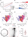
Fig. 4. Rounded HeLa cells maintain no universal pressure or tension.
a Average power-law behavior of unperturbed HeLa cells (black) in the absence and in the presence of 500 nM LatA (purple) or 50 µM CK666 (red) (n = 10 biologically different cells for each condition), corresponding mean values and standard deviations are given in Supplementary Table 2. The storage modulus Ecort is plotted as a dotted line and the loss modulus Ecort as a through line for each condition. b Applying Laplace’s law to a population of rounded cells gives two simple scenarios: i) bigger cells are stiffer, if a universal pressure exists, or ii) bigger cells are softer, if a universal tension exists. c, d Relative variations of storage and loss moduli of unperturbed (purple-gray), LatA perturbed (purple-red), and CK666 perturbed (purple-blue) HeLa cells plotted against the cell radius R and frequency f. Negative and positive relative moduli describe the cell to become softer and stiffer, respectively. Frequency-dependent variations of both moduli are best visualized by the yellow reference plane. The hypothesis of no correlation with R could not be rejected, as the t-test resulted in p-values of 0.95 and 0.92 (Methods). Red and pink planes indicate hypothetical dependencies proportional to 1/R (constant pressure) and 1/R2 (constant tension), respectively. An independency of cell size is found for every individual parameter of the power-law A, B, α, and β (Supplementary Fig. 14). e STED super resolution nanoscopy of paraformaldehyde-fixed HeLa cells with SiR-actin stained F-actin. Scale bars, 5 µm and 500 nm (inset). f For each of the 10 HeLa cells imaged by STED nanoscopy, we fitted 10 line profiles of the SiR-actin signal, to extract the cortical thickness hcort (194 ± 27 nm, mean ± S.D.). The center bar indicates the mean, the whiskers show the standard deviation. The diameter and cortex thickness measured for each cell by STED microscopy is shown in Supplementary Fig. 15.

References

    1. Bao G, Suresh S. Cell and molecular mechanics of biological materials. Nat. Mater. 2003;2:715–725. doi: 10.1038/nmat1001. - DOI - PubMed
    1. Krieg M, et al. Atomic force microscopy-based mechanobiology. Nat. Rev. Phys. 2019;1:41–57. doi: 10.1038/s42254-018-0001-7. - DOI
    1. Winder SJ, Ayscough KR. Actin-binding proteins. J. Cell Sci. 2005;118:651–654. doi: 10.1242/jcs.01670. - DOI - PubMed
    1. Mishra M, Huang J, Balasubramanian MK. The yeast actin cytoskeleton. FEMS Microbiol. Rev. 2014;38:213–227. doi: 10.1111/1574-6976.12064. - DOI - PubMed
    1. Kasza KE, et al. The cell as a material. Curr. Opin. Cell Biol. 2007;19:101–107. doi: 10.1016/j.ceb.2006.12.002. - DOI - PubMed

Publication types

LinkOut - more resources