

Comparative antler proteome of sika deer from different developmental stages
- PMID: 34006919
- PMCID: PMC8131589
- DOI: 10.1038/s41598-021-89829-6
Comparative antler proteome of sika deer from different developmental stages
Abstract
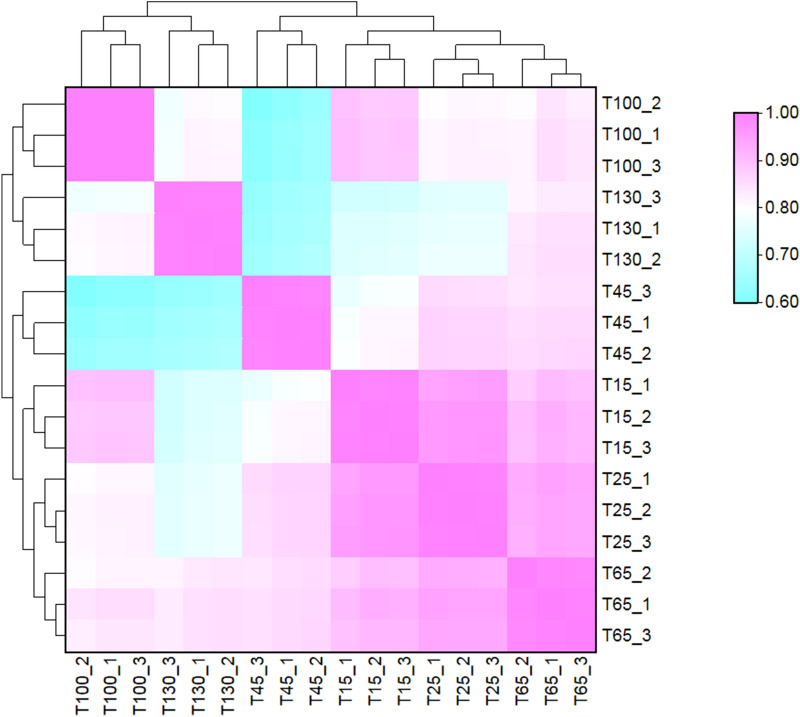
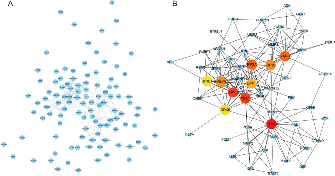
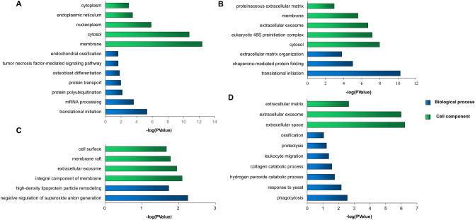
Antler is a special bone tissue that has the ability to regenerate completely periodically. It is the fastest growing bone in the animal kingdom. Antler provides a valuable research model for bone growth and mineralization. Antler grows longitudinally by endochondral ossification with their growth center located in its tip. Many scholars have carried out detailed studies on morphology and gene expression of antler tip. However, few scholars have analyzed the protein expression patterns of antler tip at different development stages. This study used label-free proteomics approach to analyze the protein expression dynamics of the antler tip in six developmental periods (15, 25, 45, 65, 100 and 130 days after the previous antler cast) and costal cartilage. In result, 2052 proteins were confidently quantified, including 1937 antler proteins and 1044 costal cartilage proteins. Moreover, 913 antler core proteins and 132 antler-special proteins were obtained. Besides, the stages special proteins and differentially expressed proteins (DEPs) in different development stages were analyzed. A total of 875 DEPs were determined by one-way AVOVA. It is found that the growth period (15, 25, 45 and 65 days) showed more up-regulated protein including several chondrogenesis-associated proteins (collagen types II, collagen types XI, HAPLN1, PAPSS1 and PAPSS2). In ossification stages, the up-regulated proteins related with lysosome (CTSD, CTSB, MMP9, CAII) indicated that the antler has higher bone remodeling activity. Given the up-regulated expression of immune-related molecules (S100A7, CATHL7, LTF, AZU1, ELANE and MPO), we speculate that the local immune system may contribute to the ossification of antler tip. In conclusion, proteomics technology was used to deeply analyze the protein expression patterns of antler at different development stages. This provides a strong support for the research on the molecular regulation mechanism of rapid growth and ossification of velvet antler.
Conflict of interest statement
The authors declare no competing interests.
Figures


Similar articles
-
Integrated Transcriptomic and Proteomic Analyses of Antler Growth and Ossification Mechanisms.Int J Mol Sci. 2024 Dec 9;25(23):13215. doi: 10.3390/ijms252313215. Int J Mol Sci. 2024. PMID: 39684926 Free PMC article.
-
Comparative transcriptome analysis of the main beam and brow tine of sika deer antler provides insights into the molecular control of rapid antler growth.Cell Mol Biol Lett. 2020 Sep 7;25:42. doi: 10.1186/s11658-020-00234-9. eCollection 2020. Cell Mol Biol Lett. 2020. PMID: 32944020 Free PMC article.
-
Comprehensive transcriptome analysis of sika deer antler using PacBio and Illumina sequencing.Sci Rep. 2022 Sep 28;12(1):16161. doi: 10.1038/s41598-022-20244-1. Sci Rep. 2022. PMID: 36171236 Free PMC article.
-
Deer antlers: a zoological curiosity or the key to understanding organ regeneration in mammals?J Anat. 2005 Nov;207(5):603-18. doi: 10.1111/j.1469-7580.2005.00478.x. J Anat. 2005. PMID: 16313394 Free PMC article. Review.
-
Histogenetic aspects of deer antler development.Front Biosci (Elite Ed). 2013 Jan 1;5(2):479-89. doi: 10.2741/e629. Front Biosci (Elite Ed). 2013. PMID: 23277003 Review.
Cited by
-
Effect of Methionine Supplementation on Serum Metabolism and the Rumen Bacterial Community of Sika Deer (Cervus nippon).Animals (Basel). 2022 Jul 31;12(15):1950. doi: 10.3390/ani12151950. Animals (Basel). 2022. PMID: 35953939 Free PMC article.
-
Sika deer velvet antler protein extract modulater bone metabolism and the structure of gut microbiota in ovariectomized mice.Food Sci Nutr. 2023 Mar 13;11(6):3309-3319. doi: 10.1002/fsn3.3316. eCollection 2023 Jun. Food Sci Nutr. 2023. PMID: 37324858 Free PMC article.
-
Use of the Phylobone database for the annotation of bone extracellular matrix proteins in reindeer (Rangifer tarandus).Sci Prog. 2024 Apr-Jun;107(2):368504241244666. doi: 10.1177/00368504241244666. Sci Prog. 2024. PMID: 38614461 Free PMC article.
-
Integrated Transcriptomic and Proteomic Analyses of Antler Growth and Ossification Mechanisms.Int J Mol Sci. 2024 Dec 9;25(23):13215. doi: 10.3390/ijms252313215. Int J Mol Sci. 2024. PMID: 39684926 Free PMC article.
-
MiRNA Profiling and Its Potential Roles in Rapid Growth of Velvet Antler in Gansu Red Deer (Cervus elaphus kansuensis).Genes (Basel). 2023 Feb 7;14(2):424. doi: 10.3390/genes14020424. Genes (Basel). 2023. PMID: 36833351 Free PMC article.
References
Publication types
MeSH terms
Substances
LinkOut - more resources
Full Text Sources
Other Literature Sources
Research Materials
Miscellaneous
