

Diffuse trophoblast damage is the hallmark of SARS-CoV-2-associated fetal demise
- PMID: 34006935
- PMCID: PMC8130566
- DOI: 10.1038/s41379-021-00827-5
Diffuse trophoblast damage is the hallmark of SARS-CoV-2-associated fetal demise
Abstract
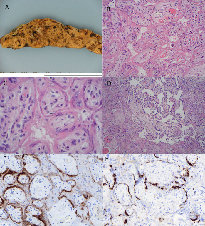
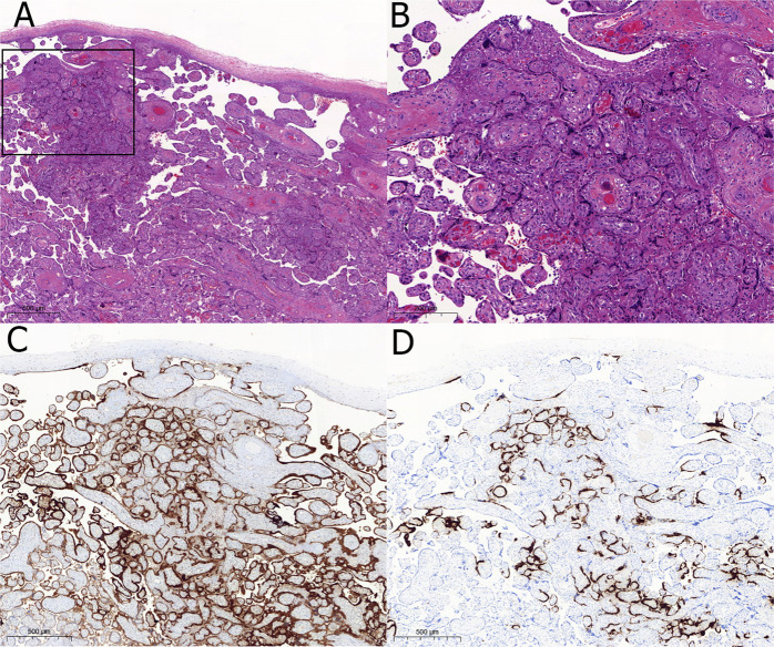
Placental pathology in SARS-CoV-2-infected pregnancies seems rather unspecific. However, the identification of the placental lesions due to SARS-CoV-2 infection would be a significant advance in order to improve the management of these pregnancies and to identify the mechanisms involved in a possible vertical transmission. The pathological findings in placentas delivered from 198 SARS-CoV-2-positive pregnant women were investigated for the presence of lesions associated with placental SARS-CoV-2 infection. SARS-CoV-2 infection was investigated in placental tissues through immunohistochemistry, and positive cases were further confirmed by in situ hybridization. SARS-CoV-2 infection was also investigated by RT-PCR in 33 cases, including all the immunohistochemically positive cases. Nine cases were SARS-CoV-2-positive by immunohistochemistry, in situ hybridization, and RT-PCR. These placentas showed lesions characterized by villous trophoblast necrosis with intervillous space collapse and variable amounts of mixed intervillous inflammatory infiltrate and perivillous fibrinoid deposition. Such lesions ranged from focal to massively widespread in five cases, resulting in intrauterine fetal death. Two of the stillborn fetuses showed some evidence of SARS-CoV-2 positivity. The remaining 189 placentas did not show similar lesions. The strong association between trophoblastic damage and placenta SARS-CoV-2 infection suggests that this lesion is a specific marker of SARS-CoV-2 infection in placenta. Diffuse trophoblastic damage, massively affecting chorionic villous tissue, can result in fetal death associated with COVID-19 disease.
© 2021. The Author(s), under exclusive licence to United States & Canadian Academy of Pathology.
Figures
Comment in
-
Response to "Diffuse trophoblast damage is the hallmark of SARS-CoV-2-associated fetal demise.".Mod Pathol. 2022 Jun;35(6):850-851. doi: 10.1038/s41379-021-00975-8. Epub 2021 Nov 29. Mod Pathol. 2022. PMID: 34845304 Free PMC article. No abstract available.
-
Authors' response to "Response to Diffuse Trophoblast Damage is the Hallmark of SARS-CoV-2-associated fetal demise".Mod Pathol. 2022 Jun;35(6):852-853. doi: 10.1038/s41379-022-01064-0. Epub 2022 Mar 23. Mod Pathol. 2022. PMID: 35322194 Free PMC article. No abstract available.
Similar articles
-
Placental lesions and SARS-Cov-2 infection: Diffuse placenta damage associated to poor fetal outcome.Placenta. 2021 Sep 1;112:97-104. doi: 10.1016/j.placenta.2021.07.288. Epub 2021 Jul 15. Placenta. 2021. PMID: 34329973 Free PMC article.
-
Stillbirth due to SARS-CoV-2 placentitis without evidence of intrauterine transmission to fetus: association with maternal risk factors.Ultrasound Obstet Gynecol. 2022 Jun;59(6):813-822. doi: 10.1002/uog.24906. Ultrasound Obstet Gynecol. 2022. PMID: 35353936 Free PMC article.
-
Placental Pathology of COVID-19 with and without Fetal and Neonatal Infection: Trophoblast Necrosis and Chronic Histiocytic Intervillositis as Risk Factors for Transplacental Transmission of SARS-CoV-2.Viruses. 2020 Nov 15;12(11):1308. doi: 10.3390/v12111308. Viruses. 2020. PMID: 33203131 Free PMC article. Review.
-
Intrauterine Fetal Demise After Uncomplicated COVID-19: What Can We Learn from the Case?Viruses. 2021 Dec 19;13(12):2545. doi: 10.3390/v13122545. Viruses. 2021. PMID: 34960815 Free PMC article.
-
Meta-analysis on COVID-19-pregnancy-related placental pathologies shows no specific pattern.Placenta. 2022 Jan;117:72-77. doi: 10.1016/j.placenta.2021.10.010. Epub 2021 Oct 19. Placenta. 2022. PMID: 34773743 Free PMC article.
Cited by
-
SARS-CoV-2 Infection in Late Pregnancy and Childbirth from the Perspective of Perinatal Pathology.J Dev Biol. 2023 Nov 16;11(4):42. doi: 10.3390/jdb11040042. J Dev Biol. 2023. PMID: 37987372 Free PMC article. Review.
-
Acute placental insufficiency two weeks after an asymptomatic COVID-19 maternal infection: the deleterious effects of the SARS-CoV-2 placentitis.Arch Gynecol Obstet. 2024 Feb;309(2):723-726. doi: 10.1007/s00404-023-06991-0. Epub 2023 Mar 13. Arch Gynecol Obstet. 2024. PMID: 36912992 Free PMC article. No abstract available.
-
Stillbirth after COVID-19 in Unvaccinated Mothers Can Result from SARS-CoV-2 Placentitis, Placental Insufficiency, and Hypoxic Ischemic Fetal Demise, Not Direct Fetal Infection: Potential Role of Maternal Vaccination in Pregnancy.Viruses. 2022 Feb 23;14(3):458. doi: 10.3390/v14030458. Viruses. 2022. PMID: 35336864 Free PMC article.
-
SARS-CoV-2 Infection: A Clinical and Histopathological Study in Pregnancy.Biology (Basel). 2023 Jan 21;12(2):174. doi: 10.3390/biology12020174. Biology (Basel). 2023. PMID: 36829453 Free PMC article.
-
Maternal Fever and Reduced Fetal Movement as Predictive Risk Factors for Adverse Neonatal Outcome in Cases of Congenital SARS-CoV-2 Infection: A Meta-Analysis of Individual Participant Data from Case Reports and Case Series.Viruses. 2023 Jul 24;15(7):1615. doi: 10.3390/v15071615. Viruses. 2023. PMID: 37515301 Free PMC article. Review.
References
-
- Smithgall MC, Liu-Jarin X, Hamele-Bena D, Cimic A, Mourad M, Debelenko L, et al. Third-trimester placentas of severe acute respiratory syndrome coronavirus 2 (SARS-CoV-2)-positive women: histomorphology, including viral immunohistochemistry and in-situ hybridization. Histopathology. 2020;77:994–999. doi: 10.1111/his.14215. - DOI - PMC - PubMed
MeSH terms
LinkOut - more resources
Full Text Sources
Other Literature Sources
Medical
Miscellaneous
