

Hepatic parenchyma and vascular blood flow changes after TIPS with spectral CT iodine density in HBV-related liver cirrhosis
- PMID: 34006977
- PMCID: PMC8131370
- DOI: 10.1038/s41598-021-89764-6
Hepatic parenchyma and vascular blood flow changes after TIPS with spectral CT iodine density in HBV-related liver cirrhosis
Abstract
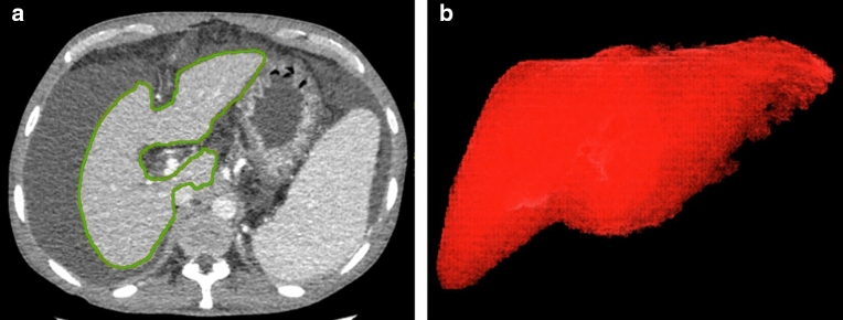
To compare changes in spectral CT iodine densities of hepatic parenchyma and vessels before and after transjugular intrahepatic portosystemic shunt (TIPS) in hepatitis B virus (HBV)-related liver cirrhosis. Twenty-five patients with HBV-related liver cirrhosis who received TIPS for gastroesophageal varices bleeding were recruited. Each patient underwent three phases contrast CT before and after TIPS within 4 weeks, with the raw data reconstructed at 1.25-mm-thick slices. Iodine density (in milligrams per milliliter) was measured on iodine-based material decomposition image. Multiple regions of interest (ROIs) in liver parenchyma, aorta and portal vein were selected from three slices of images. Portal vein trunk was set as the central one, and mean liver parenchymal iodine densities from arterial phase (AP), venous phase (VP) and equilibrium phase (EP) were recorded. Quantitative indices of iodine density (ID), including normalized ID in liver parenchyma for arterial phase (NIDLAP), ID of liver parenchyma for venous phase (IDLVP), ID of portal vein in venous phase (IDPVP) and liver arterial iodine density fraction (AIF), were measured and compared before and after TIPS. Based on Child-Pugh stage, 4, 12 and 9 patients were classified as grade A, B, and C, respectively. Liver volume was comparable before and after TIPS (1110.5 ± 287.4 vs. 1092.0 ± 276.3, P = 0.28). After TIPS, ID was decreased in aorta (146.0 ± 34.5 vs. 120.9 ± 30.7, P < 0.01) whereas increased in liver parenchyma at arterial phase, as demonstrated by IDAP (9.3 ± 3.1 vs. 13.4 ± 4.4 mg/mL) and AIF (0.40 ± 0.11 vs. 0.58 ± 0.11, P < 0.01). For venous or equilibrium phase, quantitative indices remained stable (23.1 ± 4.5 vs. 23.0 ± 5.3, 19.8 ± 4.1 vs. 19.4 ± 4.6) mg/mL (Ps > 0.05). For portal vein, ID and NID were increased after TIPS (23.1 ± 11.7 vs. 36.5 ± 13.0, 16.4 ± 8.5 vs. 31.8 ± 12.8) (P < 0.01). No positive correlation between iodine density and preoperative Child-Pugh score was observed. Based on iodine density measurement, spectral CT as a noninvasive imaging modality may assess hepatic parenchyma and vascular blood flow changes before and after TIPS in HBV-related liver cirrhosis.Clinical registration number: ChiCTR- DDC-16009986.
Conflict of interest statement
The authors declare no competing interests.
Figures

References
MeSH terms
Substances
LinkOut - more resources
Full Text Sources
Other Literature Sources
Medical
