. 2021 May 18;11(1):10540.
doi: 10.1038/s41598-021-89997-5.

BMP signaling alters aquaporin-4 expression in the mouse cerebral cortex

Affiliations

BMP signaling alters aquaporin-4 expression in the mouse cerebral cortex

Kazuya Morita et al. Sci Rep. .

Abstract

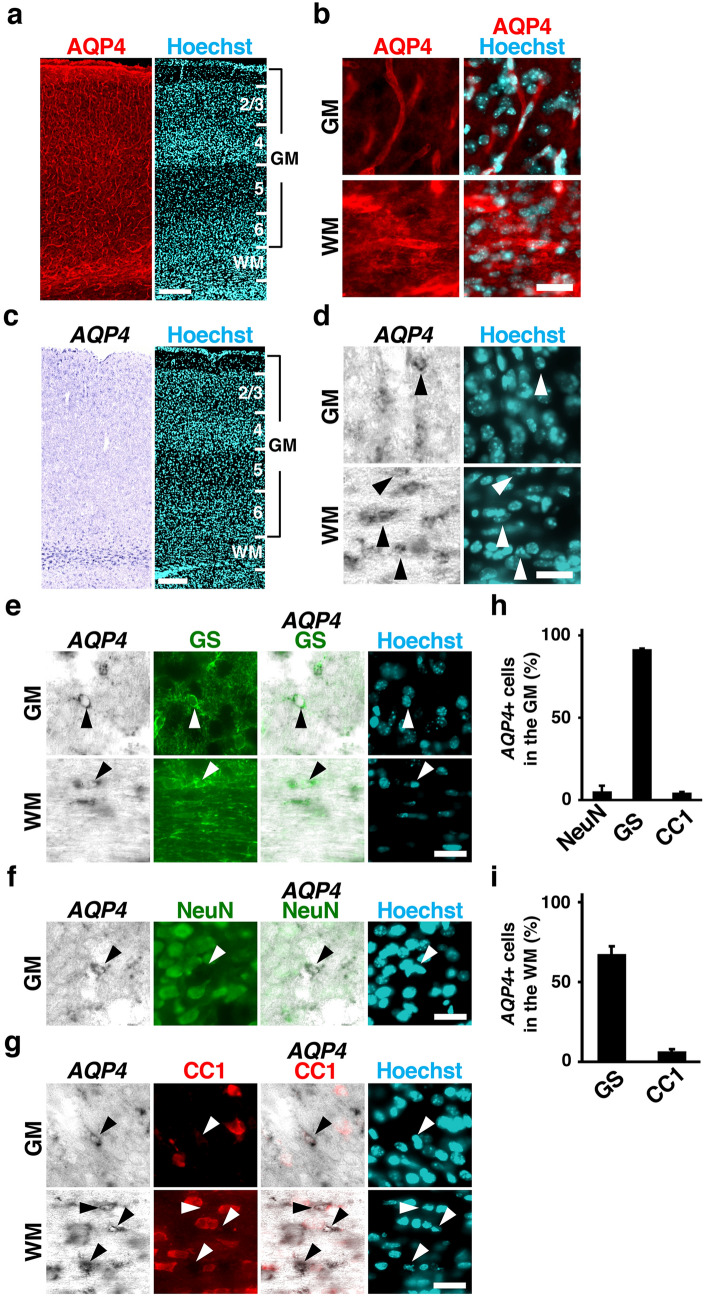
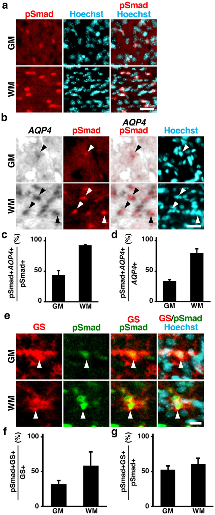
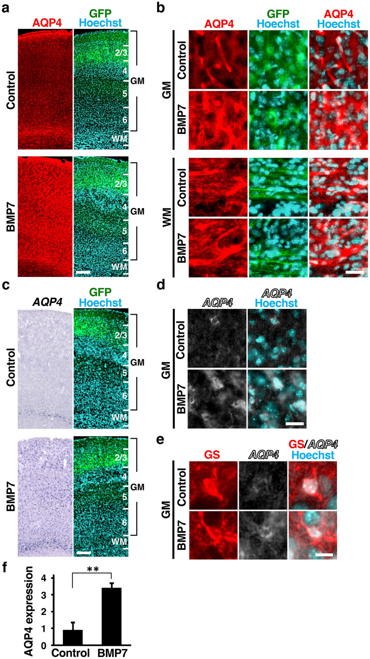
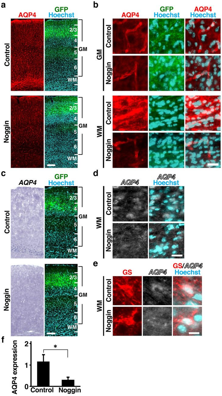
Aquaporin-4 (AQP4) is a predominant water channel expressed in astrocytes in the mammalian brain. AQP4 is crucial for the regulation of homeostatic water movement across the blood-brain barrier (BBB). Although the molecular mechanisms regulating AQP4 levels in the cerebral cortex under pathological conditions have been intensively investigated, those under normal physiological conditions are not fully understood. Here we demonstrate that AQP4 is selectively expressed in astrocytes in the mouse cerebral cortex during development. BMP signaling was preferentially activated in AQP4-positive astrocytes. Furthermore, activation of BMP signaling by in utero electroporation markedly increased AQP4 levels in the cerebral cortex, and inhibition of BMP signaling strongly suppressed them. These results indicate that BMP signaling alters AQP4 levels in the mouse cerebral cortex during development.


Conflict of interest statement

The authors declare no competing interests.

Figures

Figure 1
Figure 1
Expression patterns of AQP4 in the developing mouse cerebral cortex. (a,b) Sections of the mouse cerebral cortex at P16 were subjected to AQP4 immunohistochemistry and Hoechst 33342 staining. Low magnification images (a) and high magnification images (b) corresponding to the gray matter (GM) and the white matter (WM) are shown. AQP4 was expressed throughout the developing mouse cerebral cortex. The expression level of AQP4 was higher in the WM than in the GM. (c,d) Sections of the mouse cerebral cortex at P16 were subjected to in situ hybridization for AQP4 and Hoechst 33342 staining. Low magnification images (c) and high magnification images (d) corresponding to the GM and the WM are shown. AQP4 mRNA was preferentially distributed in the WM but was also observed in the GM. (e–g) Sections of the mouse cerebral cortex at P16 were subjected to in situ hybridization for AQP4 and immunohistochemistry for either GS (e), NeuN (f) or CC1 (g). High-magnification images of the GM and the WM are shown. AQP4 was preferentially expressed in GS-positive cells (e, arrowheads), while AQP4 signals were not colocalized with NeuN-positive cells or CC1-positive cells (f and g, arrowheads). (h,i) The percentages of AQP4-positive cells co-expressing NeuN, GS, and CC1 in the GM (h) and the WM (i). n = 3 animals for each condition. Bars represent mean ± SD. Numbers indicate the corresponding layers in the cerebral cortex. GM, gray matter; WM, white matter. Scale bars 200 µm (a,c) and 25 µm (b,d–g).
Figure 2
Figure 2
BMP signaling is activated in AQP4-positive astrocytes in the developing mouse cerebral cortex. (a) Sections of the mouse cerebral cortex at P16 were subjected to immunohistochemistry for pSmad and Hoechst 33342 staining. pSmad signals were more predominant in the WM compared with the GM. (b) Sections were subjected to immunohistochemistry for pSmad and in situ hybridization for AQP4. High magnification images are shown. Many AQP4-positive astrocytes were also labeled with pSmad (arrowheads). (c) The percentages of pSmad-positive cells co-expressing AQP4 in the GM and the WM. (d) The percentages of AQP4-positive cells co-labeled with pSmad in the GM and the WM. (e) Sections of the mouse cerebral cortex at P16 were subjected to immunohistochemistry for pSmad and GS. High magnification images are shown. pSmad signals were observed in GS-positive cells (arrowheads). (f) The percentages of GS-positive cells co-labeled with pSmad in the GM and the WM. (g) The percentages of pSmad-positive cells co-expressing GS in the GM and the WM. GM, gray matter; WM, white matter. n = 3 animals for each condition. Bars represent mean ± SD. Scale bars 25 μm (a,b) and 10 µm (e).
Figure 3
Figure 3
Activation of BMP signaling increases AQP4 levels in the mouse cerebral cortex. CAG-EGFP plus either pCAG-BMP7 or pCAG control vector was electroporated at E14, and the brains were dissected at P16. (a,b) Coronal sections stained with anti-AQP4 antibody, anti-GFP antibody and Hoechst 33342. Low magnification images (a) and high magnification images of the GM and the WM (b) are shown. The immunoreactivity of AQP4 was markedly increased by activation of BMP signaling. (c,d) Sections were subjected to in situ hybridization for AQP4 and Hoechst 33342 staining. Low magnification images (c) and high magnification images (d) are shown. Activation of BMP signaling significantly increased mRNA expression levels of AQP4. (e) Sections were subjected to in situ hybridization for AQP4 and immunohistochemistry for GS. High magnification images are shown. Activation of BMP signaling increased mRNA expression levels of AQP4 in GS-positive cells. (f) Quantification of AQP4 immunoreactivity in the cerebral cortex. Activation of BMP signaling significantly increased AQP4 immunoreactivity. n = 3 animals for each condition. Bars represent mean ± SD. **p < 0.01, Student’s t-test. Numbers indicate the corresponding layers in the cerebral cortex. GM, gray matter; WM, white matter. Scale bars 200 µm (a,c), 25 µm (b,d) and 10 µm (e).
Figure 4
Figure 4
Inhibition of BMP signaling reduces AQP4 levels in the mouse cerebral cortex. pCAG-EGFP plus either pCAG-noggin or pCAG control vector was electroporated at E14, and the brains were dissected at P16. (a,b) Coronal sections stained with anti-AQP4 antibody, anti-GFP antibody and Hoechst 33342. Low magnification images (a) and high magnification images (b) of the GM and the WM are shown. The immunoreactivity of AQP4 in the WM was markedly decreased by noggin. (c,d) Sections were subjected to in situ hybridization for AQP4 and Hoechst 33342 staining. Low magnification images (c) and high magnification images of the WM (d) are shown. Noggin significantly decreased mRNA expression levels of AQP4 in the WM. (e) Sections are subjected to in situ hybridization for AQP4 and immunohistochemistry for GS. High magnification images of the WM are shown. (f) Quantification of AQP4 immunoreactivity in the WM. Inhibition of BMP signaling by noggin significantly decreased AQP4 immunoreactivity in the WM. n = 3 animals for each condition. Bars represent mean ± SD. *p < 0.05, Student’s t-test. Numbers indicate the corresponding layers in the cerebral cortex. GM, gray matter; WM, white matter. Scale bars 200 µm (a,c), 25 µm (b,d) and 10 µm (e).

Similar articles

Cited by

References

    1. Verkman AS, Binder DK, Bloch O, Auguste K, Papadopoulos MC. Three distinct roles of aquaporin-4 in brain function revealed by knockout mice. Biochim. Biophys. Acta. 2006;1758:1085–1093. doi: 10.1016/j.bbamem.2006.02.018. - DOI - PubMed
    1. Nielsen S, et al. Specialized membrane domains for water transport in glial cells: High-resolution immunogold cytochemistry of aquaporin-4 in rat brain. J. Neurosci. 1997;17:171–180. doi: 10.1523/JNEUROSCI.17-01-00171.1997. - DOI - PMC - PubMed
    1. Rash JE, Yasumura T, Hudson CS, Agre P, Nielsen S. Direct immunogold labeling of aquaporin-4 in square arrays of astrocyte and ependymocyte plasma membranes in rat brain and spinal cord. Proc. Natl. Acad. Sci. USA. 1998;95:11981–11986. doi: 10.1073/pnas.95.20.11981. - DOI - PMC - PubMed
    1. Manley GT, et al. Aquaporin-4 deletion in mice reduces brain edema after acute water intoxication and ischemic stroke. Nat. Med. 2000;6:159–163. doi: 10.1038/72256. - DOI - PubMed
    1. Haj-Yasein NN, et al. Glial-conditional deletion of aquaporin-4 (Aqp4) reduces blood-brain water uptake and confers barrier function on perivascular astrocyte endfeet. Proc. Natl. Acad. Sci. USA. 2011;108:17815–17820. doi: 10.1073/pnas.1110655108. - DOI - PMC - PubMed

Publication types

LinkOut - more resources