

Spaceflight and hind limb unloading induces an arthritic phenotype in knee articular cartilage and menisci of rodents
- PMID: 34006989
- PMCID: PMC8131644
- DOI: 10.1038/s41598-021-90010-2
Spaceflight and hind limb unloading induces an arthritic phenotype in knee articular cartilage and menisci of rodents
Abstract
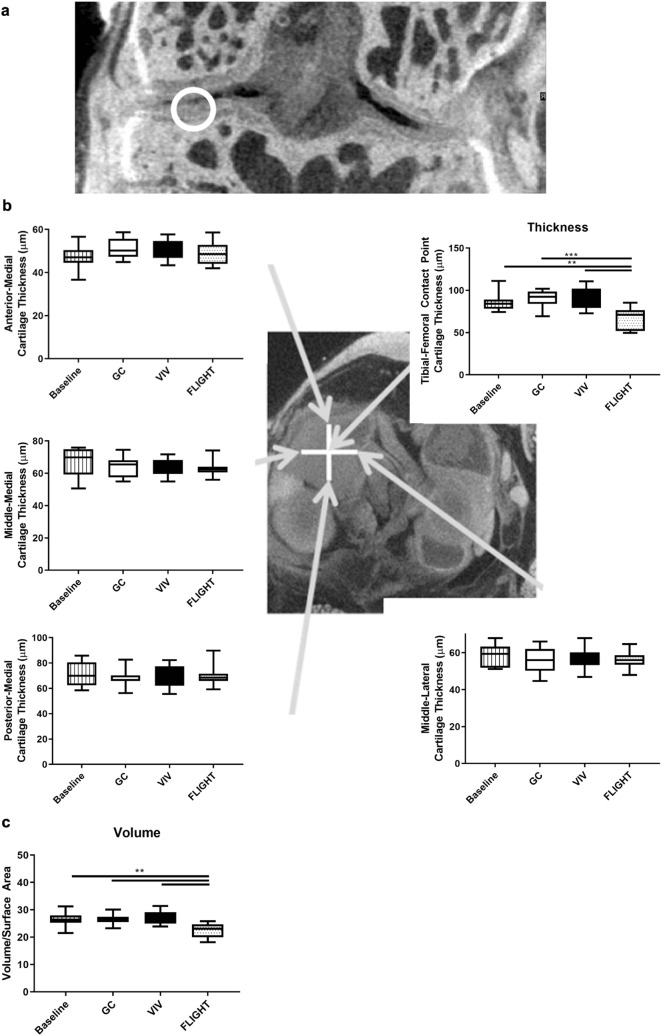
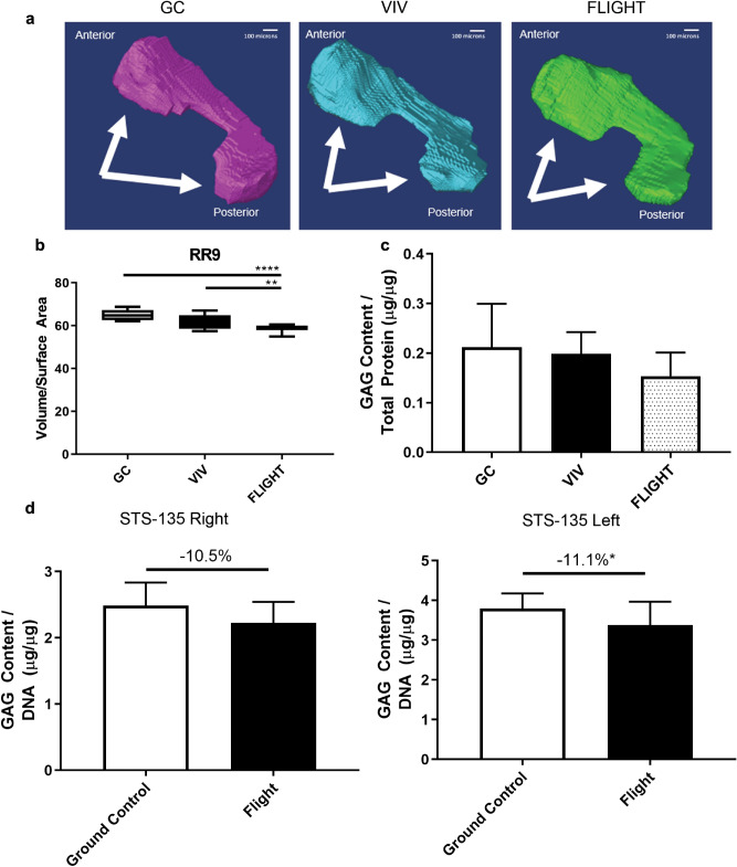
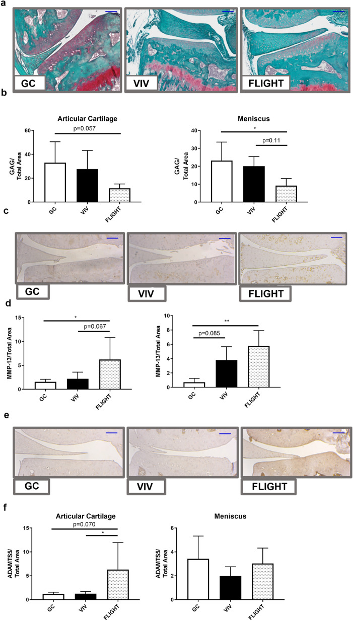
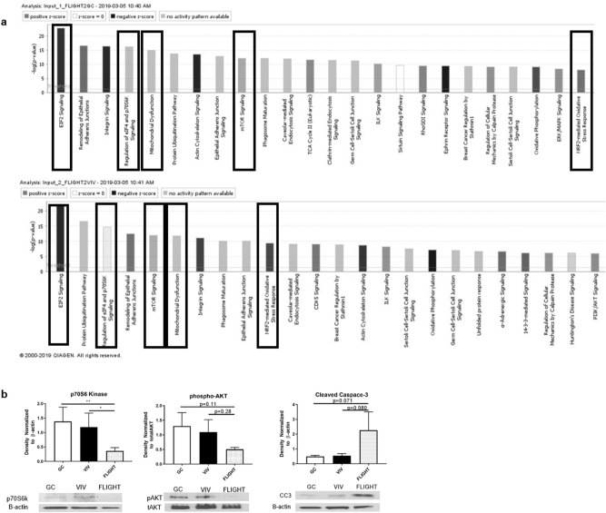
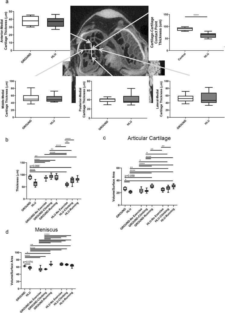
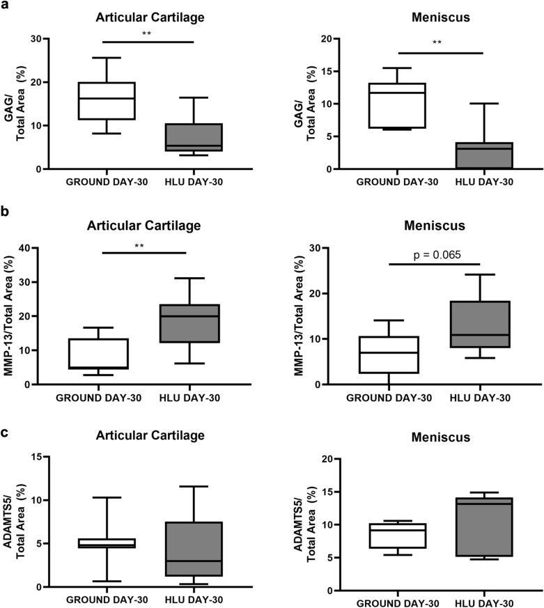
Reduced knee weight-bearing from prescription or sedentary lifestyles are associated with cartilage degradation; effects on the meniscus are unclear. Rodents exposed to spaceflight or hind limb unloading (HLU) represent unique opportunities to evaluate this question. This study evaluated arthritic changes in the medial knee compartment that bears the highest loads across the knee after actual and simulated spaceflight, and recovery with subsequent full weight-bearing. Cartilage and meniscal degradation in mice were measured via microCT, histology, and proteomics and/or biochemically after: (1) ~ 35 days on the International Space Station (ISS); (2) 13-days aboard the Space Shuttle Atlantis; or (3) 30 days of HLU, followed by a 49-day weight-bearing readaptation with/without exercise. Cartilage degradation post-ISS and HLU occurred at similar spatial locations, the tibial-femoral cartilage-cartilage contact point, with meniscal volume decline. Cartilage and meniscal glycosaminoglycan content were decreased in unloaded mice, with elevated catabolic enzymes (e.g., matrix metalloproteinases), and elevated oxidative stress and catabolic molecular pathway responses in menisci. After the 13-day Shuttle flight, meniscal degradation was observed. During readaptation, recovery of cartilage volume and thickness occurred with exercise. Reduced weight-bearing from either spaceflight or HLU induced an arthritic phenotype in cartilage and menisci, and exercise promoted recovery.
Conflict of interest statement
The authors declare no competing interests.
Figures
Similar articles
-
Spaceflight-Relevant Challenges of Radiation and/or Reduced Weight Bearing Cause Arthritic Responses in Knee Articular Cartilage.Radiat Res. 2016 Oct;186(4):333-344. doi: 10.1667/RR14400.1. Epub 2016 Sep 7. Radiat Res. 2016. PMID: 27602483
-
Treatment with a superoxide dismutase mimetic for joint preservation during 35 and 75 days in orbit aboard the international space station, and after 120 days recovery on Earth.Life Sci Space Res (Amst). 2025 Feb;44:67-78. doi: 10.1016/j.lssr.2024.10.009. Epub 2024 Oct 28. Life Sci Space Res (Amst). 2025. PMID: 39864914
-
Knee and Hip Joint Cartilage Damage from Combined Spaceflight Hazards of Low-Dose Radiation Less than 1 Gy and Prolonged Hindlimb Unloading.Radiat Res. 2019 Jun;191(6):497-506. doi: 10.1667/RR15216.1. Epub 2019 Mar 29. Radiat Res. 2019. PMID: 30925135 Free PMC article.
-
The Effects of Space Microgravity on Hip and Knee Cartilage: A New Frontier in Orthopaedics.Surg Technol Int. 2019 Nov 10;35:421-425. Surg Technol Int. 2019. PMID: 31687778
-
As Goes the Meniscus Goes the Knee: Early, Intermediate, and Late Evidence for the Detrimental Effect of Meniscus Tears.Clin Sports Med. 2020 Jan;39(1):29-36. doi: 10.1016/j.csm.2019.08.001. Clin Sports Med. 2020. PMID: 31767108 Review.
Cited by
-
Moderate exercise protects against joint disease in a murine model of osteoarthritis.Front Physiol. 2022 Dec 5;13:1065278. doi: 10.3389/fphys.2022.1065278. eCollection 2022. Front Physiol. 2022. PMID: 36545287 Free PMC article.
-
Joint Cartilage in Long-Duration Spaceflight.Biomedicines. 2022 Jun 8;10(6):1356. doi: 10.3390/biomedicines10061356. Biomedicines. 2022. PMID: 35740378 Free PMC article. Review.
-
Changes in the Serum Metabolome in an Inflammatory Model of Osteoarthritis in Rats.Int J Mol Sci. 2024 Mar 9;25(6):3158. doi: 10.3390/ijms25063158. Int J Mol Sci. 2024. PMID: 38542132 Free PMC article.
-
Organs in orbit: how tissue chip technology benefits from microgravity, a perspective.Front Lab Chip Technol. 2024;3:1356688. doi: 10.3389/frlct.2024.1356688. Epub 2024 Mar 7. Front Lab Chip Technol. 2024. PMID: 38915901 Free PMC article.
-
Impacts of Delivery Mode and Maternal Factors on Neonatal Oral Microbiota.Front Microbiol. 2022 Jun 27;13:915423. doi: 10.3389/fmicb.2022.915423. eCollection 2022. Front Microbiol. 2022. PMID: 35832807 Free PMC article.
References
-
- https://www.boneandjointburden.org/fourth-edition/viiie1/total-economic-..., T. B. o. M. D. i. t. U. S. (acessed 9/15/2019).
Publication types
MeSH terms
Substances
Grants and funding
LinkOut - more resources
Full Text Sources
Other Literature Sources
