. 2021 Dec;35(12):3509-3525.
doi: 10.1038/s41375-021-01271-9. Epub 2021 May 18.

JunB is a key regulator of multiple myeloma bone marrow angiogenesis

Affiliations

JunB is a key regulator of multiple myeloma bone marrow angiogenesis

Fengjuan Fan et al. Leukemia. 2021 Dec.

Erratum in

  • Publisher Correction: JunB is a key regulator of multiple myeloma bone marrow angiogenesis.
    Fan F, Malvestiti S, Vallet S, Lind J, Garcia-Manteiga JM, Morelli E, Jiang Q, Seckinger A, Hose D, Goldschmidt H, Stadlbauer A, Sun C, Mei H, Pecherstorfer M, Bakiri L, Wagner EF, Tonon G, Sattler M, Hu Y, Tassone P, Jaeger D, Podar K. Fan F, et al. Leukemia. 2021 Dec;35(12):3628. doi: 10.1038/s41375-021-01367-2. Leukemia. 2021. PMID: 34489554 Free PMC article. No abstract available.

Abstract

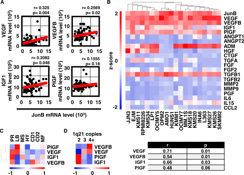
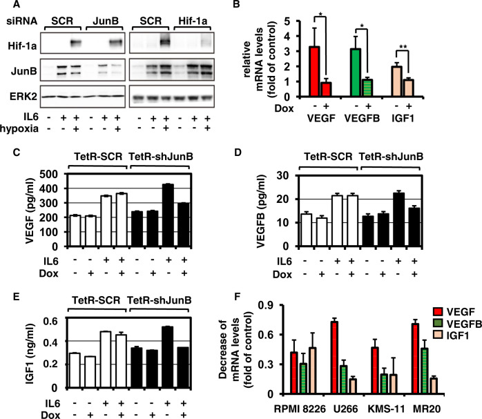
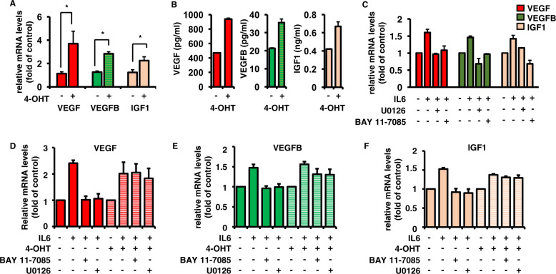
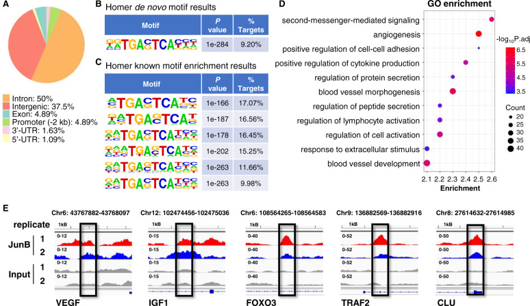
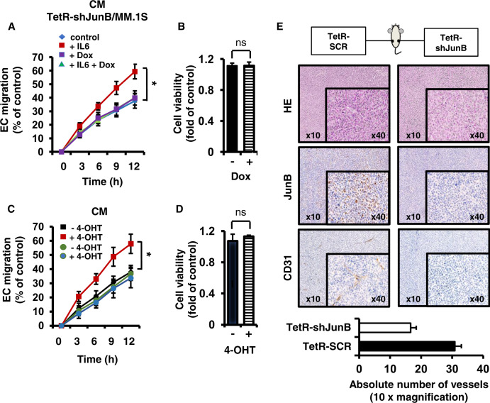
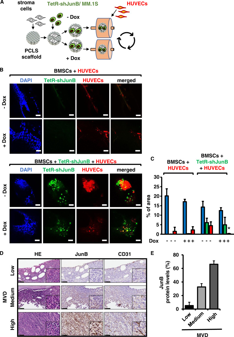
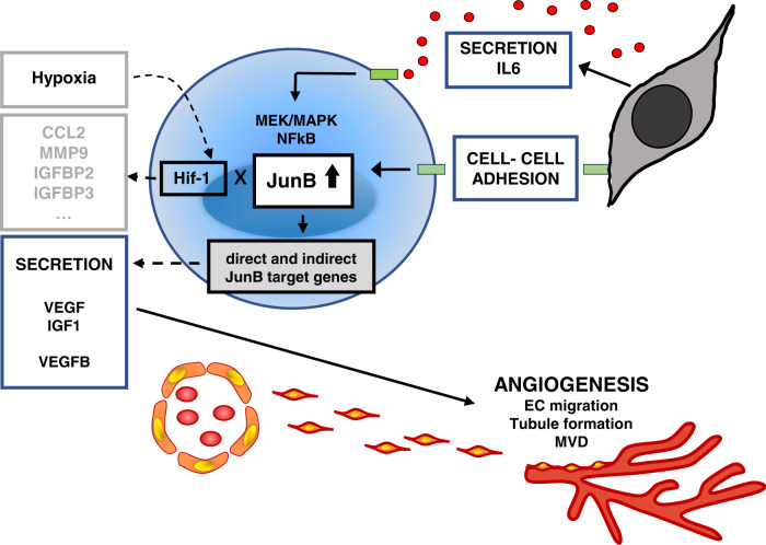
Bone marrow (BM) angiogenesis significantly influences disease progression in multiple myeloma (MM) patients and correlates with adverse prognosis. The present study shows a statistically significant correlation of the AP-1 family member JunB with VEGF, VEGFB, and IGF1 expression levels in MM. In contrast to the angiogenic master regulator Hif-1α, JunB protein levels were independent of hypoxia. Results in tumor-cell models that allow the induction of JunB knockdown or JunB activation, respectively, corroborated the functional role of JunB in the production and secretion of these angiogenic factors (AFs). Consequently, conditioned media derived from MM cells after JunB knockdown or JunB activation either inhibited or stimulated in vitro angiogenesis. The impact of JunB on MM BM angiogenesis was finally confirmed in a dynamic 3D model of the BM microenvironment, a xenograft mouse model as well as in patient-derived BM sections. In summary, in continuation of our previous study (Fan et al., 2017), the present report reveals for the first time that JunB is not only a mediator of MM cell survival, proliferation, and drug resistance, but also a promoter of AF transcription and consequently of MM BM angiogenesis. Our results thereby underscore worldwide efforts to target AP-1 transcription factors such as JunB as a promising strategy in MM therapy.


Conflict of interest statement

SV received speaker’s honoraria from Bristol Myers Squibb, MSD, Pfizer, and consultancy fees from Roche, Eusa, MSD, and Merck. DJ received consultant honoraria from Bayer, Amgen, MSD, CureVac, Roche, and BMS. KP has received speaker’s honoraria from Celgene, Amgen Inc., and Janssen Pharmaceuticals, consultancy fees from Celgene, Takeda, and Janssen Pharmaceuticals, and research support from Roche Pharmaceuticals. The authors have no other relevant affiliations or financial involvement with any organization or entity with a financial interest in or financial conflict with the subject matter or materials discussed in the manuscript apart from those disclosed. The remaining authors declare no conflict of interest.

Figures

Fig. 1
Fig. 1. JunB expression correlates with expression profiles of angiogenic factors in MM cell line and primary cells.
A Correlation of JunB and angiogenic factor (AF) expression in primary samples of NDMM patients. Data analyses of samples within the GSE6477 dataset of NDMM patients indicate a statistically significant association for JunB with VEGF, VEGFB, and IGF1 but not with PIGF [26]. The Pearson correlation coefficient was calculated to evaluate the correlation between JunB and indicated AFs. The minimal level of significance was p < 0.05. B Heatmap of mRNA expression values (log2 and Z-score scaled across genes) for the indicated genes in MM cell line cells was obtained from data extracted from the Cancer Cell Line Encyclopedia (CCLE) database. The Pearson correlation coefficient was calculated to evaluate the correlation between JunB and indicated AFs. The minimal level of significance was p < 0.05. C Correlation of JunB and AF expression differs in the GSE2658 dataset of NDMM patients dependent on the molecular risk group. Heatmap of r values calculated on samples with known molecular information obtained from the GSE2658 dataset [26]. HYperdiploid (HY) group 4, Low Bone disease (LB) group 2, CD1 group 5, CD2 group 6, PRoliferation (PR) group 1, MMSET (MS) group 3, MAF/MAFB (MF) group 7. D Correlation of JunB and AF expression differs dependent on the number of 1q21 copies. Heatmap of r values calculated on samples with known molecular information obtained from the GSE2658 dataset [24] of NDMM patients.
Fig. 2
Fig. 2. IL6 but not hypoxia is inducing JunB-dependent production and secretion of angiogenic factors.
A Impact of hypoxia on JunB versus Hif-1α expression. MM.1S cells were transiently transfected with non-targeting control (SCR) siRNA, siJunB, or siHif-1α and then treated with IL6 (25 ng/ml) and 1% O2 (hypoxia) or left untreated. After 9 h, cell lysates were immunoblotted with antibodies against Hif-1α and JunB. ERK2 served as a loading control. B Doxycycline (Dox)-mediated JunB knockdown in TetR-shJunB/MM.1S inhibits IL6-induced production of AFs. TetR-shJunB/MM cells were cultured in RPMI-1640 medium with IL6 in the presence or absence of doxycycline (1 μg/ml). Expression profiles of VEGF, VEGFB, and IGF1 in TetR-shJunB/MM cells were determined using RT-qPCR. Data represent mean ± SD for triplicate samples of three independent experiments. *p < 0.05; **p < 0.01. CE Doxycycline-mediated JunB knockdown in TetR-shJunB/MM.1S inhibits IL6-induced secretion of AFs. TetR-SCR/MM.1S or TetR-shJunB/MM.1S cells were cultured in RPMI-1640 medium with or without IL6 in the presence or absence of doxycycline (1 μg/ml). Supernatants from equal numbers of cells (1.2 × 106) were collected after 18 h and analyzed for VEGF (C), VEGFB (D), and IGF1 (E) protein levels by ELISA. Supernatants from IL6-stimulated TetR-SCR/MM.1S cells served as a control. Data are expressed a mean ± SD of culture triplicates. F siRNA-mediated JunB knockdown in RPMI 8226, U266, KMS-11, and MR20 downregulates IL6-induced production of VEGF, VEGFB, and IGF1. MM.1S cells were transiently transfected with mock (200 nM) and JunB siRNA (200 nM). Expression profiles of VEGF, VEGFB, and IGF1 in indicated MM cell lines were determined using RT-qPCR. Data represent the decrease of VEGF, VEGFB, and IGF1 expression levels in MM cells transfected with siJunB versus mock control and represent mean ± SD for triplicate samples of three independent experiments.
Fig. 3
Fig. 3. 4-OHT-induced JunB activity in JunB-ER/MM.1S cells increases production and secretion of angiogenic factors.
A 4-OHT-induced JunB activation in JunB-ER/MM.1S cells induces expression of VEGF, VEGFB, and IGF1. JunB-ER/MM.1S cells were cultured in RPMI-1640 medium and treated without or with 4-OHT (100 nM) for 16 h. Expression profiles of VEGF, VEGFB, and IGF1 in JunB-ER/MM.1S cells were determined using RT-qPCR. Data represent mean ± SD for triplicate samples of three independent experiments. *p < 0.01. B 4-OHT-induced JunB activity enhances the secretion of VEGF, VEGFB, and IGF1 in JunB-ER/MM.1S cells. JunB-ER/MM.1S cells were treated without or with 4-OHT, as indicated. Supernatants from equal numbers of cells (1.2 × 106) were collected after 48 h and analyzed for protein levels of VEGF, VEGFB, and IGF1 by ELISA. Data represent mean ± SD for triplicate samples of three independent experiments. C IL6-induced expression of VEGF, VEGFB, and IGF1 is MEK/MAPK- and NFκB-dependent. MM.1S cells were pretreated with U0126 and BAY 11-7085 or left untreated, and then cultured in RPMI-1640 medium with or without IL6. Expression profiles of VEGF, VEGFB, and IGF1 in MM.1S cells were determined by RT-qPCR with B2M as an endogenous control. Data represent mean ± SD for triplicate samples of three independent experiments. DF 4-OHT-induced JunB activation rescues BAY 11-7085- and U0126-mediated inhibition of VEGF, VEGFB, and IGF1 mRNA levels. JunB-ER/MM.1S cells were cultured alone or with IL6 for 4 h with or without U0126 and BAY 11-7085; and 4-OHT (200 nM) for an additional 2 h. Cells were then harvested, lysed, and expression levels of VEGF (D), VEGFB (E), and IGF1 (F) were determined by RT-qPCR with B2M as an endogenous control. Each value is shown as mean ± SD of three independent experiments.
Fig. 4
Fig. 4. VEGF and IGF1 are direct transcriptional targets of JunB in MM cells.
AC Motif analysis of JunB-enriched sites in MM.1S cells treated with IL6 identified by chromatin immunoprecipitation sequencing (ChIP-seq). A Peak locations across the genome. The number indicates percent of ChIP-seq distribution. B Homer de novo motif results. C Homer known motif enrichment results. D Enriched Gene Ontology (GO) analysis of JunB target genes by Metascape. JunB direct target genes in MM.1S cells treated with IL6 were identified by ChIP-seq and subjected to GO enrichment analysis. E Representative ChIP-seq peaks located on JunB direct target genes VEGF, IGF1, FOXO3, TRAF2, and CLU, visualized by genome browser Integrative Genomics Viewer (IGV). Biological duplicates were performed for the ChIP-seq.
Fig. 5
Fig. 5. JunB knockdown in MM cells results in inhibition of in vitro and in vivo angiogenesis.
A Supernatant derived from MM cells upon JunB knockdown inhibits endothelial cell (EC) migration. TetR-shJunB/MM cells were cultured in RPMI-1640 medium plus IL6 in the presence or absence of doxycycline (1 μg/ml). Conditioned media (CM) were collected after 24 h and wound healing was determined at indicated time points (closure of EC layer after mechanical disruption; mean width of initial cell gap = 200 μm). Data represent mean ± SD for triplicate samples. *p < 0.01. B Cell viability of MM cells treated as described in (A) after 24 h. Data represent mean ± SD for triplicate samples. ns non-significant. C Supernatant derived from MM cells after 4-OHT-induced JunB activation stimulates in vitro angiogenesis. JunB-ER/MM cells were cultured in RPMI-1640 medium and treated with 4-OHT (100 nM). CM were collected after 24 h and wound healing was determined as described in A. Black and red squares, JunB-ER/MM.1S; green and blue circles, IRES-GFP/MM.1S cells. *p < 0.01. D Cell viability of JunB-ER/MM cells after 4-OHT treatment for 24 h. ns non-significant. E Induced JunB silencing inhibits angiogenesis in a murine xenograft MM model. NOD/SCID mice were injected subcutaneously with TetR-SCR/MM.1S and TetR-shJunB/MM.1S together with human-derived BMSCs and matrigel into the left and right flank, respectively, and fed with doxycycline in their drinking water for 5 weeks. Representative microscopic images of tumor sections stained with antibodies against JunB and CD31 (10× and 40× magnification) (upper panel). In vivo angiogenesis was quantified by counting the number of vessels in 15 random view fields at 10× magnification. Values represent average ± SEM (95% CI of difference: 8.60–19.94) (lower panel).
Fig. 6
Fig. 6. Positive expression of JunB in MM cells correlates with vessel density in an innovative 3D MM model and in patient-derived bone marrow biopsies.
AC Positive expression of JunB in MM cells correlates with bone marrow microvessel density in a 3D MM model. A Generation of a 3D MM microenvironment. KM-105 stroma cells were pre-seeded overnight onto poly-ε-caprolactone scaffolds (PCLS). TetR-shJunB/MM.1S cells were then added in the presence or absence of doxycycline, transferred into the bioreactor together with fluorescent-tagged-HUVE cells (CellTracker™ Deep Red dye) co-cultured for up to 72 h in RPMI-1640 media with 2% FBS. B Representative Z-stack confocal images of GFP+ TetR-shJunB/MM.1S cells (green) together with stroma cells, alone, or together with fluorescence-tagged HUVE cells (red). Scale bars = 100 μm. C Quantification of DAPI-positive, GFP+ TetR-shJunB/MM.1S and fluorescence-tagged HUVE cells in Z-stack confocal images of the 3D cultures. Image processing and analysis was performed with FiJi ImageJ. All cells (DAPI stain, blue), TetR-shJunB/MM.1S cells (green), HUVECs (red); *p < 0.005. D-Positive expression of JunB in MM cells correlates with vessel density in patient-derived bone marrow biopsies. D Hematoxylin and eosin (HE) (left panels), JunB (middle vertical panels), and CD31 (right panels) immunohistochemical staining of BM sections from three representative MM patients with low (negative) JunB expression and MVD (upper panel), intermediate JunB expression and MVD (middle panels), and high JunB expression and MVD (lower panels), respectively (40× magnification). Scale bars = 50 μm. The quantification was performed as described in “Materials and Methods”; results for each BM sample are listed in Supplementary Table 3. E The percentage of JunB-positive cells is shown as mean ± SD and is associated with increased BM MVD. p = 0.0036 using one-way ANOVA analysis followed by Tukey’s multiple comparisons test.
Fig. 7
Fig. 7. JunB regulates expression and secretion of angiogenic factors and thereby BM angiogenesis in MM.
In contrast to Hif-1α which is induced by hypoxia in MM BM microenvironment, JunB upregulation in MM cell is triggered predominantly by IL-6 secreted from bone marrow stroma cells (BMSC) and also by direct MM: BMSC cell-cell contact. Importantly, induced upregulation of JunB is a critical, MEK/MAPK- and NFκB- dependent but Ras status- independent mediator for the transcription of key angiogenic factors VEGF, VEGFB and IGF1, and thereby MM BM angiogenesis, particularly in early MM.

References

    1. Vacca A, Ribatti D. Bone marrow angiogenesis in multiple myeloma. Leukemia. 2006;20:193–9. - PubMed
    1. Vacca A, Ribatti D, Roncali L, Ranieri G, Serio G, Silvestris F, et al. Bone marrow angiogenesis and progression in multiple myeloma. Br J Haematol. 1994;87:503–8. - PubMed
    1. Vacca A, Ria R, Semeraro F, Merchionne F, Coluccia M, Boccarelli A, et al. Endothelial cells in the bone marrow of patients with multiple myeloma. Blood. 2003;102:3340–8. - PubMed
    1. Nosàs-Garcia S, Moehler T, Wasser K, Kiessling F, Bartl R, Zuna I, et al. Dynamic contrast-enhanced MRI for assessing the disease activity of multiple myeloma: a comparative study with histology and clinical markers. J Magn Reson Imaging. 2005;22:154–62. - PubMed
    1. Vacca A, Ribatti D, Presta M, Minischetti M, Iurlaro M, Ria R, et al. Bone marrow neovascularization, plasma cell angiogenic potential, and matrix metalloproteinase-2 secretion parallel progression of human multiple myeloma. Blood. 1999;93:3064–73. - PubMed

Publication types

MeSH terms