. 2021 Apr 28:2021:5577875.
doi: 10.1155/2021/5577875. eCollection 2021.

Qindan Capsule Attenuates Myocardial Hypertrophy and Fibrosis in Pressure Overload-Induced Mice Involving mTOR and TGF- β 1/Smad Signaling Pathway Inhibition

Affiliations

Qindan Capsule Attenuates Myocardial Hypertrophy and Fibrosis in Pressure Overload-Induced Mice Involving mTOR and TGF- β 1/Smad Signaling Pathway Inhibition

Wenwu Bai et al. Evid Based Complement Alternat Med. .

Abstract

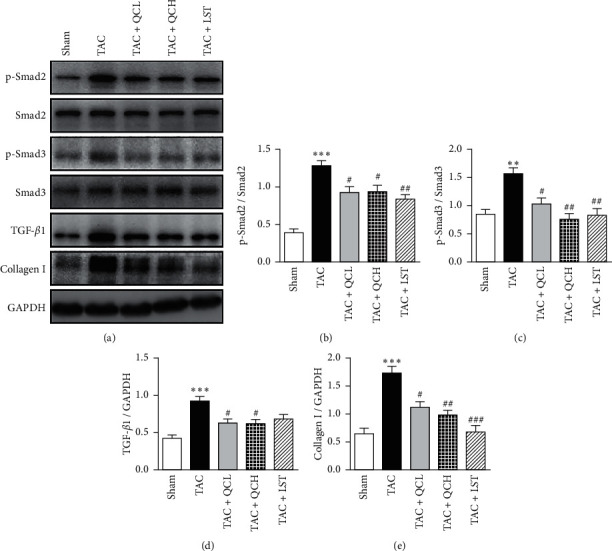
Qindan capsule (QC), a traditional Chinese medicine compound, has been used to treat hypertension in the clinic for over 30 years. It is still not known about the effects of QC on pressure overload-induced cardiac remodeling. Hence, this study aims to investigate the effects of QC on pressure overload-induced cardiac hypertrophy, fibrosis, and heart failure in mice and to determine the possible mechanisms. Transverse aortic constriction (TAC) surgery was used to induce cardiac hypertrophy and heart failure in C57BL/6 mice. Mice were treated with QC or losartan for 8 weeks after TAC surgery. Cardiac function indexes were evaluated with transthoracic echocardiography. Cardiac pathology was detected using HE and Masson's trichrome staining. Cardiomyocyte ultrastructure was detected using transmission electron microscopy. Hypertrophy-related fetal gene expression was investigated using real-time RT-PCR. The expression of 8-OHdG and the concentration of MDA and Ang-II were assessed by immunohistochemistry stain and ELISA assay, respectively. The total and phosphorylated protein levels of mTOR, p70S6K, 4EBP1, Smad2, and Smad3 and the expression of TGF-β1 and collagen I were measured using western blot. The results showed that low- and high-dose QC improved pressure overload-induced cardiac hypertrophy, fibrosis, and dysfunction. QC inhibited ANP, BNP, and β-MHC mRNA expression in failing hearts. QC improved myocardial ultrastructure after TAC surgery. Furthermore, QC downregulated the expression of 8-OHdG and the concentration of MDA, 15-F2t-IsoP, and Ang-II in heart tissues after TAC surgery. We also found that QC inhibited the phosphorylation of mTOR, p70S6K, and 4EBP1 and the expression of TGF-β1, p-Smad2, p-Smad3, and collagen I in pressure overload-induced failing hearts. These data indicate that QC has direct benefic effects on pressure overload-induced cardiac hypertrophy, fibrosis, and dysfunction. The protective effects of QC involve prevention of increased oxidative stress injury and Ang-II levels and inhibition of mTOR and TGF-β1/Smad pathways in failing hearts.


Conflict of interest statement

The authors declare that there are no conflicts of interest associated with the manuscript.

Figures

Figure 1
Figure 1
Effects of QC on cardiac function after TAC surgery in mice. (a) Transthoracic echocardiography of mice in the indicated groups 8 weeks after TAC surgery. (b) Fractional shortening (FS%), (c) ejection fraction (EF%), and (d) LV posterior wall at diastole (LVPWd) were evaluated. ∗∗∗P < 0.001 vs. sham group. #P < 0.05 and ##P < 0.01 vs. TAC group. Data are presented as mean ± SEM and n = 12.
Figure 2
Figure 2
Effects of QC on myocardial hypertrophy and fibrosis after TAC surgery in mice. (a) Representative images of hearts, HE stain, and Masson's trichrome staining of mice left ventricles at the end of 8 weeks after TAC surgery. Scale bar, 50 μm. (b) Ratios of heart weight to tibial length (HW/TL) and (c) ratios of lung weight to tibial length (LW/TL) were measured 8 weeks after TAC surgery. (d) Cross-sectional area of cardiomyocytes was quantified with HE sections. (e) Cardiac fibrosis area was quantified with Masson's trichrome-stained sections. Relative mRNA levels of (f) ANP, (g) BNP, and (h) β-MHC were evaluated using RT-PCR. ∗∗∗P < 0.001 vs. sham group. #P < 0.05 and ##P < 0.01 vs. TAC group. Data are presented as mean ± SEM and n = 5.
Figure 3
Figure 3
Effects of QC on myocardial ultrastructure after TAC surgery in mice. (a) TEM of cardiomyocytes from the indicated groups. Scale bar, 2 μm. (b) Quantification of mitochondrial volume density (MitoVD). ∗∗∗P < 0.001 vs. sham group. #P < 0.05 and ##P < 0.01 vs. TAC group. Data are presented as mean ± SEM and n = 3.
Figure 4
Figure 4
Effects of QC on cardiac oxidative stress injury and Ang-II content after TAC surgery in mice. (a) IHC-stained sections of 8-OHdG; scale bar, 50 μm. (b) Quantification of 8-OHdG-positive cells in indicated groups. (c) Levels of malondialdehyde (MDA), (d) 15-F2t-IsoP, and (e) Ang-II were quantified in heart tissues of mice. ∗∗P < 0.01 and ∗∗∗P < 0.001 vs. sham group; #P < 0.05 and ##P < 0.01 vs. TAC group. Data are presented as mean ± SEM and n = 5.
Figure 5
Figure 5
Effects of QC on the cardiac mTOR signaling pathway after TAC surgery in mice. (a) Representative results of protein levels and phosphorylation levels of mTOR, 70S6K, and 4EBP1. Quantification of ratios of p-mTOR to mTOR (b), p-70S6K to 70S6K (c), and p-4EBP1 to 4EBP1 (d). ∗∗P < 0.01 and ∗∗∗P < 0.001 vs. sham group. #P < 0.05, ##P < 0.01, and ###P < 0.001 vs. TAC group. Data are presented as mean ± SEM and n = 3.
Figure 6
Figure 6
Effects of QC on the cardiac TGF-β1/Smad pathway after TAC surgery in mice. (a) Representative results of the protein levels of TGF-β1, collagen I, and GAPDH and phosphorylation levels of Smad2 and Smad3. Quantification of ratios of p-Smad2 to Smad2 (b), p-Smad3 to Smad3 (c), TGF-β1 to GAPDH (d), and collagen I to GAPDH (e). ∗∗P < 0.01 and ∗∗∗P < 0.001 vs. sham group. #P < 0.05, ##P < 0.01, and ###P < 0.001 vs. TAC group. Data are presented as mean ± SEM and n = 3.

References

    1. Messerli F. H., Rimoldi S. F., Bangalore S. The transition from hypertension to heart failure. JACC: Heart Failure. 2017;5(8):543–551. doi: 10.1016/j.jchf.2017.04.012. - DOI - PubMed
    1. Frangogiannis N. G. The extracellular matrix in ischemic and nonischemic heart failure. Circulation Research. 2019;125(1):117–146. doi: 10.1161/circresaha.119.311148. - DOI - PMC - PubMed
    1. Ponikowski P., Voors A. A., Anker S. D., et al. 2016 ESC guidelines for the diagnosis and treatment of acute and chronic heart failure. Revista Española de Cardiología (English Edition) 2016;69(12):p. 1167. doi: 10.1016/j.rec.2016.11.005. - DOI - PubMed
    1. Wang S. M., Ye L. F., Wang L. H. Shenmai injection improves energy metabolism in patients with heart failure: a randomized controlled trial. Frontiers in Pharmacology. 2020;11:p. 459. doi: 10.3389/fphar.2020.00459. - DOI - PMC - PubMed
    1. Sun X., Chen G., Xie Y., et al. Qiliqiangxin improves cardiac function and attenuates cardiac remodelling in doxorubicin-induced heart failure rats. Pharmaceutical Biology. 2020;58(1):417–426. doi: 10.1080/13880209.2020.1761403. - DOI - PMC - PubMed

LinkOut - more resources