

Early manifestation of Moyamoya syndrome in a 2-year-old child with Down syndrome
- PMID: 34007395
- PMCID: PMC8111440
- DOI: 10.1016/j.radcr.2021.04.017
Early manifestation of Moyamoya syndrome in a 2-year-old child with Down syndrome
Abstract
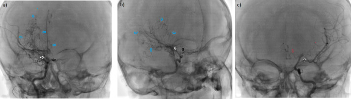
Moyamoya is a rare occlusive cerebrovascular disease characterized by progressive stenosis of the terminal portion of the internal carotid artery and the circle of Willis. Over time, collateral arteries are usually formed at the basal ganglia, the so-called Moyamoya vessels. The exact cause of Moyamoya disease is unknown, while Moyamoya syndrome refers to Moyamoya-like vasculopathy due to autoimmune diseases, neurofibromatosis type I, sickle cell disease, radiation, or rarely Down syndrome. Down syndrome is one of the most common genetic conditions, characterized by typical physical traits, associated with intellectual disability and a heterogeneous group of structural defects that may vulnerable the patient for the development of Moyamoya syndrome. The reported case is an unusual case of a 2-year-old boy with Down syndrome who presented to the hospital with seizures and right-side weakness. Brain MRI shows acute as well as old lacunar infarctions in both cerebral hemispheres. Catheter angiography of the patient demonstrates severe stenosis and occlusion of the large vessels of the circle of Willis, predominantly on the right side. The collateral vessels with the typical pattern of "puff of smoke" were also depicted in the right basal ganglia, which is a characteristic imaging finding for Moyamoya. The patient was managed conservatively and eventually discharged with a minimal improvement of the right-sided weakness. This case report is noteworthy because of the rarity of Moyamoya syndrome as a cause of a stroke as well as its possible association with Down syndrome.
Keywords: 3D-TOF, 3D time-of-flight; ACA, anterior cerebral artery; Angiography; CSF, cerebrospinal fluid; CT, computed tomography; CTA, computed tomography angiography; DWI, diffusion-weighted imaging; Down syndrome; ECA, external carotid artery; FLAIR, fluid-attenuated inversion recovery; ICA, internal carotid artery; Lacunar infarction; MCA, middle cerebral artery; MMD, Moyamoya disease; MMS, Moyamoya syndrome; MRA, magnetic resonance angiography; MRI; Moyamoya syndrome; PCA, posterior cerebral artery; SWI, susceptibility-weighted imaging; TIAs, transient ischemic attacks.
© 2021 The Authors. Published by Elsevier Inc. on behalf of University of Washington.
Figures


References
-
- Mimi L, Alami B, Baptiste AJ, Quenum L, Lamrani YA, Boubbou M. Moya Moya disease: about 3 cases and a review of literature. Diagn Interven Imag J. 2019;2(1):22–31. doi: 10.1016/j.jidi.2018.11.005. - DOI
Publication types
LinkOut - more resources
Full Text Sources
Other Literature Sources
Research Materials
Miscellaneous
