. 2021 Apr 28:2021:8849568.
doi: 10.1155/2021/8849568. eCollection 2021.

Synthesis and Biological Screening of New 4-Hydroxycoumarin Derivatives and Their Palladium(II) Complexes

Affiliations

Synthesis and Biological Screening of New 4-Hydroxycoumarin Derivatives and Their Palladium(II) Complexes

Edina H Avdović et al. Oxid Med Cell Longev. .

Abstract

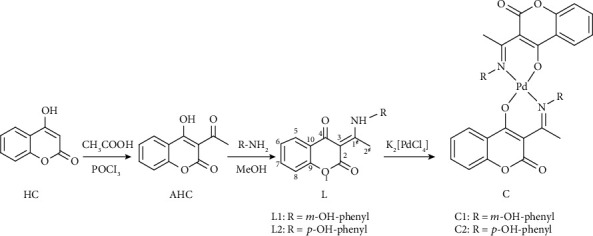
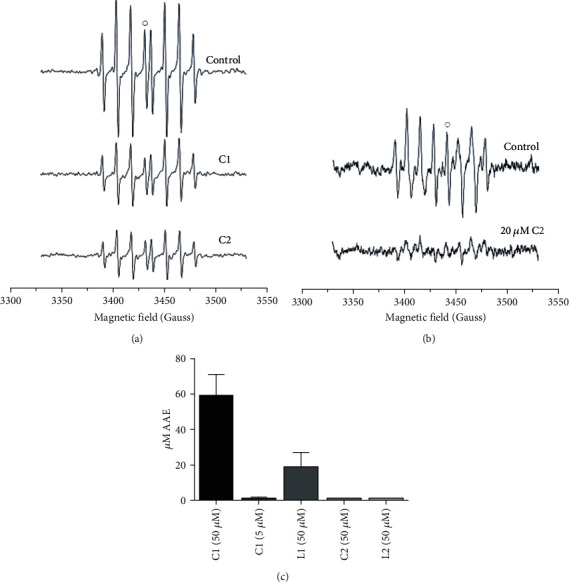
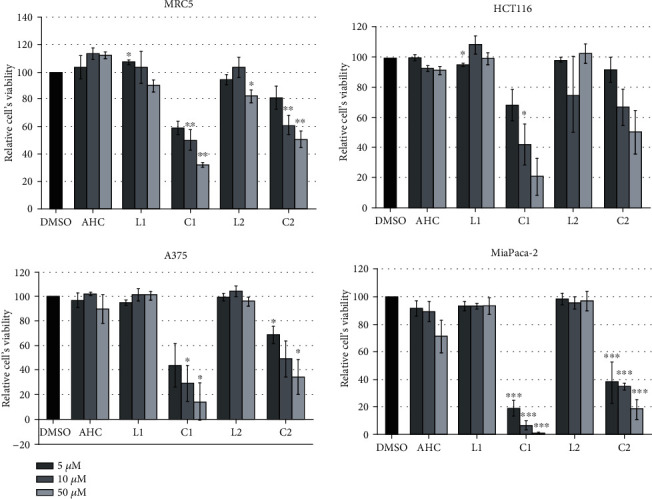
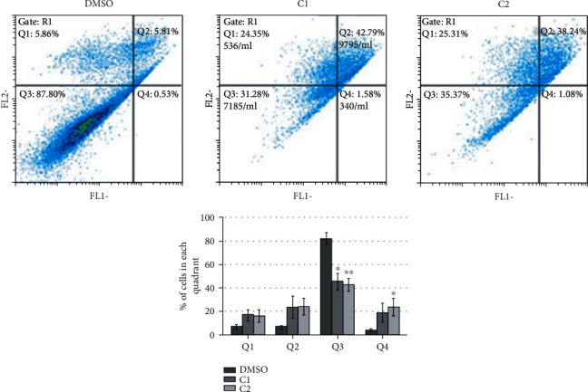
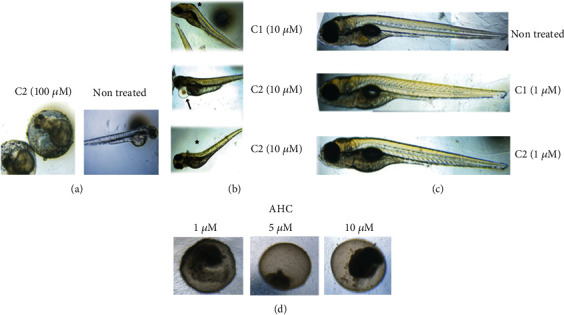
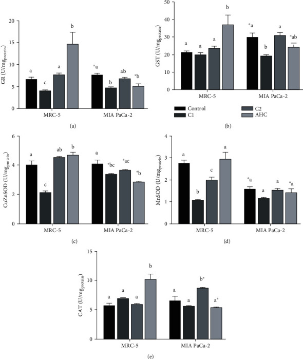
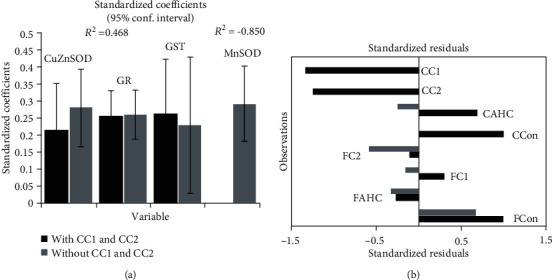
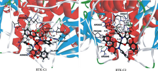
Two newly synthesized 4-hydroxycoumarin bidentate ligands (L1 and L2) and their palladium(II) complexes (C1 and C2) were screened for their biological activities, in vitro and in vivo. Structures of new compounds were established based on elemental analysis, 1H NMR, 13C NMR, and IR spectroscopic techniques. The obtained compounds were tested for their antioxidative and cytotoxic activities and results pointed to selective antiradical activity of palladium(II) complexes towards OH and -•OOH radicals and anti-ABTS (2,2'-Azino-bis(3-ethylbenzothiazoline-6-sulfonic acid) cation radical) activity comparable to that of ascorbate. Results indicated the effect of C1 and C2 on the enzymatic activity of the antioxidative defense system. In vitro cytotoxicity assay performed on different carcinoma cell lines (HCT166, A375, and MIA PaCa-2), and one healthy fibroblast cell line (MRC-5) showed a cytotoxic effect of both C1 and C2, expressed as a decrease in carcinoma cells' viability, mostly by induction of apoptosis. In vivo toxicity tests performed on zebrafish embryos indicated different effects of C1 and C2, ranging from adverse developmental effect to no toxicity, depending on tested concentration. According to docking studies, both complexes (C1 and C2) showed better inhibitory activity in comparison to other palladium(II) complexes.


Conflict of interest statement

The authors declare that there is no conflict of interest regarding the publication of this paper.

Figures

Scheme 1
Scheme 1
The general procedure for the synthesis of the ligands L1 and L2 and complexes C1 and C2.
Figure 1
Figure 1
EPR spectra of DEPMPO/OH spin-adduct upon addition of 5 μM C1 and C2 (a) and DEPMPO-OOH adduct upon addition of 20 μM C2 (b). The antioxidant activity is calculated relative to the heights of the peaks marked with open circles; ABTS quenching activity of C1, L1, C2, and L2. Concentrations tested are given in brackets (c).
Figure 2
Figure 2
In vitro cytotoxicity assay. Healthy fibroblasts (MRC-5) and three different carcinoma cell lines (HCT 116, A375, and MIA PaCa-2) were exposed to DMSO as a negative control, AHC as a starting coumarin compound, its derivates L1 and L2, and the corresponding palladium(II) complexes C1 and C2. Relative cells' viability was calculated as a percentage of DMSO treated cell viability that was set as 100%. Data are presented as the means ± S.E.M. (standard error mean) of at least three independent experiments performed in 3-plicates for each concentration. Mean values of relative cell viability were compared with Student's t-test, and P values are presented as P ≤ 0.05, ∗∗P ≤ 0.01, and ∗∗∗P ≤ 0.001. Each color corresponds to a bar presented on the histogram.
Figure 3
Figure 3
Apoptosis assay. MIA PaCa-2 cells were treated with DMSO as a negative control and coumarin complexes C1 and C2. 24 h upon treatment, cells were collected, and Annexin V-FITC assay was performed. The percentage of cells in each quadrant is presented as the means ± S.E.M. (standard error mean) of at least three independent experiments. Mean values were compared with Student's t-test, and P values that were evaluated to DMSO treatment are presented as P ≤ 0.05, ∗∗P ≤ 0.01, and ∗∗∗P ≤ 0.001. Each color corresponds to a bar presented on the histogram. Q1: necrotic cells; Q2: late apoptosis; Q3: living cells; Q4: early apoptosis.
Figure 4
Figure 4
Zebrafish toxicity testing. (a) Representative image of unsuccessful hatching upon treatment of zebrafish embryos with 100 μM of coumarin complex C2 (left panel) compared to successful hatching of nontreated embryos (right panel). (b) Skeletal (asterisk) and cardiovascular abnormalities (arrow) detected at 10 μM concentration of either C1 or C2. (c) Representative images of normal development of nontreated embryos or treated with 1 μM of C1 or C2. (d) Lethal effect of starting coumarin compound (AHC) detected within first 24 h, at any tested concentration.
Figure 5
Figure 5
Effect of the selected compounds (AHC and complexes C1 and C2) on the activity of enzymes of the antioxidative defense system: GR (a), GST (b), CuZnSOD (c), MnSOD (d), and CAT (e) in a healthy fibroblast cell line (MRC-5) and carcinoma cell line (MIA PaCa-2). Effects of the selected compounds on the same cell line that are significantly different (P < 0.05) are marked with different letters (a, b, c). A statistically significant difference (P < 0.05) between effects of the same treatment on MRC-5 and MIA PaCa-2 cell lines is marked with an asterisk ().
Figure 6
Figure 6
Quality of the Partial least square regression (PLSR) model describing the dependence of the cytotoxic effects of the investigated compounds (AHC and complexes C1 and C2) on the activity of the antioxidative defense system enzymes (GST, GR, MnSOD, CuZnSOD, with (black bars) and without (grey bars) CC1 and CC2 observations) present in a healthy fibroblast cell line (FAHC, FC1, and FC2, respectively) and pancreatic carcinoma cell line (CAHC, CC1, and CC2, respectively) (a). PLSR standardized regression coefficients. The bars indicate 95% confidence intervals based on jackknifing. Bar chart of the standardized residuals (b).
Figure 7
Figure 7
The hydrogen bond (green dotted lines) and hydrophobic (rose pink dotted lines) docking interactions of the most stable conformation of C1 and C2 with RTK protein.

References

    1. Emami S., Dadashpour S. Current developments of coumarin-based anti-cancer agents in medicinal chemistry. European Journal of Medicinal Chemistry. 2015;102:611–630. doi: 10.1016/j.ejmech.2015.08.033. - DOI - PubMed
    1. Kruk J., Aboul-Enein H. Y. Reactive oxygen and nitrogen species in carcinogenesis: implications of oxidative stress on the progression and development of several cancer types. Mini Reviews in Medicinal Chemistry. 2017;17(11):904–919. doi: 10.2174/1389557517666170228115324. - DOI - PubMed
    1. Kaur M., Kohli S., Sandhu S., Bansal Y., Bansal G. Coumarin: a promising scaffold for anticancer agents. Anti-Cancer Agents in Medicinal Chemistry. 2015;15(8):1032–1048. doi: 10.2174/1871520615666150101125503. - DOI - PubMed
    1. Carocho M., Ferreira I. C. F. R. The role of phenolic compounds in the fight against cancer – a review. Anti-Cancer Agents in Medicinal Chemistry. 2013;13(8):1236–1258. doi: 10.2174/18715206113139990301. - DOI - PubMed
    1. Winocur G., Vardy J., Binns M., Kerr L., Tannock I. The effects of the anti-cancer drugs, methotrexate and 5-fluorouracil, on cognitive function in mice. Pharmacology Biochemistry and Behavior. 2006;85(1):66–75. doi: 10.1016/j.pbb.2006.07.010. - DOI - PubMed

LinkOut - more resources