

Point-of-care ultrasonography in the initial characterization of patients with COVID-19
- PMID: 34007877
- PMCID: PMC8120770
- DOI: 10.1016/j.medcle.2020.12.016
Point-of-care ultrasonography in the initial characterization of patients with COVID-19
Abstract
Background: There is growing evidence regarding the imaging findings of coronavirus disease 2019 (COVID-19) in chest X-rays and computed tomography scans; however, their availability during this pandemic outbreak might be compromised. Currently, the role of point-of-care ultrasonography (POCUS) has yet to be explored.
Objectives: To describe the POCUS findings of COVID-19 in patients with the disease admitted to the emergency department (ED), correlating them with vital signs, laboratory and radiologic results, therapeutic decisions, and the prognosis.
Methods: Prospective study performed in the ED of 2 academic hospitals. Patients with highly suspected or confirmed COVID-19 underwent a lung ultrasonography (lung POCUS), focused cardiac ultrasound (FOCUS), and inferior vena cava (IVC) exam.
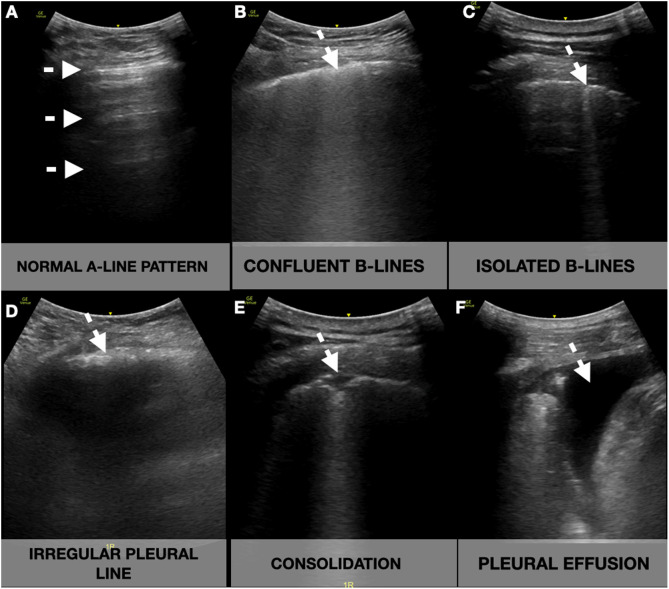
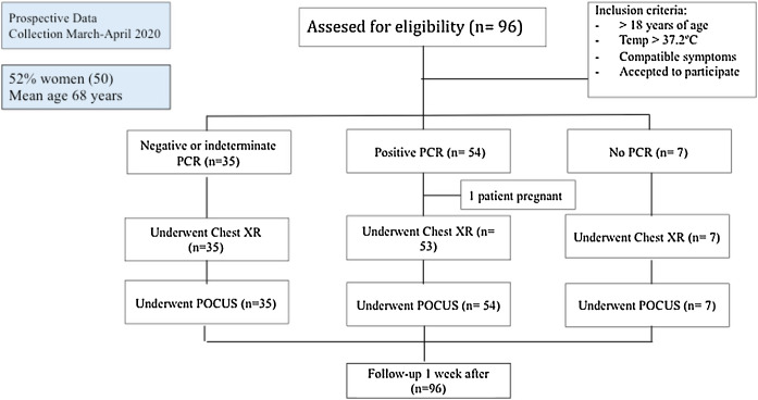
Results: Between March and April 2020, 96 patients were enrolled. The mean age was 68.2 years (SD 17.5). The most common findings in the lung POCUS were an irregular pleural line (63.2%), bilateral confluence (55.2%), and isolated B-lines (53.1%), which were associated with a positive RT-PCR (odds ratio 4.327; 95% CI 1.216-15.401; p < .001), and correlated with IL-6 levels (rho = 0.622; p = .002). The IVC negatively correlated with levels of expiratory pO2 (rho = -0.539; p = .014) and inspiratory pO2 (rho = -0.527; p = 0.017), and expiratory diameter positively correlated with troponin I (rho = 0.509; p = .03). After the POCUS exam, almost 20% of the patients had an associated condition that required a change in their treatment or management.
Conclusions: POCUS parameters have the potential to impact the diagnosis, management, and prognosis of patients with confirmed or suspected COVID-19.
Antecedentes: Existe una evidencia creciente con respecto a los hallazgos por imagen de la COVID-19, tanto en radiografías de tórax como en tomografía computarizada; sin embargo, la disponibilidad de estas técnicas durante la pandemia podría verse comprometida.
Objetivos: Describir los hallazgos en la ecografía en el punto de atención (POCUS) en pacientes con COVID-19 que consultaron en el servicio de urgencias (SU), correlacionándolos con signos vitales, resultados analíticos y radiológicos, decisiones terapéuticas y pronóstico.
Métodos: Estudio prospectivo realizado en los SU de dos hospitales académicos. Los pacientes con COVID-19 con alta sospecha o confirmada se sometieron a una ecografía pulmonar (POCUS pulmonar), una ecocardioscopia y una ecografía de la vena cava inferior (VCI).
Resultados: Entre marzo y abril del 2020, se reclutaron 96 pacientes. La edad media fue de 68,2 años (DE 17,5). Los hallazgos más comunes en el POCUS pulmonar fueron la línea pleural irregular (63,2%), las líneas B confluyentes bilateral (55,2%) y aisladas (53,1%), que se vincularon con una RT-PCR (odds ratio 4,327; IC 95% 1,216 a 15,401; p < 0,001), y se asoció con los niveles de interleucina-6 (IL-6) (ρ = 0,622; p = 0,002). La VCI se correlacionó negativamente con los niveles de pO2 espiratorio (ρ = − 0,539; p = 0,014) y pO2 inspiratorio (ρ = − 0,527; p = 0,017), y el diámetro espiratorio se relacionó positivamente con la troponina I (ρ = 0,509; p = 0, 03). Después del examen POCUS, casi el 20% de los pacientes tenían una condición asociada que requería un cambio en el tratamiento o manejo previo.
Conclusiones: Los parámetros POCUS tienen el potencial de afectar el diagnóstico, manejo y pronóstico de pacientes con sospecha o confirmación de COVID-19.
Keywords: Coronavirus disease 2019 (COVID-19); Focused cardiac ultrasonography (FOCUS).; Point-of-care ultrasonography (POCUS); Severe acute respiratory syndrome coronavirus 2 (SARS-CoV-2).
© 2021 Elsevier España, S.L.U. All rights reserved.
Figures
Similar articles
-
Point-of-care ultrasonography in the initial characterization of patients with COVID-19.Med Clin (Barc). 2021 May 21;156(10):477-484. doi: 10.1016/j.medcli.2020.12.007. Epub 2021 Jan 28. Med Clin (Barc). 2021. PMID: 33593636 Free PMC article.
-
Correlation between Chest Computed Tomography and Lung Ultrasonography in Patients with Coronavirus Disease 2019 (COVID-19).Ultrasound Med Biol. 2020 Nov;46(11):2918-2926. doi: 10.1016/j.ultrasmedbio.2020.07.003. Epub 2020 Jul 13. Ultrasound Med Biol. 2020. PMID: 32771222 Free PMC article.
-
Prospective Longitudinal Evaluation of Point-of-Care Lung Ultrasound in Critically Ill Patients With Severe COVID-19 Pneumonia.J Ultrasound Med. 2021 Mar;40(3):443-456. doi: 10.1002/jum.15417. Epub 2020 Aug 14. J Ultrasound Med. 2021. PMID: 32797661 Free PMC article.
-
Point-of-care ultrasound use in COVID-19: a narrative review.Ann Transl Med. 2024 Feb 1;12(1):13. doi: 10.21037/atm-23-1403. Epub 2023 Jul 13. Ann Transl Med. 2024. PMID: 38304913 Free PMC article. Review.
-
Thoracic imaging tests for the diagnosis of COVID-19.Cochrane Database Syst Rev. 2020 Sep 30;9:CD013639. doi: 10.1002/14651858.CD013639.pub2. Cochrane Database Syst Rev. 2020. Update in: Cochrane Database Syst Rev. 2020 Nov 26;11:CD013639. doi: 10.1002/14651858.CD013639.pub3. PMID: 32997361 Updated.
Cited by
-
The correlation between point-of-care ultrasound and digital tomosynthesis when used with suspected COVID-19 pneumonia patients in primary care.Ultrasound J. 2022 Feb 22;14(1):11. doi: 10.1186/s13089-022-00257-7. Ultrasound J. 2022. PMID: 35192076 Free PMC article.
-
Past and Present of Point-of-Care Ultrasound (PoCUS): A Narrative Review.Cureus. 2023 Dec 8;15(12):e50155. doi: 10.7759/cureus.50155. eCollection 2023 Dec. Cureus. 2023. PMID: 38192958 Free PMC article. Review.
-
Importance of Lung Ultrasound Follow-Up in Patients Who Had Recovered from Coronavirus Disease 2019: Results from a Prospective Study.J Clin Med. 2021 Jul 20;10(14):3196. doi: 10.3390/jcm10143196. J Clin Med. 2021. PMID: 34300362 Free PMC article.
-
A Case of Restrictive Lung Disease Masquerading as SARS-CoV-2 Pneumonia: the Complexity of Integrating Lung Ultrasound in Clinical Management.SN Compr Clin Med. 2022;4(1):45. doi: 10.1007/s42399-022-01122-3. Epub 2022 Jan 24. SN Compr Clin Med. 2022. PMID: 35098035 Free PMC article.
-
One-month outcomes of patients with SARS-CoV-2 infection and their relationships with lung ultrasound signs.Ultrasound J. 2021 Apr 9;13(1):19. doi: 10.1186/s13089-021-00223-9. Ultrasound J. 2021. PMID: 33835273 Free PMC article.
References
-
- World Health Organization. Rolling updates on coronavirus (COVID-19); 2020. WHO characterizes COVID-19 as a pandemic. Available from: https://www.who.int/emergencies/diseases/novel-coronavirus-2019/events-a... [accessed 31.07.20].
-
- Johns Hopkins Coronavirus Resource Center; 2020. Available from https://coronavirus.jhu.edu/map.html [accessed 12.10.20].
-
- Guidance for Corona Virus Disease 2019: Prevention, Control, Diagnosis and Management National Health Commission (NHC) of the PRC, General Office; National Administration of Traditional Chinese Medicine of the PRC, General Office; 2020. Available from: http://www.pmph.com/ [accessed 31.07.20].
LinkOut - more resources
Full Text Sources
Other Literature Sources
Miscellaneous