Review
. 2022 Jan 12;43(1):61-90.
doi: 10.1210/endrev/bnab017.

Osteogenesis Imperfecta: Mechanisms and Signaling Pathways Connecting Classical and Rare OI Types

Affiliations
Review

Osteogenesis Imperfecta: Mechanisms and Signaling Pathways Connecting Classical and Rare OI Types

Milena Jovanovic et al. Endocr Rev. .

Abstract

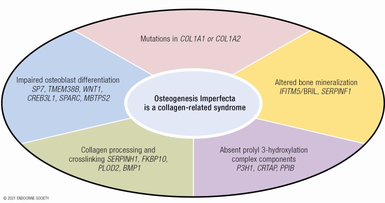
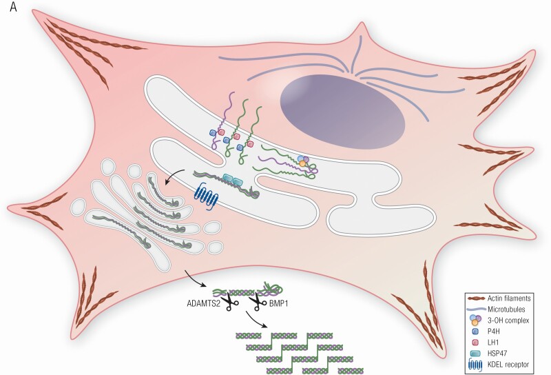
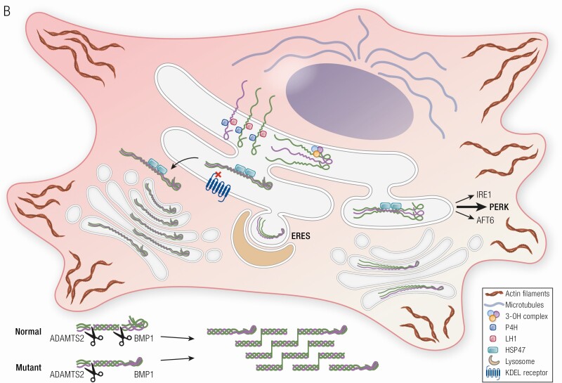
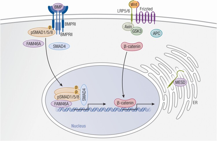
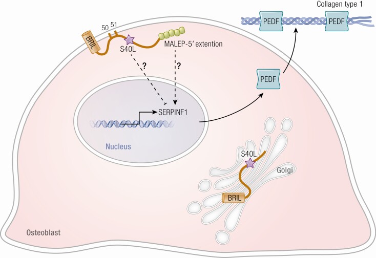
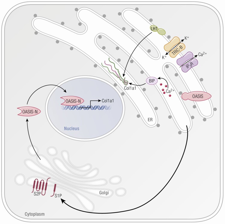
Osteogenesis imperfecta (OI) is a phenotypically and genetically heterogeneous skeletal dysplasia characterized by bone fragility, growth deficiency, and skeletal deformity. Previously known to be caused by defects in type I collagen, the major protein of extracellular matrix, it is now also understood to be a collagen-related disorder caused by defects in collagen folding, posttranslational modification and processing, bone mineralization, and osteoblast differentiation, with inheritance of OI types spanning autosomal dominant and recessive as well as X-linked recessive. This review provides the latest updates on OI, encompassing both classical OI and rare forms, their mechanism, and the signaling pathways involved in their pathophysiology. There is a special emphasis on mutations in type I procollagen C-propeptide structure and processing, the later causing OI with strikingly high bone mass. Types V and VI OI, while notably different, are shown to be interrelated by the interferon-induced transmembrane protein 5 p.S40L mutation that reveals the connection between the bone-restricted interferon-induced transmembrane protein-like protein and pigment epithelium-derived factor pathways. The function of regulated intramembrane proteolysis has been extended beyond cholesterol metabolism to bone formation by defects in regulated membrane proteolysis components site-2 protease and old astrocyte specifically induced-substance. Several recently proposed candidate genes for new types of OI are also presented. Discoveries of new OI genes add complexity to already-challenging OI management; current and potential approaches are summarized.

Keywords: IFITM5/BRIL; MBTPS2; PEDF; bone mass; bone mineralization; collagen synthesis; osteogenesis imperfecta; regulated intramembrane proteolysis.


Figures

Graphical Abstract
Graphical Abstract
Figure 1.
Figure 1.
Collagen structure, folding, modification and processing in OI. (A) Procollagen is a heterotrimer consisting of two proα1 (I) and one proα2 (I) chains, which undergo posttranslational modifications of proline and lysine residues during helix folding by prolyl-4-hydroxylase and lysyl hydroxylase 1, respectively. The prolyl 3-OH complex (P3H1, cartilage-associated protein, and peptidylprolyl isomerase B) serves as a folding chaperone; the modification of substrate P986 residue finetunes collagen alignment for cross-linking. The triple helix is further stabilized by Hsp47, an ER chaperone. Once secreted into extracellular space, N- and C-propeptides of procollagen are cleaved by ADAMTS-2 (a disintegrin and metalloproteinase with thrombospondin motifs) and BMP1 enzymes, respectively, and mature type I collagen is released and incorporated into extracellular matrix. A newly discovered OI-causing gene KDELR2, encodes KDEL receptor 2 that together with Hsp47 facilitates intracellular recycling of ER-resident proteins. (B) In OI pathophysiology, mutations cause misfolding and overmodification of procollagen chains that may increases protein accumulation in the ER, resulting in ER stress. ER stress causes alterations in cytoskeleton proteins (actin filaments, microtubules), induction of UPR pathways [PERK (especially), inositol requiring enzyme 1, ATF6] to accommodate the chronic stress. For some mutations, ER stress capacity is exceeded, and mutant collagens are labeled with autophagy proteins for lysosomes degradation via microautophagy process at the ER exit sites (ERES). Mutations in KDELR2 unable binding of Hsp47 to KDEL receptor 2, thus Hsp47 remains bound to collagen molecules extracellularly disrupting collagen fibers formation.
Figure 1.
Figure 1.
Collagen structure, folding, modification and processing in OI. (A) Procollagen is a heterotrimer consisting of two proα1 (I) and one proα2 (I) chains, which undergo posttranslational modifications of proline and lysine residues during helix folding by prolyl-4-hydroxylase and lysyl hydroxylase 1, respectively. The prolyl 3-OH complex (P3H1, cartilage-associated protein, and peptidylprolyl isomerase B) serves as a folding chaperone; the modification of substrate P986 residue finetunes collagen alignment for cross-linking. The triple helix is further stabilized by Hsp47, an ER chaperone. Once secreted into extracellular space, N- and C-propeptides of procollagen are cleaved by ADAMTS-2 (a disintegrin and metalloproteinase with thrombospondin motifs) and BMP1 enzymes, respectively, and mature type I collagen is released and incorporated into extracellular matrix. A newly discovered OI-causing gene KDELR2, encodes KDEL receptor 2 that together with Hsp47 facilitates intracellular recycling of ER-resident proteins. (B) In OI pathophysiology, mutations cause misfolding and overmodification of procollagen chains that may increases protein accumulation in the ER, resulting in ER stress. ER stress causes alterations in cytoskeleton proteins (actin filaments, microtubules), induction of UPR pathways [PERK (especially), inositol requiring enzyme 1, ATF6] to accommodate the chronic stress. For some mutations, ER stress capacity is exceeded, and mutant collagens are labeled with autophagy proteins for lysosomes degradation via microautophagy process at the ER exit sites (ERES). Mutations in KDELR2 unable binding of Hsp47 to KDEL receptor 2, thus Hsp47 remains bound to collagen molecules extracellularly disrupting collagen fibers formation.
Figure 2.
Figure 2.
Wnt and Bmp signaling pathways disrupted in osteogenesis imperfecta. (Left) BMP ligand binds to BMPRI and BMPRII receptors and induces phosphorylation of SMAD 1/5/8. Terminal nucleotidyltransferase 5A (FAM46A) binds to phosphorylated SMAD 1/5/8 and protects it from ubiquitination. Together SMAD 1/5/8, FAM46A, and SMAD4 form a complex that translocates to the nucleus where it induces transcription of BMP target genes. (Right) Wnt signaling is activated by binding of WNT1 ligand to the Frizzled receptor and LRP5/6 co-receptors, which stabilizes β-catenin levels by inhibiting the degradation complex consisting of axin, adenomatous polyposis coli (APC), and glycogen synthase kinase 3 (GSK3). Subsequently, β-catenin migrates to the nucleus where it activates transcription of Wnt target genes. The recently proposed causative gene for OI, MESD, is an ER chaperone that facilitates maturation and trafficking of LRP5/6 co-receptors.
Figure 3.
Figure 3.
Defects in BRIL and PEDF, altering bone mineralization, cause OI. BRIL has an important role in the regulation of SERPINF1 transcription in the nuclei and resultant production of the protein PEDF. In a normal osteoblast, BRIL is attached to the cell membrane by palmytoilation sites. In the mutation causing Type V OI, 5 aa resides are added to the 5’ end of BRIL. This in turn increases the transcription of SERPINF1 and its protein PEDF. In the presence of a BRIL Serine40Leucine substitution, the palmytoilation process is impaired, and BRIL is trapped in the Golgi apparatus. Clinically, BRIL S40L patients have features of severe type VI OI (type atypical VI OI) rather than type V OI. PEDF binding to collagen in matrix is critical for its anti-angiogenic function. Osteoid is increased in PEDF null bone tissue by an unknown mechanism.
Figure 4.
Figure 4.
OASIS, RIP, and TRIC-B role in bone formation. The ER serves as a major storage site for intracellular Ca+2. Type I collagen is synthesized within this compartment and Ca+2 is a cofactor for many enzymes involved in collagen folding and modification. Upon receiving extracellular stimuli, the ER lumen releases Ca+2 to the cytoplasm through IP3R. TRIC-B is involved indirectly in the kinetics of Ca+2 entry and released from the ER by mediating K+ flux; this maintains the electroneutrality through the ER membrane. When TRIC-B is deficient, altered Ca flux interferes with multiple Ca+2 binding chaperones such as BiP and modifying enzymes such as LH1, thus disrupting the synthesis and secretion of collagen. Subsequent to cell stress, regulatory proteins in the ER membrane, such as OASIS, are transported from the ER to the Golgi membrane for RIP. After sequential cleavage by S1P and S2P, transmembrane proteases in the Golgi, the released N-terminal portion of OASIS can then translocate into the nucleus and activate transcription of a set of matrix-related genes, including collagen type I alpha/alpha 21 chain.

Similar articles

Cited by

References

    1. Gatti D, Rossini M, Viapiana O, et al. Teriparatide treatment in adult patients with osteogenesis imperfecta type I. Calcif Tissue Int. 2013;93(5):448-452. - PubMed
    1. Orwoll ES, Shapiro J, Veith S, et al. Evaluation of teriparatide treatment in adults with osteogenesis imperfecta. J Clin Invest. 2014;124(2):491-498. - PMC - PubMed
    1. Nicol L, Srikanth P, Henriksen K, et al. Widespread disturbance in extracellular matrix collagen biomarker responses to teriparatide therapy in osteogenesis imperfecta. Bone. 2021;142:115703. - PubMed
    1. Forlino A, Marini JC. Osteogenesis imperfecta. Lancet. 2016;387(10028):1657-1671. - PMC - PubMed
    1. Kuivaniemi H, Tromp G, Prockop DJ. Mutations in collagen genes: causes of rare and some common diseases in humans. Faseb J. 1991;5(7):2052-2060. - PubMed

Publication types