. 2021 Sep;10(9):1329-1342.
doi: 10.1002/sctm.20-0501. Epub 2021 May 18.

Facile bead-to-bead cell-transfer method for serial subculture and large-scale expansion of human mesenchymal stem cells in bioreactors

Affiliations

Facile bead-to-bead cell-transfer method for serial subculture and large-scale expansion of human mesenchymal stem cells in bioreactors

Shangwu Chen et al. Stem Cells Transl Med. 2021 Sep.

Abstract

The conventional planar culture of adherent cells is inefficient for large-scale manufacturing of cell and gene therapy products. We developed a facile and efficient bead-to-bead cell-transfer method for serial subculture and large-scale expansion of human mesenchymal stem cells (hMSCs) with microcarriers in bioreactors. We first compared culture medium with and without nucleosides and found the former maintained the expression of surface markers of hMSCs during their prolonged culture and enabled faster cell proliferation. Subsequently, we developed our bead-to-bead cell transfer method to subculture hMSCs and found that intermittent agitation after adding fresh microcarriers to cell-populated microcarriers could promote spontaneous cell migration to fresh microcarriers, reduce microcarrier aggregation, and improve cell yield. This method enabled serial subculture of hMSCs in spinner flasks from passage 4 to passage 9 without using proteolytic enzymes, which showed faster cell proliferation than the serial planar cultures undergoing multiple enzyme treatment. Finally, we used the medium containing nucleosides and our bead-to-bead cell transfer method for cell culture scale-up from 4- to 50-L cultures in single-use bioreactors. We achieved a 242-fold increase in the number of cells to 1.45 × 10¹⁰ after 27-day culture and found that the cells harvested from the bioreactors maintained proliferation ability, expression of their surface markers, tri-lineage differentiation potential and immunomodulatory property. This study shows the promotive effect of nucleosides on hMSC expansion and the potential of using our bead-to-bead transfer method for larger-scale manufacturing of hMSCs for cell therapy.

Keywords: bead-to-bead transfer; bioreactor; mesenchymal stem cell; microcarrier; nucleosides; scale up.


Conflict of interest statement

The authors declared no potential conflicts of interest.

Figures

FIGURE 1
FIGURE 1
Effect of nucleosides in culture medium on proliferation and surface marker expression of hMSCs. A, Number of cells harvested from tissue culture flasks (175 cm2). Cells from two donors were cultured from passage 4 to passage 6 in ɑMEM/10% FBS with and without nucleosides. Dash lines represent number of cells seeded (5.25 × 105) per flask. B, Expression of surface markers on passage 6 cells cultured without and with nucleosides. Red solid lines represent cells treated with negative and positive antibody cocktails and dashed lines represent cells treated with negative and positive isotype controls. C, Proliferation of cells with Cytodex 1 microcarriers in 100‐mL spinner flasks. Experiments were run in triplicate; mean ± SD. Asterisk * indicates significant statistical difference (P < .01, Tukey post hoc tests after two‐way ANOVA)
FIGURE 2
FIGURE 2
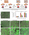
Effect of agitation mode on bead‐to‐bead cell transfer after microcarrier addition to spinner flasks. A, Different agitation modes used during bead‐to‐bead cell transfer. B–H, Staining of cells (live cells: green; dead: red) on beads after microcarrier addition. B, Samples taken immediately after microcarrier addition (day 0). C–E, Samples taken on day 1 after microcarrier addition. F–H, Samples taken on day 7 after microcarrier addition. C and F are from intermittent 24 hours group, D and G are from intermittent 6 hours group, and E and H are from continuous group. Arrows in H show large microcarrier aggregates. Scale bar = 200 μm. I, Ratio of cell‐populated microcarriers in cultures that underwent different agitation modes. Samples were taken on days 0, 1, and 7 after microcarrier addition. Experiments were run in triplicate; mean ± SD; *P < .01, Tukey post hoc tests after one‐way ANOVA
FIGURE 3
FIGURE 3
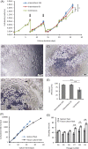
Proliferation and harvest of hMSCs cultured with different agitation modes during passaging and comparison of suspension culture with planar culture. A, hMSC proliferation in cultures underwent three agitation modes. Cells were cultured with microcarriers for 26 days during which fresh microcarrier suspensions were added on days 7 and 13 (marked with arrows). Data points represent average cell densities of triplicate spinner flask cultures (N = 3). B‐D, Phase contrast images taken after enzyme treatment of cells subcultured with intermittent 24 hours, intermittent 6 hours, and continuous agitation modes, respectively. Arrows in D mark cell clumps trapped in aggregated microcarriers. E, Harvest efficiency of cells from microcarriers. F–G, Cumulative fold increase and cell doubling times of serial subcultures in spinner flasks and tissue culture flasks. Experiments were run in triplicate; *P < .01; **P < .001; Tukey post hoc tests after one‐way ANOVA (A and E) or two‐way ANOVA (G)
FIGURE 4
FIGURE 4
Serial subculture from 4‐ to 50‐L cultures in bioreactors. A, Subculture of hMSCs in stirred‐tank bioreactors (STR). B, Fluorescent microscopy of cells (live cells: green; dead cells: red) on microcarriers sampled from 4‐, 20‐, and 50‐L cultures in bioreactors. Left: samples taken on days 1 and 9 from 4‐L culture; middle: samples taken on days 10 and 13 from 20‐L culture; right: samples taken on days 14 and 27 from 50‐L culture. Scale bar = 200 μm. Fresh microcarriers were added on days 9 and 13 followed by 24‐hour intermittent agitation to facilitate bead‐to‐bead cell transfer
FIGURE 5
FIGURE 5
Cell density (A) and calculated total cell number (B) during serial culture in bioreactors. Days 0‐9:4‐L culture; days 9‐13:20‐L culture; days 13‐27:50‐L culture. Arrows mark addition of fresh microcarrier suspension. Data points represent average cell density or cell number calculated from three samples
FIGURE 6
FIGURE 6
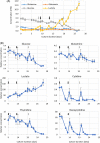
Concentration of metabolites measured using two medium analyzers. A, BioProfile FLEX Automated Cell Culture Analyzer, B, Cell Culture Media Analysis Platform. Days 0‐9:4‐L culture; days 9‐13:20‐L culture; days 13‐27:50‐L culture. Black arrows mark addition of fresh microcarrier suspension (microcarrier concentration = 5000 cm2/L). Gray arrows mark 50% medium exchange. Data points represent average metabolite concentrations measured from three medium samples
FIGURE 7
FIGURE 7
Harvest of cells from microcarriers and characterization of harvested cells. A, Microscopic images of microcarriers before and after cell harvest. B, Proliferation of harvested cells in tissue culture flask. C, Flow cytometry analysis of surface markers of harvested cells. Red solid lines represent cells treated with negative and positive antibody cocktails and dashed lines represent cells treated with negative and positive isotype controls. D, Staining of adipogenic, osteogenic, and chondrogenic differentiation of harvested cells. Scale bar = 100 μm. E, The number of T cells per well after culturing PBMCs with different ratio of hMSCs (PBMCs:hMSCs = 100 to 1, 20 to 1, 8 to 1, 4 to 1, and 2 to 1). PBMCs cultured without hMSCs were used as a control group. Experiments were run in triplicate; Mean ± SD. **P < .001, Tukey post hoc tests after one‐way ANOVA

References

    1. Kwon HM, Hur S‐M, Park K‐Y, et al. Multiple paracrine factors secreted by mesenchymal stem cells contribute to angiogenesis. Vascul Pharmacol. 2014;63:19‐28. - PubMed
    1. Osugi M, Katagiri W, Yoshimi R, Inukai T, Hibi H, Ueda M. Conditioned media from mesenchymal stem cells enhanced bone regeneration in rat calvarial bone defects. Tissue Eng Part A. 2012;18:1479‐1489. - PMC - PubMed
    1. Chen L, Tredget EE, Wu PYG, Wu Y. Paracrine factors of mesenchymal stem cells recruit macrophages and endothelial lineage cells and enhance wound healing. PLoS One. 2008;3:e1886. - PMC - PubMed
    1. Rajan TS, Giacoppo S, Diomede F, et al. The secretome of periodontal ligament stem cells from MS patients protects against EAE. Sci Rep. 2016;6:38743. - PMC - PubMed
    1. Lee J‐K, Park S‐R, Jung B‐K, et al. Exosomes derived from mesenchymal stem cells suppress angiogenesis by down‐regulating VEGF expression in breast cancer cells. PLoS One. 2013;8:e84256. - PMC - PubMed

Publication types